Effect of COVID‑19 a Public Health Emergency of International Concern (PHEIC) on a National programme.
- Prabhashree C. , Assistant Professor, Department of Pathology, Adichunchanagiri Institute of Medical Sciences, India.
- Bharati M Bhavikatti , Associate Professor, Department of Pathology, Karnataka Medical College and Research Institute, Hubballi, India.
- Purushotham Reddy , Professor, Department of Pathology, Karnataka Medical College and Research Institute, Hubballi, India.
Article Information:
Abstract:
Introduction: The declaration of COVID‑19 as a Public Health Emergency of International Concern (PHEIC) in early 2020 reshaped global health priorities, and its ripple effects were strongly felt in India’s National Leprosy Eradication Programme (NLEP). , During the pandemic years (2020–2022), lockdowns, restricted mobility, and diversion of frontline health workers toward COVID‑19 duties disrupted these detection mechanisms. Objectives: 1.To assess the impact of the COVID‑19 pandemic (2020–2022) on leprosy detection, reporting. 2. To analyze whether pandemic‑related disruptions influenced clinical presentation, epidemiological trends, and histopathological recognition of leprosy cases. Materials and Methods: The skin biopsies of cases who were clinically diagnosed or suspected of Hansen’s disease submitted to the Department of Pathology, over a period of 3 years (1 year retrospective and 2years prospective study) between July 2019 to June 2022, were studied with their clinical features. Results: The present investigation was designed as a retrospective–prospective study spanning a total duration of three years (one year retrospective and two years prospective), conducted between July 2019 and June 2022. Over the course of this period, 284 patients were clinically diagnosed with leprosy and included for analysis. From July 2019 to December 2019 which was considered pre covid era a total of 44 leprosy cases were observed out of these 49 case 2 (4.54%) cases were below the age of 15 years. Of these 44 cases 31(70.45%) cases were male and remaining 18 (40.90%) cases were females making male to female ratio as 1.7:1. Of 44 cases 34 (77.2%) cases presented with multiple lesions over body with hypopigmented patch as most common lesion in 24 (54.54%) cases. On histopathological examination borderline tuberculoid leprosy was the most common variant accounting to 34 (77.7%) cases as shown in table 1. However, type1 and type 2 reaction cases were 1 and 2 in number forming 2.2% and 4.5% of cases respectively on presentation. Conclusion: The present study highlights the adverse impact of the COVID‑19 pandemic on leprosy detection and control in India emphasising need for strengthening post‑COVID strategies by revitalizing active case detection, integrating leprosy services into primary healthcare, and prioritizing vulnerable groups such as children.
Keywords:
Article :
INTRODUCTION:
The declaration of COVID‑19 as a Public Health Emergency of International Concern (PHEIC) in early 2020 reshaped global health priorities, and its ripple effects were strongly felt in India’s National Leprosy Eradication Programme (NLEP). Traditionally, NLEP has relied on active door‑to‑door screenings, contact tracing, and community campaigns such as the Leprosy Case Detection Campaign (LCDC), Focused Leprosy Campaign (FLC), and Sparsh awareness drive to ensure early detection and reduce transmission. These strategies, supported by ASHA‑based surveillance (ABSULS) and integration with Ayushman Bharat primary health hubs, have been central to identifying new cases, especially among children and household contacts. However, during the pandemic years (2020–2022), lockdowns, restricted mobility, and diversion of frontline health workers toward COVID‑19 duties disrupted these detection mechanisms.
Routine contact tracing and prophylaxis programs were delayed, while fear of infection reduced community participation in house‑to‑house surveys.1,2 As a result, new leprosy case reporting declined, leading to diagnostic delays and increased risk of disability progression. Beyond programmatic disruption, the pandemic raises deeper epidemiological and clinical concerns. Did the reduction in active surveillance alter the true incidence and prevalence of leprosy in endemic regions? Were clinical presentations—already diverse and often misleading—further obscured by delayed consultations? How did the pandemic affect the detection of rare histopathological variants such as histoid leprosy, which require careful clinicopathological correlation? And critically, did interruptions in case detection contribute to shifts in the spectrum of leprosy types being reported post‑pandemic?
These questions underscore the need to re‑evaluate leprosy control strategies in the post‑COVID era, ensuring that neglected tropical disease programs regain momentum and adapt to the vulnerabilities exposed during the global emergency. This article aims at finding answers to these questions and helping in planning control strategies.
OBJECTIVES
1. To assess the impact of the COVID‑19 pandemic (2020–2022) on leprosy detection, reporting.
2. To analyze whether pandemic‑related disruptions influenced clinical presentation, epidemiological trends, and histopathological recognition of leprosy cases.
MATERIALS AND METHODS:
The present study is a 3 years Retro-prospective study (1 year retrospective and 2years prospective study between July 2019 to June 2022) taken in Department of Pathology, Karnataka Medical College and Research Institute, Hubballi (KMCRI), Hubli. The clinical and histopathological data of retrospective cases were retrieved from departmental records, tissue blocks and slides. During the prospective study period, clinical history, physical examination will be noted. Skin biopsies for study were obtained either by punch biopsy or incisional biopsy performed by Dermatologist and submitted to Pathology department.
The specimen so obtained is described under heading: size, type, borders, colour and shape. The specimen is then subjected for processing. Paraffin blocks are prepared followed by staining the 5-micron thickness sections with haematoxylin and eosin with standard techniques 6,7as observed in Department of Pathology, KMCRI, Hubli. Special stains (Fite-Faraco stain) are done. Histopathological diagnosis and Bacterial index are noted. Various histopathological findings like epidermal thinning, grenz zone, inflammatory cell are noted. In India, covid 19 was reported somewhere in mid 2019 so this time period can be taken as the "pre COVID-19" period, 2020 through 2022 as the acute "During COVID-19" phase marked by severe waves and lockdowns thus the data from July 2019 to December 2019 is taken as pre covid era and data during 2020 to June 2022 as covid 19 era.
INCLUSION CRITERIA: All clinically diagnosed or suspected and histologically proven cases of leprosy submitted to Department of Pathology, KMCRI, Hubballi.
EXCLUSION CRITERIA: • Inadequate samples. • Clinical data not available. • Biopsies which do not reveal histology of the leprosy.
RESULTS:
The present investigation was designed as a retrospective–prospective study spanning a total duration of three years (one year retrospective and two years prospective), conducted between July 2019 and June 2022. Over the course of this period, 284 patients were clinically diagnosed with leprosy and included for analysis. From July 2019 to December 2019 which was considered pre covid era a total of 44 leprosy cases were observed out of these 49 case 2 (4.54%) cases were below the age of 15 years. Of these 44 cases 31(70.45%) cases were male and remaining 18 (40.90%) cases were females making male to female ratio as 1.7:1. Of 44 cases 34 (77.2%) cases presented with multiple lesions over body with hypopigmented patch as most common lesion in 24 (54.54%) cases. On histopathological examination borderline tuberculoid leprosy was the most common variant accounting to 34 (77.7%) cases as shown in table 1. However, type1 and type 2 reaction cases were 1 and 2 in number forming 2.2% and 4.5% of cases respectively on presentation.
Table 1: Showing distribution of cases in different variants of leprosy during pre-covid 19 period.
|
Type |
Percentage of cases |
|
IL |
4.54 |
|
TT |
2.27 |
|
BT |
77.27 |
|
MB |
2.27 |
|
BL |
4.54 |
|
LL |
2.27 |
|
HL |
0 |
|
T1 |
2.27 |
|
T2 |
4.54 |
Between January 2020 and June 2022, that is during covid a total of 240 leprosy cases were documented. Among these, 49 cases involved individuals below 15 years of age, with 16 cases (6.66%) falling into this paediatric category. Of the overall cohort, 153 patients (63.75%) were male, while 87 patients (36.2%) were female, yielding a male‑to‑female ratio of 1.7:1. Of 240 cases 191(79.1%) cases presented with multiple lesions over body with hypopigmented patch as most common lesion in 99 (41.25%) cases. On histopathological examination borderline tuberculoid leprosy was the most common variant accounting to 98 (48.8%) cases as shown in table 2. However, type1 and type 2 reaction cases were 8 and 13 in number forming 3.3% and 5.4% of cases respectively on presentation.
Table 2: Showing distribution of cases in different variants of leprosy during covid 19 period.
|
Type |
Percentage |
|
IL |
17.91 |
|
TT |
9.58 |
|
BT |
40.83 |
|
MB |
2.91 |
|
BL |
8.75 |
|
LL |
7.5 |
|
HL |
3.75 |
|
T1 |
3.33 |
|
T2 |
5.41 |
Overall, the comparison highlights that during the COVID‑19 pandemic, leprosy detection patterns shifted toward a higher proportion of pediatric cases as shown in graph 1, more patients presenting with multiple lesions, and a relative decline in borderline tuberculoid forms. These findings suggest that pandemic‑related disruptions in surveillance and clinicopathological correlation may have contributed to delayed detection, under‑reporting of early paucibacillary cases, and a greater burden of advanced disease at presentation.
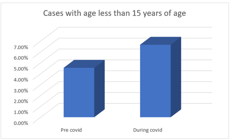
Graph 1: Showing distribution of leprosy cases among children less than 15 years of age during pre-covid 19 and covid 19 period.
Reactional cases deserve particular attention. While the absolute number of Type 1 and Type 2 reactions increased during the pandemic period, their relative proportions showed variation compared to the pre‑COVID era as shown in graph 1. This may reflect delayed diagnosis, immune stressors associated with infection or vaccination, and interruptions in treatment continuity. The presence of reactions at presentation underscores the likelihood that patients were detected at a more advanced stage of disease, reinforcing the concern that COVID‑19 disrupted early detection and timely management of leprosy.
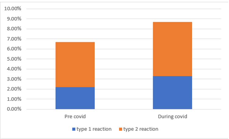
Graph 2: Showing relative distribution of lepra rection cases during pre-covid 19 and covid 19 period.
DISCUSSION:
The COVID‑19 pandemic posed significant challenges to the implementation of the National Leprosy Eradication Programme (NLEP) in India. National surveillance data revealed that following the imposition of lockdown measures, there was a marked decline in case detection. Specifically, during the first quarter of 2020 (April–June), the number of reported leprosy cases fell by 29% compared to the same period in 2019, underscoring the extent to which pandemic‑related restrictions disrupted routine screening, contact tracing, and early diagnosis efforts.
In the present study, male predominance was observed with 185 (65.14%) cases as males and remaining 99 (34.85%) cases as female. Male to female ratio was 1.9:1. These findings were similar to observations done by Soni et al.62 (2019) and Tiwari M et al.63 (2019) as shown in table 3 and table 4.
Table 3: Comparison of male to female ratio of leprosy cases in different studies.
|
Male to female ratio |
Studies |
||
|
Soni et al.8 (2019) n=41 |
Tiwari M et al.9(2019) n=62 |
Present study n=284 |
|
|
1.6:1 |
1.4:1 |
1.9:1 |
|
Table 4: Comparison of gender distribution of leprosy cases in different studies.
|
Gender |
Studies |
|||
|
Soni et al.8 (2019) n=41 |
Present study n=284 |
|||
|
Number of cases |
% |
Number of cases |
% |
|
|
Male |
25 |
60.9 |
185 |
65.1 |
|
Female |
16 |
39.02 |
99 |
34.9 |
More number of male cases could be attributed to their greater mobility and increased chances of exposure to disease. Another reason which could be attributed for male preponderance is that males are more willing in reporting to health care facilities and for seeking treatment.
Detection of leprosy in children carries serious implications, as it not only reflects ongoing transmission within households but also raises the risk of spread among peers in school settings. Importantly, the occurrence of childhood leprosy in the post‑elimination era signals persistent community transmission (Nair et al., 2017). Recent data highlight this concern: in 2020, the global childhood leprosy rate was reported at 6.2% (Global Leprosy Update, 2020), while national figures for India stood at 5.77% (NLEP Annual Report, 2020–21). Various regional studies have documented rates ranging between 5.1% and 11.43%, underscoring the variability and continued burden of pediatric leprosy across different populations. In our study among 49 cases 16 cases (6.66%) involved individuals below 15 years of age, which was falling within this range.
Colour and texture of skin lesions In the present study most common skin colour and texture observed was hypopigmentation accounting for 152 (53.5%) cases, similar to findings in study done by Soni et al.62 (2019) as shown in table 5.
Table 5: Comparison of different type of skin lesion of leprosy in different studies.
|
Type of lesion |
Studies |
|||
|
Soni et al.8(2019) n=41 |
Present study n=284 |
|||
|
Number of cases |
% |
Number of cases |
% |
|
|
Patch |
2 |
4.9 |
147 |
51.7 |
|
Plaque |
6 |
14.6 |
76 |
26.7 |
|
Nodule |
12 |
29.3 |
45 |
15.8 |
|
Diffuse infiltration |
- |
- |
10 |
3.5 |
|
Macule |
17 |
41.5 |
6 |
2.1 |
In our study, lepra reactions (LRs) were documented in 8.7% of cases, a proportion lower than that reported by Gupta et al. (2019) at 34.9%, Chhabra et al. (2015) at 37.4%, and Relhan et al. (2016) at 23.4%. Elevated rates of LRs generally reflect a greater burden of multibacillary (MB) disease, attributable to higher bacterial loads, however the rate so obtained in our study is much higher compared to previous fingings in our study during pre covid 19 period. Furthermore, during the COVID‑19 pandemic (C19P), patients with more severe manifestations of LRs were more likely to seek care at tertiary centers, whereas asymptomatic or early leprosy cases often remained undetected.
Chhabra et al.10 (2015) reported a predominance of Type 1 reactions (T1R), while Jacob et al.11
(2008) observed an equal distribution of T1R and Type 2 reactions (T2R). In contrast, our findings revealed a higher frequency of T2R, corroborating the results of Gupta et al.12 (2019). The predominance of T2R in our cohort can be attributed to the increased proportion of lepromatous leprosy (LL) and borderline lepromatous (BL) cases, consistent with the epidemiological trends noted by Gupta et al.
These findings suggest a potential association between COVID‑19 infection or vaccination and the increased incidence of lepra reactions (LRs) in the post‑pandemic period. This observation is consistent with other studies, who also reported a rise in deformities and reactional states following COVID‑19. During the COVID‑19 pandemic, global leprosy reporting was significantly affected, with only 127 countries submitting data in 2020, compared to 160 in 2019, highlighting the disruption in surveillance and control efforts.
This study highlights the negative impact of the COVID‑19 pandemic on leprosy control efforts. The disruption stemmed from public health measures implemented to curb coronavirus transmission, including social distancing, restrictions on patient flow in outpatient clinics, and reduced accessibility to health services. While these recommendations were essential to limit the spread of COVID‑19, they inadvertently hindered routine activities of the National Leprosy Programme, thereby compromising early detection and continuity of care. The findings emphasize that, alongside infection‑control strategies, practical measures must be adopted to sustain leprosy control activities and safeguard patients, ensuring that progress toward elimination is not reversed.
Limitations: Limitations of our study are small sample size, retrospective design and lack of data on COVID‑19 coinfection and vaccination status.
CONCLUSION:
The present study highlights the adverse impact of the COVID‑19 pandemic on leprosy detection and control in India. A clear reduction in early case identification was observed. The predominance of Type 2 reactions rates reflect that patients often presented at more advanced stages, a consequence of restricted surveillance, reduced outpatient services, and barriers to healthcare access during the pandemic. India must strengthen post‑COVID strategies by revitalizing active case detection, integrating leprosy services into primary healthcare, and prioritizing vulnerable groups such as children. Addressing coinfections, immune dysregulation, and treatment continuity will be essential to prevent disability progression and sustain progress toward elimination. The pandemic has revealed critical vulnerabilities in the NLEP, and addressing these gaps is vital to ensure that India does not lose momentum in its long‑standing fight against leprosy.
REFERENCES:
1. WHO. Novel coronavirus—China. Jan 12, 2020. http://www.who. int/csr/don/12-january-2020-novel-coronavirus-china/en/ 2
2. World Health Organization. Coronavirus Disease 2019 (COVID 19): Situation Report- 51. Geneva: World Health Organization, 2020. https://www.who.int/docs/default-source/coronaviruse/situa tion-reports/20200311-sitrep-51-covid-19.pdf?sfvrsn=1ba62e57_ 10
3. Arif T, Amin SS, Adil M et al (2018). Leprosy in the post-elimination era: a clinico-epidemiological study from a northern Indian tertiary care hospital. Act Dermato Venerol. 28 : 7-10.
4. Brandsma JW, Brakel WHV (2003). WHO disability grading - operational definitions. Lepr Rev. 74: 366-373.
5. John AS, Rao PS, Kundu R, Raju MS. Leprosy among adolescents in Kolkata, India. Indian J Lepr. 2005;77(3):247–53.
6. McAdam AJ, Milner DA, Sharpe AH. Infectious Diseases. In: Kumar V, Abbas AK, Aster JC. Robbins and Cotran Pathologic Basis of Disease. South Asian Ed. New Delhi: Elsevier; 2014; 1:377-378.
7. Bancroft JD, Layton C, The hematoxylins and eosin, Editor(s): Suvarna SK, Layton C, Bancroft JD. Bancroft's Theory and Practice of Histological Techniques. Eighth Edition. Elsevier; 2019: 126-138.Soni S, Shah N, Bhalodia J. Clinicopathological correlation in leprosy. Int J Med Sci Public Health 2019;8(6):459-464.
8. Tiwari M, Ranabhat S, Maharjan S. Clinico-histopathological correlation of leprosy: A retrospective study of skin biopsy specimens in Chitwan medical college. Int J Med Sci Res Pract2015;2:8-11.
9. Dayal R, Hashmi NA, Mathur PP, Prasad R. Leprosy in childhood. Indian Pediatr 1990;27:170-80.
10. Chhabra N, Grover C, Singal A, Bhattacharya SN, Kaur R. Leprosy scenario at a tertiary level hospital in Delhi: A 5-year retrospective study. Indian J Dermatol 2015;60:55-9.
11. Jacob JT, Kozarsky P, Dismukes R et al (2008). Short report: five-year experiences with type 1 and type 2 reactions in Hansen disease at us travel clinic. Am J Trop Med Hyg. 79: 452–454.
12. Gupta R, Sinha R, Pradhan S et al (2019). Clinico epidemiological profile of leprosy in post elimination era: a hospital based study. Indian J Lepr. 91: 197-205.
13. RaoAG. Studyofleprosy in children. Indian J Lepr. 2009;81(4):195 7.
14. Hariharasudhan S, Durgavathi C, Priyavathani Annie Malathy R, Daniel SJ, Sampath V. A clinicoepidemiological study of leprosy among children and adolescents in a tertiary care centre in the post elimination era. IP Indian J Clin Exp Dermatol 2024;10(1):1-10.
15. National Leprosy Eradication Programme Progress Report for 2014-15. New Delhi; 2015.
16. Kumar B, Rani R, Kaur I. Childhood leprosy in Chandigarh; clinico-histopathological correlation. Int J Lepr Other Mycobact Dis 2000;68:330-1.
17. Prabhakaran K. Metabolism of M. leprae separated from human nodules. Int J Lepr. 1967;35:34-41.
18. Verma G, Kumari S, Negi A, Rattan R, Gupta M, Chauhan U, et al. Clinico-Epidemiological Trends of Leprosy in 21st Century and During COVID-19 Pandemic. Indian J Leprosy 2022;94:299-308.
19. Matos TS, Do Nascimento VA, Do Carmo RF, Moreno de Oliveira Fernandes TR, De Souza CD, Da Silva TF. Impact of the COVID-19 Pandemic on the Diagnosis of New Leprosy Cases in Northeastern Brazil, 2020. Int J Dermatol 2021;60:1003-6.Stoner GL. Importance of the neural predilection of Mycobacterium leprae in leprosy. Lancet. 1979;10:994-6.
20. Rambukkana A, Salzer JL, Yurchenco PD, et al. Neural targeting of Mycobacterium leprae mediated by the G domain of the laminin α2 chain. Cell. 1977;88:811-21.
21. Cole ST, Eiglmeier K, Parkhill J, et al. Massive gene decay in the leprosy bacillus. Nature. 2001;409(6823):1007-11.