Evaluating Ridley–Jopling Classification: Practical Challenges Under the Microscope in a Tertiary Centre.
- Prabhashree C. , Assistant Professor, Department of Pathology, Adichunchanagiri Institute of Medical Sciences, India.
- Bharati M Bhavikatti , Associate Professor, Department of Pathology, Karnataka Medical College and Research Institute, Hubballi, India.
- Purushotham Reddy , Professor, Department of Pathology, Karnataka Medical College and Research Institute, Hubballi, India.
Article Information:
Abstract:
Introduction: Leprosy is among the most ancient diseases known to humankind. It is a chronic, debilitating granulomatous condition caused by Mycobacterium leprae, which primarily affects the cooler regions of the body, notably the skin and peripheral nerves, while also involving the muscles, eyes, bones, testes, and internal organs.1 However various histopathological findings are overlapping among these different classes and also with other disease. In this study we will discuss about various microscopic finding and their impact on classifying different types of leprosy. Objectives: 1. To study the histopathological spectrum of skin lesions in Hansen’s disease and classify according to Ridley-Jopling classification. 2.To study bacterial index in different types of leprosy. Methods: The present study is a 3 years Retro-prospective study (1 year retrospective and 2years prospective study between July 2019 to June 2022) taken in Department of Pathology, Karnataka Medical College and Research Institute, Hubballi (KMCRI), Hubli. Skin biopsies for study were obtained either by punch biopsy or incisional biopsy performed by Dermatologist and submitted to Pathology department. Results: During this period, 284 patients were clinically diagnosed with leprosy and included for analysis. In present study epidermis was unremarkable in majority, accounting to 128 (39.2%) cases. The grenz zone was preserved in one end of the spectrum of leprosy cases. It was preserved in most cases of lepromatous leprosy that is 10 (52.6%) cases, followed by HL, T1, BL, BB, T2, IL, TT and BT in order. Bacterial index was observed to increase from tuberculoid pole to lepromatous pole. Majority of cases belonging to TT, BT, BB, BL, IL, T1 and T2 had BI as 0. BI of majority of cases of TT was between 0-1+ but for majority of LL cases it was 3+ and above. Cases lying in borderline groups that is BT, BB, BL had their BI between 0 to 4+. BI was highest for HL cases where it was 5+ and 6+. Conclusion: present study concludes that the histopathological findings are not hard and fast rule for diagnosis thus emphasizing on holistic approach for diagnosis of different categories of leprosy.
Keywords:
Article :
INTRODUCTION:
Leprosy is among the most ancient diseases known to humankind. It is a chronic, debilitating granulomatous condition caused by Mycobacterium leprae, which primarily affects the cooler regions of the body, notably the skin and peripheral nerves, while also involving the muscles, eyes, bones, testes, and internal organs.1 Since antiquity, leprosy has been referred to as “Kushtaroga.” The causative organism, M. leprae, was first identified in 1873 by Armauer Hansen.2,3 Despite its early discovery, the bacterium has not yet been successfully cultured in vitro. Adequate clinical information combined with histopathology and bacteriological index help in diagnosis and classification of different types of leprosy there by in management of cases.4 Ridley-Jopling classification which was initially considered as classification for research purpose in now being used widely even in clinical practice.
Along with five types of leprosy classified under Ridley-Jopling classification differentiation of Indeterminate leprosy, histoid leprosy and reactions associated with leprosy and its treatment are also important.5 In majority of situations Ridely- Jopling classification is used to classify leprosy, it is referred to as immunological classification. 6 Ridely- Jopling classification is based on the fact that bacteriological, immunological, histopathological and clinical features are interwoven. 6 Advantages of Ridely- Jopling classification: 6 Easier to comprehend and helps in better understanding of disease. Strengthens the polar and spectral concept of the disease. Under this classification leprosy is classified as Tuberculoid Leprosy (TT), Borderline Tuberculoid Leprosy (BT), Mid borderline (BB), Borderline Lepromatous (BL), Lepromatous Leprosy (LL). However various histopathological findings are overlapping among these different classes and also with other disease. In this study we will discuss about various microscopic finding and their impact on classifying different types of leprosy.
OBJECTIVES
1. To study the histopathological spectrum of skin lesions in Hansen’s disease and classify according to Ridley-Jopling classification.
2. To study bacterial index in different types of leprosy.
MATERIALS AND METHODS:
The present study is a 3 years Retro-prospective study (1 year retrospective and 2years prospective study between July 2019 to June 2022) taken in Department of Pathology, Karnataka Medical College and Research Institute, Hubballi (KMCRI), Hubli. Skin biopsies for study were obtained either by punch biopsy or incisional biopsy performed by Dermatologist and submitted to Pathology department.
The specimen so obtained is described under heading: size, type, borders, colour and shape. The specimen is then subjected for processing. Paraffin blocks are prepared followed by staining the 5-micron thickness sections with haematoxylin and eosin with standard techniques 7,8 as observed in Department of Pathology, KMCRI, Hubli. Special stains (Fite-Faraco stain) are done. Histopathological diagnosis and Bacterial index are noted. Various histopathological findings like epidermal thinning, grenz zone, inflammatory cell are noted.
INCLUSION CRITERIA: All clinically diagnosed or suspected and histologically proven cases of leprosy submitted to Department of Pathology, KMCRI, Hubballi.
EXCLUSION CRITERIA: • Inadequate samples. • Clinical data not available. • Biopsies which do not reveal histology of the leprosy.
RESULTS:
The present study was conducted as a retrospective–prospective investigation spanning a total of three years (one year retrospective and two years prospective), between July 2019 and June 2022. During this period, 284 patients were clinically diagnosed with leprosy and included for analysis.
In present study epidermis was unremarkable in majority, accounting to 128 (39.2%) cases, followed by thinned out epidermis in 104 (31.9%) cases, acanthosis in 41 (12.5%) cases, hyper keratosis in 33 (10.1%) and other features like focal papillomatosis, blunting of retention ridges and parakeratosis was seen in 19 (6.6%) cases. Unremarkable epidermis was most common feature in TT, IL, LL, T1 and T2. Thinned out epidermis was more common in BB, BL and HL. BT had equal number of unremarkable and thinned out epidermisas shown in table 1 and graph1.
Table 1: Showing relationship between epidermal changes with type of leprosy in the present study
|
Epidermal Feature |
TT |
BT |
BB |
BL |
LL |
IL |
T1 |
T2 |
HL |
Total n (%) |
|
|
Unremarkable epidermis |
20 |
48 |
3 |
8 |
9 |
21 |
6 |
11 |
2 |
128, (39.2) |
|
|
Epiderm is with changes |
Thinned out |
0 |
48 |
5 |
13 |
8 |
17 |
2 |
5 |
5 |
103, (31.9) |
|
Acanthosis |
1 |
27 |
1 |
2 |
1 |
2 |
0 |
1 |
1 |
35, (12.3) |
|
|
Hyperkeratosis |
3 |
7 |
2 |
1 |
1 |
2 |
1 |
0 |
1 |
18, (6.3) |
|
|
Total |
24 |
148 |
13 |
27 |
20 |
54 |
11 |
18 |
11 |
|
|
*Following percentages are calculated for total number of cases in particular type of leprosy as many cases presented with more than 1 type of epidermal features.
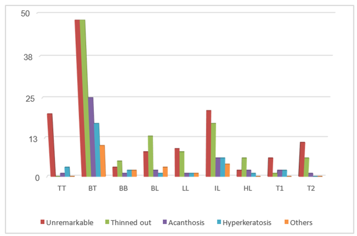
Graph1: Showing relationship between epidermal changes with type of leprosy in the present study.
The grenz zone was preserved in one end of the spectrum of leprosy cases. It was preserved in most cases of lepromatous leprosy that is 10 (52.6%) cases, followed by HL, T1, BL, BB, T2, IL, TT and BT in order as shown in table 2 and graph 2. While the grenz zone was absent/destroyed in BT accounting to 128 (98.4%) cases, followed by TT, IL, T2, BB, BL, T1, HL and LL in order. This zone was infiltrated by inflammatory cell infiltrate in these cases. In present study it was observed that with increase in extent of involvement of both papillary and reticular dermis dermis by inflammatory cells there was decrease in grenz zone and vice versa, as shown in graph2.
Table 2: Showing presence grenz zone and extent of dermal involvement by inflammatory cells.
|
|
TT n (%) |
BT n (%) |
BB n (%) |
BL n (%) |
LL n (%) |
IL n (%) |
HL n (%) |
T2 n (%) |
T1 n (%) |
Total n (%) |
|
GZ |
1 (4.2) |
2 (1.5) |
2 (18.2) |
5 (21.7) |
10 (52.6) |
4 (9.5) |
4 (44.4) |
2 (11.7) |
2 (22.2) |
32 (11.2) |
|
LG |
23 (95.8) |
128 (98.4) |
9 (81.8) |
18 (78.2) |
9 (47.3) |
38 (90.4) |
5 (55.5) |
15 (88.2) |
7 (77.7) |
252 (88.7) |
|
PD |
2 (8.3) |
20 (15.3) |
1 (9.0) |
2 (8.6) |
1 (5.2) |
8 (19.0) |
1 (11.1) |
2 (11.7) |
1 (11.1) |
38 (13.3) |
|
RD |
0 |
0 |
0 |
0 |
0 |
0 |
0 |
0 |
0 |
0 |
|
PD+ RD |
22 (91.6) |
110 (84.6) |
10 (90.9) |
21 (91.3) |
18 (94.7) |
34 (80.9) |
8 (88.8) |
15 (88.2) |
8 (88.8) |
246 (86.6) |
GZ- Grenz zone; LG- Loss of subepidermal clear zone; PD- Papillary dermis; RD-Reticular dermis; PD+RD- both papillary dermis and reticular dermis.
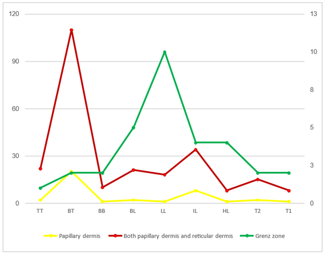
Graph 2: Showing presence grenz zone and extent of dermal involvement by inflammatory cells (the second y axis is drawn for grenz zone).
Various dermal changes in individual subtypes of leprosy.
The dermal changes were analysed in detail in present study. The type of inflammatory infiltrate, presence or absence of well-formed granulomas and involvement of the adnexal structures and dermal nerves were studied. The analysis in each type of leprosy is detailed below.
1. Tuberculoid leprosy: Among 24 cases of tuberculoid leprosy, 18 (75%) cases showed dermal abundant lymphocytic infiltrate followed by moderate lymphocytic infiltrate in 6 (25%) cases with well-formed granulomas in all 24 (100%) cases. These inflammatory cells and granulomas were seen occupying both papillary and reticular dermis in 22 (91.6%) cases followed involvement of only papillary dermis in 2 (8.3%) cases. Scant number of Langhans type of giant cell was seen in 1 (4.1%) case. Necrotic areas in dermis were seen in 22 (91.6%) cases. Foamy histiocytes and plasma cells were seen in 5 (20.8%) cases and 2 (8.3%) cases respectively. Cases of leprosy had lymphocytic and granulomatous infiltration in periadnexal, perivascular and perineural regions these findings were seen overlapping in many cases that is more than one region was involved in many cases.
2. Borderline tuberculoid leprosy: Out of 130 cases of BT, all 130 (100%) cases showed epithelioid cell which were loosely distributed in dermis followed by 25 (19.2%) cases forming granulomas. Majority of cases showed moderate lymphocytic infiltration accounting to 127 (97.6%) cases, followed by scant lymphocytic infiltration in 2 (1.5%) cases and abundant lymphocytic infiltration in 1 (0.7%) case. Cases of leprosy had lymphocytic and granulomatous infiltration in periadnexal, perivascular and perineural regions these findings were seen overlapping in many cases that is more than one region was involved in many cases. Lymphocytic infiltration with admixture of foamy histiocytes and macrophages was seen in 83 (63.8%) cases and 57 (43.8%) cases respectively. These inflammatory cells involved both papillary and reticular dermis in 110 (84.6%) cases, followed by involvement of only papillary dermis alone in remaining 20 (15.3%) cases.
3. Mid Borderline leprosy: In the present study, out of 11 cases of BB, 8 (72.7%) cases and 4 (36.3%) cases showed foamy histiocytes and macrophages respectively as predominant cells with admixture of epithelioid cells in all 11 (100%) cases. Scant lymphocytic infiltrate was seen in 10 (90%) cases, followed by moderate amount of lymphocytic infiltration in 1 (10%) case. Cases of leprosy had lymphocytic and granulomatous infiltration in periadnexal, perivascular and perineural regions these findings were seen overlapping in many cases that is more than one region was involved in many cases. Lymphocytic infiltration was predominantly seen in peri adnexal region, accounting to 10 (90%) cases, followed by perineural region in 6 (54.5%) cases and perivascular region in 4 (52.3%) cases. Involvement of both papillary and reticular dermis by inflammation was seen in 10 (90.9%) cases, followed by involvement of papillary dermis alone in remaining 1 (9.09%) case with dermal necrotic areas and plasma cells in 2 (18.18%) cases each. Borderline
4. Lepromatous Leprosy: In the present study out of 23 cases of BL, most common finding was presence of foamy histiocytes, accounting to 19 (82.6%) cases and macrophages in 7 (30.4%) cases, with scant lymphocytic infiltrate in 22 (95.6%) cases. Moderate amount of lymphocytic infiltrate and eosinophils was seen in 1 (4.3%) case each. These inflammatory cells were seen predominantly in peri adnexal area in 21 (91.3%) cases, followed by perineural in 9 (39.1%) and perivascular area in 7 (30.4%) cases. Cases of leprosy had lymphocytic and granulomatous infiltration in periadnexal, perivascular and perineural regions these findings were seen overlapping in many cases that is more than one region was involved in many cases. Involvement of both papillary and reticular dermis by inflammation was seen in 21 (91.3%) cases, followed by involvement of papillary dermis alone in remaining 2 (8.6%) cases.
5. Lepromatous Leprosy: In the present study out of 19 cases of LL, majority of cases that is 10 (52.63%) cases had preserved grenz zone and in remaining 9 (47.3%) cases subepidermal zone was infiltrated by inflammatory infiltrates. Seventeen cases (89.4%) showed dense infiltration by macrophages, making it majority. Foamy histiocytes and plasma cells were seen in 4 (21%) cases and 2 (10.5%) cases respectively. All 19 (100%) cases showed scant lymphocytic infiltration in dermis. Cases of leprosy had lymphocytic and granulomatous infiltration in periadnexal, perivascular and perineural regions these findings were seen overlapping in many cases that is more than one region was involved in many cases. These cells were seen predominantly around adnexa in 18 (94.7%) cases followed by perivascular in 10 (52.6%) cases and perineural in 6 (31.5%) cases.
6. Indeterminate Leprosy: In present study, all 42 (100%) cases presented with scant lymphocytic infiltrate.Cases of leprosy had lymphocytic and granulomatous infiltration in periadnexal, perivascular and perineural regions these findings were seen overlapping in many cases that is more than one region was involved in many cases. These inflammatory cells were predominantly seen in peri adnexal area in 27 (64.2%) cases followed by perivascular in 23 (54.7%) and perineural area in 19 (45.2%) cases. Plasma cells were seen in 6 (14.2%) cases in few cases. Grenz zone was preserved in 4 (9.52%) cases. Involvement of both papillary and reticular dermis by inflammation was seen in 34 (80.95%) cases, followed by involvement of papillary dermis alone in remaining 8 (19%) cases. Thickened nerve fibers were seen in 2 (4.7%) cases.
7. Histoid Leprosy: In present study out of 9 cases, 4 (44.4%) cases had preservedgrenz zone, remaining 5 (55.5%) cases showed narrowing of it by inflammatory cells. All 9 (100%) cases had scant lymphocytic infiltration, distributed predominantly in peri adnexal area, accounting to 6 (66.6%) cases followed by perineural area in 4 (44.4%) cases. Foamy histiocytes and spindle shaped histiocytes were present in all 9 (100%) cases. pseudo capsule formation was seen in 7 (77.7%) cases. Plasma cell and macrophages were present in 1 (11.1%) case and 2 (22.2%) cases respectively. Cases of leprosy had lymphocytic and granulomatous infiltration in periadnexal, perivascular and perineural regions these findings were seen overlapping in many cases that is more than one region was involved in many cases. Involvement of both papillary and reticular dermis by inflammation was seen in 8 (88.8%) cases, followed by involvement of papillary dermis alone in remaining 1 (11.1%) case. Dermal nerve erosion was seen in 4 (44.4%) cases.
8. Type 1 Lepra Reaction: In present study, out of 9 cases of T1 lepra reaction moderate lymphocytic infiltration was most commonly seen in 7 (77.7%) cases followed by scant and abundant amount of lymphocytic infiltration in 1 (11.1) case each. Scattered epithelioid cells were seen in all 7 (77.7%) cases and well-formed granuloma of epithelioid cells in 2 (22.2%) cases. Also seen were foamy histiocytes in 7 (77.7%) cases, macrophages in 2 (22.2%) cases, plasma cells in 2 (22.2%) cases, giant cells in 1 (11.1%) cases and neutrophils in 5 (55.5%) cases in varying frequencies. These inflammatory cells were predominantly seen in peri adnexal area in 9 (100%) cases followed by perivascular in 5 (55.5%) cases and perineural area in 4 (44.4%) cases. Involvement of both papillary and reticular dermis by inflammation was seen in 8 (88.8%) cases, followed by involvement of papillary dermis alone in remaining 1 (11.1%) case. Necrosis was present in 6 (66.6%) cases and subcutaneous inflammatory cell infiltrate in 1 (11.1%) case. Grenz zone was preserved in 2 (22.2%) cases and in remaining 7 (77.7%) cases it was infiltrated by inflammatory cells. Eroded dermal nerves were seen in 1 (11.1%) case. Edema in between granuloma was present in all 9 (100%) cases. Fibroblasts and infiltration of subcutaneous tissue by inflammatory cells was seen in 1 (11.1%) case.
9. Type 2 Lepra Reaction: In present study, out of 17 cases of T2 lepra reaction majority of cases presented with scant lymphocytic infiltration,accounting to 15 (88.2%) cases followed by moderate amount of lymphocytic infiltration in 2 (11.7%) cases. Scattered epithelioid cells in 7 (41.1%) cases, foamy histiocytes in 14 (82.3%) cases,macrophages in 7 (41.1%) cases, plasma cells in 7 (41.1%) cases, eosinophils in 1 (5.8%) case, fibroblast in 1 (5.8%) case and neutrophils in 13 (76.4%) cases were also seen in varying frequencies among T2 cases. These inflammatory cells were predominantly seen in peri adnexal area accounting to 15 (88.2%) cases followed by perivascular area in 14 (82.3%) cases and perineural area in 8 (47%) cases. Involvement of both papillary and reticular dermis by inflammation was seen in 15 (88.2%) cases, followed by involvement of papillary dermis alone in remaining 2 (11.7%) cases. Necrosis was present in 2 (11.7%) cases and subcutaneous inflammatory cell infiltrate in 4 (23.5%) cases. Vasculitis was present in all 17 (100%) cases with edema in papillary dermis in 7 (41.1%) cases.
Bacterial index was observed to increase from tuberculoid pole to lepromatous pole. Majority of cases belonging to TT, BT, BB, BL, IL, T1 and T2 had BI as 0. BI of majority of cases of TT was between 0-1+ but for majority of LL cases it was 3+ and above. Cases lying in borderline groups that is BT, BB, BL had their BI between 0 to 4+. BI was highest for HL cases where it was 5+ and 6+.
DISCUSSION:
In the present study epidermis was unremarkable in majority of cases, accounting to 128 (39.2%) cases. This was similar to observation by Yadav N et al.9 (2019), Banushree CS et al.10 (2016) and Pathak D et at (2013).11 However thinned out epidermis was most common pathology found in all the studies. Acanthosis and hyperkeratosis were other epidermal changes noted in the present study similar to study by Pathak D et at (2013).11 as shown in table 3.
Table 3: Comparison of epidermal changes in leprosy in different studies.
|
|
Studies |
||||||||
|
Banushree CS et al.10 (2016) n= 107 |
Yadav N et al.9(2019) n= 62 |
Pathak D et at (2013).11 n= 71 |
Present study n=284 |
|
|||||
|
No. of cases |
% |
No. of cases |
% |
No. of cases |
% |
No. of cases |
% |
|
|
|
Unremarkable |
49 |
45.8 |
58 |
93.5 |
50 |
70.4 |
128 |
39.2 |
|
|
Thinned out |
32 |
29.9 |
2 |
3.2 |
8 |
11.3 |
104 |
31.9 |
|
|
Acanthosis |
- |
- |
- |
- |
5 |
7 |
41 |
12.5 |
|
|
Hyperkeratosis |
- |
- |
- |
- |
7 |
9.8 |
33 |
10.1 |
|
|
Others |
26 |
24.3 |
2 |
1.6 |
1 |
1.4 |
20 |
7.1 |
|
Dermal changes
Tuberculoid leprosy: Among 24 cases of tuberculoid leprosy all 24 (100%) cases showed epithelioid cell granuloma, which was similar to study done by Yadav N et al.9 (2019) and Banushree CS et al.10 (2016) who had epithelioid cell granuloma in 21 (100% of total TT cases) and 8 (100% of total TT cases) cases respectively. In the present study majority of cases showed dense lymphocytic infiltrate accounting to 18 (75%) cases, in peri adnexal area among 23 (95.8%) cases. Giant cells and grenz zone were seen in 1 (4.1%) case each. These findings were almost similar to studies done by Yadav N et al.9 (2019) and Banushree CS et al.10 (2016). The study done by Yadav N et al.9 (2019) predominantly had moderate lymphocytic infiltrate accounting to 3 (60%) cases, in peri adnexal area among 3 (60%) cases. Giant cells were seen in 4 (80%) cases. Both the studies done by Yadav N et al.9 (2019) and Banushree CS et al.10 (2016) showed no Grenz zone.
Borderline tuberculoid leprosy: In the present study 127 (97.6%) cases out of 130 cases showed moderate lymphocytic infiltrate, located mainly around the skin adnexal structures accounting to 124 (95.3%) cases. Epithelioid cell granulomas were seen in 25 (19.2%) cases, along with scattered epithelioid cells in 130 (100%) cases, foamy histiocytes in 83 (63.8%) cases, macrophages in 57 (43.8%) cases and 2 (1.5%) cases showed presence of giant cells and grenz zone. These findings were almost similar to studies done by Yadav N et al.9 (2019) and Banushree CS et al.10 (2016).
Mid Borderline leprosy: In present study, among 11 cases of BB majority of cases presented with foamy histiocytes accounting to 8 (72.7%) cases and scant lymphocytic infiltrate in 10 (90.9%) cases, in peri adnexal area accounting to10 (90.9%) cases involving both upper and lower dermis in 10 (90.9%) cases. Macrophages were seen in 4 (36.3%) cases and 11 (100%) cases showed scattered epithelioid cells. Grenz zone was seen in 2 (18.1%) cases with none showing giant cell formation. Valand et al.12 (2017) study had 8 cases of BB in which presence of foamy macrophages, epithelioid cells and scant lymphocytic infiltrate were the predominant features which was similar to the present study.
Borderline Lepromatous Leprosy: BL leprosy most commonly presented with scant lymphocytic infiltrate accounting for 22 (95.6%) cases, in peri adnexal area in 21 (91.3%) cases, involving both upper and lower dermis in 21 (91.3%) cases. Along with Foamy histiocytes in 19 (82.6%) cases and macrophages in 7 (30.4%) cases, scattered epithelioid cells in 20 (8.9%) cases were also seen on histopathology. Grenz zone was seen in 5 (21.7%) cases. Yadav N et al.9 (2019) in his study observed11 BL cases, most commonly presenting with moderate lymphocytic infiltrate accounting for 5 (45.4%) cases, in peri adnexal area in 4 (36.3%) cases, involving both upper and lower dermis in 6 (54.5%) cases. Along with Foamy histiocytes andmacrophagesin all11 (100%) cases. Epithelioid cells were also seen in 20 (8.9%) cases. Grenz zone was seen in 8 (72.7%) cases. Banushree CS et al.10 (2016) had 10 cases of BLin their study with periadnexal and perineural lymphocytic infiltration and macrophages in all 10 (100%) cases. Grenz zone was present in 8 (80%) cases
Lepromatous Leprosy: In the present study out of 19 cases of LL majority of cases that is 10 (52.6%) cases had grenz zone on histopathology. Infiltration by macrophages and foamy histiocytes were seen in all 19 (100%) cases. Scant lymphocytic infiltration of dermis was seen in 14 (73.6%) cases. This lymphocytic infiltration waspredominantlyseen around adnexa in 18 (94.7%) cases. These findings were almost similar to the study done by Yadav N et al.9 (2019) and Banushree CS et al.10 (2016). Banushree CS et al.10 (2016) had 16 cases of LLin their study with all 16 (100%) cases showing grenz zone and infiltration by macrophages similar to the present study. Perineural lymphocytic infiltration was the most common feature accounting for 9 (56.3%) cases which was contradicting with present study with peri adnexal inflammatory infiltration as more common feature.
Indeterminate Leprosy: In the present study, out of 42 case of IL,all 42 (100%) casesshowed scant lymphocytic infiltration. The lymphocytic infiltrate was predominantly seen in peri adnexal area among 27 (64.2%) cases. Grenz zone was present in 4 (9.52%) cases. Yadav N et al.9 (2019) in his study observed 21 cases of IL among these 21 cases they reported that majority of cases had scant lymphocytic infiltration accounting to 18 (85.7%) cases. The lymphocytic infiltrate was predominantly seen in perivascular area among 14 (66.7%) cases. Grenz zone was present in 3 (14.28%) cases. All these findings were almost similar to the present studies except for the fact that lymphocytic infiltration was predominantly seen inperi adnexal area in the present study.
Histoid Leprosy: In the present study out of 9 cases of HL almost half of the cases had grenz zone accounting to 4 (44.4%) cases, with pseudocapsule formation in 7 (77.7%) cases. This. Foamy histiocytes and spindle shaped histiocytes were present in all 9 (100%) cases. These findings were almost similar to the finding in studies done by Punia et al.13 (n= 385) (2017) and Mendiratta et al.14 (n=962) (2011).
Type 1 Lepra Reaction: In present study, T1 lepra reaction most commonly presented with moderate lymphocytic infiltrate accounting to 7 (77.7%) cases. This lymphocytic infiltration was seen in periadnexal area in 9 (100%) cases. Dermal edema was seen in all 9 (100%) cases. Subcutaneous inflammatory cell infiltrate and fibroblasts were present in 1 (11.1%) case each. These findings were almost similar to the finding in studies done by Shah UH et al.15 (2019) and Adhe V et al.16 (2012).
Type 2 Lepra Reaction: In the present study there were 17 cases of T2 lepra reaction out of which scant lymphocytic infiltration was seen in 15 (88.2%) cases. This lymphocytic infiltrate was predominantly seen in periadnexal area among 15 (88.2%) cases, involving both upper and lower dermis in 15 (88.2%) cases. Abundant neutrophilic infiltrate was seen in 13 (76.4%) cases. Vasculitis was present in all 17 (100%) cases. These findings were almost similar to the finding in study done by Shah UH et al.15 (2019) and Adhe V et al.16 (2012).
Bacterial index (BI): In the present study FF positivity was highest for HL accounting to 9 (100%) cases followed by LL in 18 (94.7%) cases, BL in 7 (30.4%) cases, T2 in 4 (23.5%) cases, T1 in 1 (11.1%) case, BT in 1 (4.1%) cases and IL in 1 (2.3%) case respectively. These findings were similar to findings in study done by Soni et al.17 (2019) and Yadav N et al.9 (2019).
CONCLUSION:
This observational study highlights the practical difficulties encountered in applying the Ridley–Jopling classification under routine microscopic examination in a tertiary care setting. Despite its established role in categorizing leprosy, variability in tissue morphology, overlapping histological features, and reactional states often complicate accurate interpretation. These challenges emphasize the need for careful clinicopathological correlation and the integration of molecular or immunohistochemical tools to strengthen diagnostic precision. Histopathological findings are not hard and fast rule for diagnosis thus emphasizing on holistic approach for diagnosis of different categories of leprosy. By recognizing these limitations, leprosy programs in India can improve diagnostic accuracy, ensure timely treatment, and reduce the risk of disability progression, thereby supporting national elimination goals.
REFERENCES:
1. Park J, Park K. Epidemiology of communicable diseases. Preventive and Social Medicine. 1991.
2. Rees R, Yound D. The Microbiology of leprosy. Leprosy. 1994.
3. Menghani B, Gupta A, Gupta N, Gupta R. Pattern of Leprosy: A Histomorphological Study with Clinical Correlation in Ajmer District. Ann Pathol Lab Med. 2021;8(3):100-5.
4. Sawant S, Gaikwad N, Hajirnis K, Sabnis V. Clinicopathological Correlation in Leprosy: A One Year Study. International Journal of Scientific Research 2018;7(11):36-38.
5. Vasikar MS, Patil BM, Thakur RY. A Study of Histological Types of Leprosy Along with Clinico-Histopathological Correlation in a Tertiary Centre from North Maharashtra Region. Annals of Pathology and Laboratory Medicine 2017; 4(3):321-324.
6. Desikan KV, Pandya SS. History of Leprosy in India: A Historical overview from Antiquity to the Introduction of MDT. In: Kumar B, Kar HK. IAL textbook of Leprosy. Second Ed. New Delhi: Jaypee Brothers Medical; 2017; 1.3-1.32.
7. Bancroft JD, Layton C, The hematoxylins and eosin, Editor(s): Suvarna SK, Layton C, Bancroft JD. Bancroft's Theory and Practice of Histological Techniques. Eighth Edition. Elsevier; 2019: 126-138.
8. Wolfe D. Tissue processing, Editor(s): Suvarna SK, Layton C, Bancroft JD. Bancroft's Theory and Practice of Histological Techniques. Eighth Edition. Elsevier; 2019:73-83.
9. Yadav N, Kumar B, Joshi U, Kumar M, Agarwal S. Histomorphological Study of Leprosy with Clinical Correlation in Kumaon Region. International Journal of Medical Science and Clinical Invention 2019;6(12):4657–4663.
10. Banushree Cs, Bhat R V, Udayashankar C, Clinicopathological correlation of Hansen’s disease: a retrospective study of skin biopsies. Indian J Pathol Oncol 2016;3(3):491-495.
11. Deeptara Pathak Thapa, Anil Kumar Jha: clinics- histopathological correlation in leprosy: a tertiary care hospital based study. Our Dermatol Online. 2013;4(3): 294-296.
12. Ghodke BA, Valand AG, Deka AB, Ramraje SN, Ali ZS. Clinico Histopathological Correlation Study of Leprosy. Annals of Pathology and Laboratory Medicine 2017;4(6):788-794.
13. Punia R.P.S., Dhingra H, Baliyan A, Handa U, Mohan H, Thami G.P. Clinicopathologic Spectrum of Histoid leprosy. International Journal of Current Research 2017; 9(5): 50765-9.
14. Mendiratta V, Jain A, Chander R, Khan A, Barara M. A nine-year clinico-epidemiological study of Histoid Hansen in India. J Infect Dev Ctries 2011; 5(2): 128-131.
15. Shah UH, Jadwani MM, Raju SP, Ladani PH, Bhuptani NV. Clinical and histopathological features in lepra reaction: a study of 50 cases. Int J Res Dermatol 2019;5:382-7.
16. Adhe V, Dongre A, Khopkar U. A retrospective analysis of histopathology of 64 cases of lepra reactions. Indian J Dermatol. 2012 Mar;57(2):114-7.
17. Soni S, Shah N, Bhalodia J. Clinicopathological correlation in leprosy. Int J Med Sci Public Health 2019;8(6):459-464.