Clinicopathological Pitfalls in Histoid Leprosy: Diagnostic Difficulties observed in a Tertiary Care Setting.
- Prabhashree C , Assistant Professor, Department of Pathology, Adichunchanagiri Institute of Medical Sciences.
- Bharati M Bhavikatti , Associate Professor, Department of Pathology, Karnataka Medical College and Research Institute, Hubballi.
- Purushotham Reddy , Professor, Department of Pathology, Karnataka Medical College and Research Institute, Hubballi.
Article Information:
Abstract:
Background: Histoid leprosy, a rare variant of multibacillary leprosy, is distinguished by its succulent, smooth, skin‑colored to erythematous papulonodular lesions that arise on apparently normal skin.1 These lesions most commonly occur over the extensor surfaces of the limbs, bony prominences, lower back, and buttocks. Histoid leprosy nodules may occasionally be large and pedunculated.3 The high bacillary load in such patients makes them important reservoirs of infection, and delayed diagnosis significantly increases the risk of ongoing transmission.2 Objectives: 1.To study and correlate between clinical and histopathological diagnosis in histoid leprosy. Methods: The present study is a 12 years Retro-prospective study (Retrospective: October 2010 to June 2020, Prospective: July 2020 to September 2022) taken in Department of Pathology, Karnataka Institute of Medical sciences (KIMS), Hubli. Kappa values for clinicopathological correlation was calculated with the help of graph pad software by dotmatics. Results: During the study period, a total of 854 leprosy cases were diagnosed, of which 18 (2.1%) were identified as histoid leprosy (HL). 12 (66.6%) cases showed correlation between clinical and histopathological diagnosis for HL, remaining 6 (33.33%) had sarcoidosis, dermatofibroma and other variants of leprosy as clinical diagnosis Conclusion: Strengthening surveillance and integrating HL recognition into leprosy control programs will be crucial for India’s ongoing efforts toward eradication of leprosy.
Keywords:
Article :
INTRODUCTION:
Histoid leprosy, a rare variant of multibacillary leprosy, is distinguished by its succulent, smooth, skin‑colored to erythematous papulonodular lesions that arise on apparently normal skin.1 These lesions most commonly occur over the extensor surfaces of the limbs, bony prominences, lower back, and buttocks. Histoid leprosy nodules may occasionally be large and pedunculated.3 Histological sections reveal hypercellular granulomas composed predominantly of spindle‑shaped cells, which often give the impression of centrifugal expansion, compressing adjacent fibrous tissue into a distinct “pseudocapsule”. 4The younger cells are arranged in compact bundles and whorls, closely resembling a histiocytoma, hence the term histoid.4 These cells are heavily bacillated, containing predominantly solid organisms aligned in parallel stacks, a feature classically referred to as the “histoid habitus”.3
In some cases, areas with polygonal cells resembling conventional macrophages are observed.3 A few lesions also demonstrate nests of epithelioid cells with minimal organisms, described as “epithelioid contaminants”.3,4 Histoid lesions are thought to evolve through downgrading from tuberculoid forms or as a result of localized immune modulation; however, several de novo cases have also been reported.3
Overall, histoid leprosy represents an uncommon form of the disease (1.2–3.6% of all cases), marked by distinctive clinical and histopathological features, yet capable of mimicking other dermatoses, thereby posing a significant diagnostic challenge [10].
Histopathological examination typically reveals epidermal atrophy, a well‑defined Grenz zone, and nodular aggregates of fusiform histiocytes arranged in a storiform pattern, along with numerous acid‑fast bacilli that are longer than conventional lepra bacilli and aligned in parallel bundles.1 Unlike in classical lepromatous leprosy, these bacilli do not form globi.Clinically, histoid leprosy is regarded as a great masquerader due to its variable presentation. The high bacillary load in such patients makes them important reservoirs of infection, and delayed diagnosis significantly increases the risk of ongoing transmission.2
OBJECTIVES
1. To study and correlate between clinical and histopathological diagnosis in histoid leprosy.
MATERIALS AND METHODS:
The present study is a 12 years Retro-prospective study (Retrospective: October 2010 to June 2020, Prospective: July 2020 to September 2022) taken in Department of Pathology, Karnataka Institute of Medical sciences (KIMS), Hubli. The clinical and histopsthological data of retrospective cases were retrieved from departmental records, tissue blocks and slides. During the prospective study period, clinical history, physical examination will be noted.
Skin biopsies for study were obtained either by punch biopsy or incisional biopsy performed by Dermatologist and submitted to Pathology department. The specimen so obtained is described under heading: size, type, borders, colour and shape. The specimen is then subjected for processing. Paraffin blocks are prepared followed by staining the 5-micron thickness sections with haematoxylin and eosin with standard techniques 5,6 as observed in Department of Pathology, KIMS, Hubli. Special stains (Fite-Faraco stain) are done.
Histopathological diagnosis and Bacterial index are noted. Various histopathological findings like epidermal thinning, grenz zone, inflammatory cell are noted. Clinically diagnosed cases of leprosy will be correlated with histopathological examination of respective biopsies. Kappa values for clinicopathological correlation was calculated with the help of graph pad software by dotmatics.
INCLUSION CRITERIA: All clinically diagnosed or suspected and histologically proven cases of Histoid leprosy submitted to Department of Pathology, KIMS, Hubballi.
EXCLUSION CRITERIA: Inadequate samples. Clinical data not available. Biopsies which do not reveal histology of the leprosy.
RESULTS:
During the study period, a total of 854 leprosy cases were diagnosed, of which 18 (2.1%) were identified as histoid leprosy (HL). Of the 18 HL cases, 17 (94.44%) were de novo presentations, while only 1 patient (5.5%) had a prior history of leprosy with irregular treatment. Clinically, nodules were the most common lesion, present in all cases (100%), followed by plaques in 5 patients (27.7%).
Earlobe infiltration was noted in 3 cases (16.6%), and lepra reaction in 1 case (5.5%). Histopathological examination consistently demonstrated epidermal atrophy, a distinct Grenz zone, and well‑circumscribed dermal collections of spindle‑shaped to polygonal histiocytes in all patients. The mean bacterial index was 5.52. as shown in table 1.
Table 1 - Showing different histopathology findings of leprosy in present study.
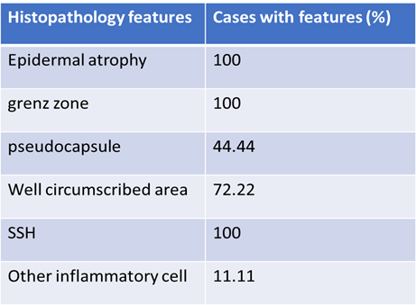
12 (66.6%) cases showed correlation between clinical and histopathological diagnosis for HL, remaining 6 (33.33%) had sarcoidosis, dermatofibroma and other variants of leprosy as clinical diagnosis as shown in table 2.
Table 2: Showing clinicopathological correlation of types of leprosy in the present study.
|
Clinical diagnosis
|
Histopathological diagnosis (Number of cases) |
|
|
|
HL |
Non HL |
|
HL |
12 |
0 |
|
Sarcoidosis |
2 |
0 |
|
Dermatofibroma |
2 |
0 |
|
TT |
1 |
0 |
|
BT |
1 |
0 |
|
Non HL |
|
836 |
|
Total |
854 |
|
Kappa values for clinicopathological correlation was calculated with the help of graph pad software by dotmatics with 95% confidence interval.
· Kappa < 0: No agreement
· Kappa between 0.00 and 0.20: Slight agreement
· Kappa between 0.21 and 0.40: Fair agreement
· Kappa between 0.41 and 0.60: Moderate agreement
· Kappa between 0.61 and 0.80: Substantial agreement
· Kappa between 0.81 and 1.00: Almost perfect agreement
The calculated kappa value for clinicopathological correlation is 0.79 showing Substantial agreement.
DISCUSSION:
Leprosy remains endemic in underdeveloped and developing nations. In India, widespread low socioeconomic conditions contribute to cycles of overcrowding, poor sanitation, and limited education, which in turn facilitate the persistence of infectious diseases such as leprosy. In the present study, histoid leprosy (HL) accounted for 2.1% of all leprosy cases, a figure comparable to the incidences reported by Mendritta et al. 7 (2011, n=11; 1.14%) and Punia et al. 8 (2017, n=13; 3.37%), though slightly lower than the 4.7% incidence noted by Girishkumar et al. 9 (2018, n=20). Collectively, these findings reaffirm the rarity of HL.
Among the 18 HL cases in this study, only one patient (5.5%) had a prior history of leprosy with irregular treatment, while the remaining were de novo presentations. This proportion is lower than that reported by Punia et al. 8, Girishkumar et al. 9 , and Mendritta et al. 7, suggesting that HL may not always arise from drug resistance. Instead, it may represent a distinct clinical entity rather than merely a variant of lepromatous leprosy, as proposed by several authors. However, discrepancies across studies could also reflect biases in patient reporting, given the social stigma surrounding leprosy and the likelihood of under‑disclosure of past medical history. Clinically, nodules were the most frequent lesion (100%), followed by plaques in 27.7% of cases, findings consistent with Punia et al. [9] and Girishkumar et al. 9.
The upper and lower limbs and trunk were the most commonly affected sites, with occasional facial involvement, in agreement with Girishkumar et al. 9, Kaveri et al. [12], and Punia et al. [10]. In contrast, Mendritta et al. 7 reported the face as the predominant site. Histopathologically, epidermal atrophy was present in all cases (100%), mirroring the findings of Punia et al. [10], but differing from Mendritta et al. 7, who observed it in only 30% of cases. The mean bacterial index (5.52) in this study was comparable to values reported by Kaveri et al. 10 (5.12) and Punia et al. 11 (5.54). Furthermore, clinicopathological correlation was achieved in 66.6% of cases, slightly higher than the 46.15% correlation reported by Punia et al. 11.
CONCLUSION:
This study reaffirms that histoid leprosy (HL) remains a rare entity, accounting for 2.1% of all leprosy cases in our cohort, consistent with previous reports from India. The predominance of de novo cases (94.4%) suggests that HL may not always be a consequence of drug resistance, but rather a distinct clinicopathological form of leprosy. Clinically, nodular lesions were universal, with plaques and involvement of limbs and trunk being common, while histopathology consistently demonstrated epidermal atrophy, Grenz zone, and spindle‑shaped histiocytes with a high bacillary load. Overall, the findings emphasize the need for heightened clinical suspicion, careful histopathological evaluation, and improved awareness among healthcare providers to ensure timely diagnosis and treatment. Strengthening surveillance and integrating HL recognition into leprosy control programs will be crucial for India’s ongoing efforts toward eradication of leprosy.
REFERENCES:
1. Kaur I, Dogra S, De D, Saikia UN. Histoid leprosy: a retrospective study of 40 cases from India. Br J Dermatol 2009; 160:305–10.
2. Sawatkar GU, Sharma A, Keshavamurthy V, Mehta H, Chatterjee D, Narang T, et al. Unusual clinical presentations in leprosy: a case series and review. Trans R Soc Trop Med Hyg 2023; 117:489–95.
3. McAdam AJ, Milner DA, Sharpe AH. Infectious Diseases. In: Kumar V, Abbas AK, Aster JC. Robbins and Cotran Pathologic Basis of Disease. South Asian Ed. New Delhi: Elsevier; 2014; 1:377-378.
4. Lockwood DNJ. Infections and Infestations, Leprosy. In: Griffiths CEM, Barker J, Bleiker T, Chalmers R, Creamer D, editor. Rook's textbook of Dermatology. John Wiley & Sons; 2016; 1:28.1-28.18.
5. Bancroft JD, Layton C, The hematoxylins and eosin, Editor(s): Suvarna SK, Layton C, Bancroft JD. Bancroft's Theory and Practice of Histological Techniques. Eighth Edition. Elsevier; 2019: 126-138. [58].
6. Wolfe D. Tissue processing, Editor(s): Suvarna SK, Layton C, Bancroft JD. Bancroft's Theory and Practice of Histological Techniques. Eighth Edition. Elsevier; 2019:73-83.
7. Mendiratta V, Jain A, Chander R, Khan A, Barara M. A nine-year clinico-epidemiological study of Histoid Hansen in India. J Infect Dev Ctries 2011; 5(2): 128-131.
8. Punia R.P.S., Dhingra H, Baliyan A, Handa U, Mohan H, Thami G.P. Clinicopathologic Spectrum of Histoid leprosy. International Journal of Current Research 2017; 9(5): 50765-9.
9. Girish kumar, Kaveri M, Wali VV. Clinico-pathological study of cases of histoid leprosy. Int J Res Dermatol 2020; 6:474-8.
10. Kaveri M, Wali VV. Clinico-pathological study of cases of histoid leprosy. Int J Res Dermatol 2020; 6:474-8.
11. Punia R.P.S., Dhingra H, Baliyan A, Handa U, Mohan H, Thami G.P. Clinicopathologic Spectrum of Histoid leprosy. International Journal of Current Research 2017; 9(5): 50765-9.
12. Venkatesan K, Deo N. Biochemical aspects of leprosy. In:Kumar B, Kar HK. IAL textbook of Leprosy. Second Ed. New Delhi: Jaypee Brothers Medical; 2017; 2.33-2.44.
13. Draper P, Kandler O, Darbre A. Peptidoglycan and arabinogalactan of Mycobacterium leprae. J Gen Microbiol. 1987;133(5):1187-94.
14. Hunter SW, Brennen PJ. A novel phenolic glycolipid from Mycobacterium leprae possibly involved in immunogenicity and pathogenicity. J Bacteriol. 1981;147(3):728-35.
15. Draper P, Payne SN, Rees RJ. Isolation of a characteristic phthiocerol dimycocerosate from Mycobacterium leprae. J Gen Microbiol. 1983;129(3):859-63. [14]. Prabhakaran K. Metabolism of M. leprae separated from human nodules. Int J Lepr. 1967;35:34-41.
16. Prabhakaran K, Braganca MB. Glutamic acid decarboxylase activity of M. leprae and occurrence of gamma-aminobutyric acid in skin lesions of leprosy. Nature. 1962;196:589-90. [16]. Skinsnes OK. Mycobacterium leprae and its affinity for nerves. Int J Lepr. 1971;39:762-5.
17. Stoner GL. Importance of the neural predilection of Mycobacterium leprae in leprosy. Lancet. 1979;10:994-6.
18. Rambukkana A, Salzer JL, Yurchenco PD, et al. Neural targeting of Mycobacterium leprae mediated by the G domain of the laminin α2 chain. Cell. 1977;88:811-21.