Assessment of Lower Limb Venous Insufficiency Using Colour Doppler Ultrasonography in Patients with Varicose Veins.
- Kakshil Patel , Third year resident doctor, Department of Radio-diagnosis, Narendra Modi Medical College, Gujarat University, Ahmedabad, INDIA
- Kavita U. Vaishnav , Associate Professor, Department of Radio-diagnosis, Narendra Modi Medical College, Gujarat University, Ahmedabad, INDIA
- Jiten Modi , Third year resident doctor, Department of Radio-diagnosis, Narendra Modi Medical College, Gujarat University, Ahmedabad, INDIA
- Hardi Patel , Third year resident doctor, Department of Radio-diagnosis, Narendra Modi Medical College, Gujarat University, Ahmedabad, INDIA
- Parin Gajjar , Second year resident doctor, Department of Radio-diagnosis, Narendra Modi Medical College, Gujarat University, Ahmedabad, INDIA
- (Maj.) Deepak K. Rajput , (Professor and Head of Department), Department of Radio-diagnosis, Narendra Modi Medical College, Gujarat University, Ahmedabad, INDIA.
Article Information:
Abstract:
Background:Varicose veins are the most common symptoms of chronic venous insufficiency, resulting from reverse blood flow through damaged valves, perforating veins, or post-thrombotic sequelae. Duplex colour Doppler imaging is the primary diagnostic tool for lower limb venous diseases, preferred by clinicians due to its non-invasive nature and high acceptability, making it an ideal choice for routine pre-operative evaluation.Materials and Methods: The study was undertaken from January 2025 to December 2025 in patients referred to the department of Radio diagnosis, Narendra Modi Medical College & L.G hospital, Ahmedabad. The study population consisted first 85 patients with symptomatic and asymptomatic visible varicose veins referred from other clinical departments were subjected to perform clinical maneuvers for complete assessment of causes of chronic venous insufficiency. Patency of the sapheno-femoral junction (SFJ) along with duration of venous reflux at SFJ was assessed. Level of incompetent perforating veins and varicosities of the superficial venous system were recorded using a Mindray Resona I9 ultrasound machine.Results: In this study of 85 patients, 67 patients were male while 18 patients were female. The most common age group affected was 41-50 years. The observed mean age was 46.5±6.22 years .The bilateral limb involvement with varicosities was 41.2% followed by 30.6% in left limb and 28.2% in right limb. Both great saphenous vein (GSV) and small saphenous vein (SSV) territories were affected in 50% of patients who had bilateral varicosities and predominantly SSV territory was affected in those having unilateral varicosity. Grade I & II incompetency at SFJ was most frequently observed in both bilateral and unilateral varicosities of lower limbs. Many patients had incompetent perforators at multiple sites with anterior thigh perforators were more common amongst the unilateral and posterior mid-calf perforator were incompetent in bilateral varicosity of lower limbs. SFJ reflux duration was more than 1000 msec in majority of the patients (75.0 % - 52 of 69 patients). Conclusion: Duplex ultrasound is a cost-effective and non-invasive modality for assessing venous insufficiency of lower limb. It helps in determining the type and level of vessel incompetence, venous reflux. The employment of various manoeuvres during the scan helps in determining the site and severity of vessel incompetence. Hence preoperative venous mapping of incompetent sites to target the sites of venous incompetence is essential to achieve increased post-operative success rate
Keywords:
Article :
INTRODUCTION:
Varicose veins are one of the most common manifestations of chronic venous disease of the lower limbs and a significant source of morbidity worldwide. They result from incompetence of venous valves, leading to retrograde blood flow, sustained venous hypertension, and progressive dilatation and tortuosity of the superficial venous system.9 Patients typically present with limb heaviness, pain, swelling, and skin changes, which in advanced cases can progress to lipodermatosclerosis and venous ulceration. Risk factors include increasing age, prolonged standing, obesity, and a positive family history.⁴
Accurate preoperative assessment is essential for identifying the site and extent of venous incompetence across the superficial, deep, and perforator systems, as incomplete evaluation is a well-recognised cause of treatment failure and recurrence.⁵ Clinical examination and handheld Doppler alone are insufficient for this purpose. CT venography, while capable of delineating venous anatomy, cannot provide real-time valvular assessment and carries limitations of radiation exposure and contrast-related contraindications.³
Magnetic Resonance Venous Imaging is useful in evaluation of the abdominal and pelvic vasculature. It can be employed for evaluation of pelvic venous outflow obstruction by providing information from the IVC through the iliac venous system. CT Venography can be used in the preoperative evaluation of varicose veins. CT Venography with 3D reconstruction can provide a thorough understanding of the of the varicose vein anatomy but has certain limitations such as it cannot provide real- time images and thus the movement of valve cannot be evaluated. Other associated limitations are radiation exposure, additional cost and contraindication in patients with renal dysfunction and allergy.
Duplex colour Doppler ultrasonography has become the investigation of choice for the evaluation of lower limb venous disease. It provides simultaneous anatomical and functional assessment including real-time identification of reflux, grading of junction incompetence, and precise localisation of incompetent perforating veins in a non-invasive, cost-effective, and reproducible manner.¹⁵ Proper identification of the site and extent of venous incompetence is essential for selecting the appropriate therapeutic approach, including endovenous procedures or surgical intervention.
The present study was undertaken to evaluate the role of duplex colour Doppler ultrasonography in patients presenting with varicose veins, and to determine the pattern and distribution of venous incompetence as a basis for preoperative planning.
MATERIALS AND METHODS:
Study design, duration and settings
This was a hospital-based prospective observational study conducted in the Department of
Radiodiagnosis, Narendra Modi Medical College and L.G. Hospital, Maninagar, Ahmedabad, Gujarat, India between January 2025 to December 2025 for a duration of 12 months.
Equipment used
Mindray Resona I9 ultrasound machine equipped with linear transducer (7–12 MHz) and color and spectral Doppler facilities (Duplex color Doppler system).
Inclusion criteria
· Patients of any age and gender presenting with clinically suspected varicose veins of the lower limbs.
· Patients willing to participate in the study and providing written informed consent.
Exclusion criteria
· All patients who don’t give consent / assent of parent or guardian for participation in the study.
· Patients with a history of lower limb venous surgery or endovenous intervention.
Advantage
· Available more readily, cost effective and less time-consuming.
· Provides real-time hemodynamic information
· Accurately localizes venous reflux and incompetent perforators
· Essential for treatment planning and follow-up.
Limitation
· Reduced accuracy in severely obese patients or in those with extensive limb edema
· Limited visualization in deeply located pelvic veins
· Inter and intraoperation dependent.
Source of data: Patients who undergo B mode ultrasound & duplex doppler study being referred to the department of radio diagnosis at L. G. hospital, Narendra Modi Medical College, Maninagar, Ahmedabad for duration of 12 months after obtaining an informed consent.
Study type: Prospective observational single centered study.
Method of data collection:
▪SAMPLE SIZE & STUDY PERIOD: Minimum 80 cases from January 2025 to December 2025.
Method of examination:
All patients underwent duplex color Doppler ultrasonography of both lower limbs in standing and supine positions.
The following were assessed:
· Patency and compressibility of deep veins (common femoral, superficial femoral, popliteal and calf veins)
· Superficial venous system (great saphenous vein and small saphenous vein)
· Saphenofemoral junction competence
· Perforator veins
· Presence and duration of venous reflux (reflux duration >0.5 seconds considered significant) Provocative maneuvers such as distal compression and Valsalva were used to elicit reflux.
RESULTS:
TABLE 1: DISTRIBUTION OF PATIENTS ACCORDING TO GENDER (n=85)
|
Gender |
No. of patients |
Percentage |
|
Male |
67 |
78.8% |
|
Female |
18 |
21.2% |
|
Total |
85 |
100% |
Out of 85 patients included in the study, 67 were males(78.8%) & 18 were females(21.2%).
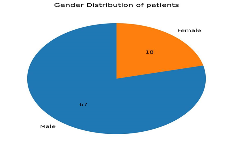
FIGURE. 1: DISTRIBUTION OF PATIENTS ACCORDING TO GENDER(n=85)
TABLE 2: DISTRIBUTION OF PATIENTS ACCORDING TO AGE(n=85)
|
AGE |
NO. OF PATIENTS |
PERCENTAGE |
|
21-30 |
8 |
9.4% |
|
31-40 |
10 |
11.8% |
|
41-50 |
33 |
38.8% |
|
51-60 |
17 |
20.0% |
|
61-70 |
9 |
10.6% |
|
71-80 |
8 |
9.4% |
|
Total |
85 |
100% |
The age of the patients ranged from 21 to 80 years, with the highest proportion falling within the 41–50-year age group, followed by those aged 51–60 years. The mean age of the cohort was 46.5 ± 6.2 years.
TABLE 3: Distribution of Limb Involvement
|
Limb involvement |
No. of patients |
Percentage |
|
Right limb |
24 |
28.2% |
|
Left limb |
26 |
30.6% |
|
Bilateral |
35 |
41.2 % |
|
Total |
85 |
100% |
Bilateral lower-limb disease was the most frequently encountered pattern, followed by unilateral involvement.
Bilateral varicosities were present in 41.2% of patients, while unilateral disease showed a slightly higher occurrence on the left side compared to the right.
TABLE 4: Competency of the Saphenofemoral Junction)
|
SFJ Status/Grade |
Bilateral (n=35) |
Right (n=24) |
Left (n=26) |
|
Competent |
6 (17.1%) |
5 (20.8%) |
4 (15.4%) |
|
Incompetent |
29 (82.9%) |
19 (79.2%) |
22 (84.6%) |
|
Grade I |
11 |
8 |
10 |
|
Grade II |
10 |
9 |
8 |
|
Grade III |
6 |
1 |
3 |
|
Grade IV |
2 |
1 |
1 |
Saphenofemoral junction (SFJ) assessment revealed a high prevalence of valvular incompetence, particularly in patients with bilateral limb involvement. Lower grades of reflux predominated across both unilateral and bilateral disease.
Grade I and Grade II reflux together accounted for the majority of SFJ incompetence across all patterns of limb involvement.
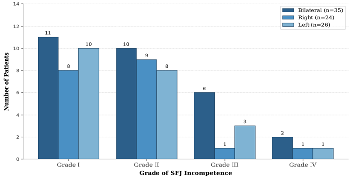
FIGURE. 2: Competency of the Saphenofemoral Junction)
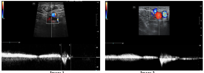
Image 1 Image 2
Image 1: Transverse Pulsed wave (PW) Doppler image showing grade I reflux seen in right SFJ on Valsalva manoeuvre on standing.
Image 2: Transverse PW Doppler image showing grade II reflux seen in right SFJ on Valsalva manoeuvre on standing and supine position.
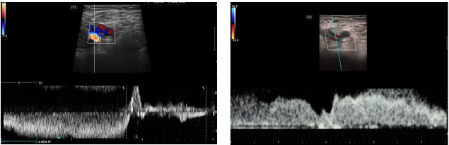
Image 3 Image 4
Image 3: Transverse PW Doppler image showing high velocity grade III reflux at left SFJ in standing position.
Image 4: Transverse PW Doppler image showing high velocity grade IV reflux at left SFJ in supine position
TABLE 5: Distribution of Varicosities by Venous Territory
|
Venous Territory |
Bilateral(n=35) |
Right(n=24) |
left(n=26) |
Overall(%) |
|
SSV Territory |
9(25.7%) |
12 |
14 |
41.2 |
|
GSV Territory |
9(25.7%) |
10 |
9 |
32.9 |
|
Both GSV and SSV |
18(51.4%) |
2 |
3 |
25.9 |
|
Total |
35 |
24 |
26 |
100 |
Territorial analysis of varicosities showed small saphenous vein (SSV) involvement to be the most common overall, particularly in unilateral disease. In contrast, combined involvement of the great saphenous vein (GSV) and SSV was most frequently observed in patients with bilateral disease.
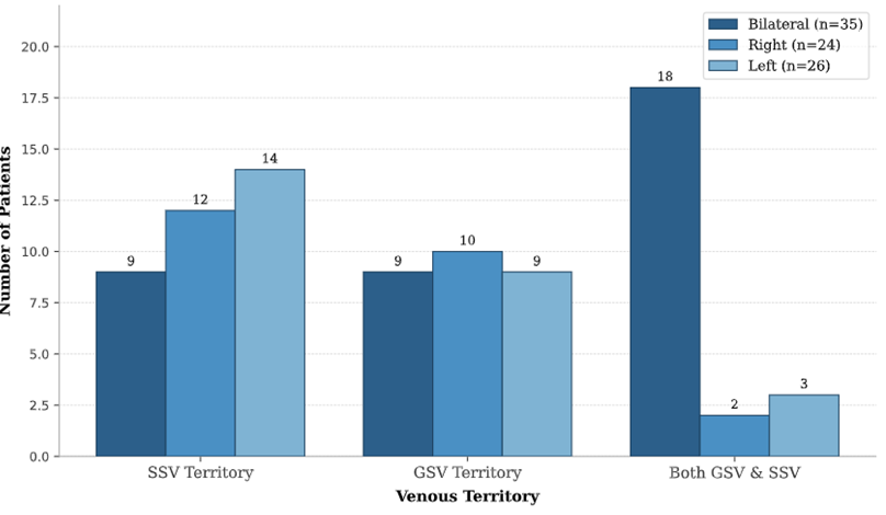
FIGURE. 3 Distribution of Varicosities by Venous Territory
TABLE 6: Anatomical Distribution of Incompetent Perforator Veins
|
Anatomical Level |
Bilateral (n=35) |
Right (n=24) |
Left (n=26) |
|
Anterior thigh |
3 |
9 |
11 |
|
Around the knee |
4 |
8 |
9 |
|
Posterior mid-calf |
16 |
6 |
8 |
|
Medial mid-leg |
10 |
7 |
6 |
|
Above the ankle |
2 |
1 |
1 |
Multiple incompetent perforators were frequently detected. In unilateral disease, perforators located in the anterior thigh were most commonly affected. Conversely, posterior mid-calf perforators predominated in bilateral disease, followed by those in the medial mid-leg region.
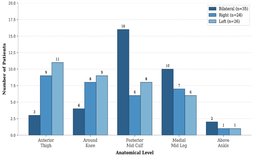
FIGURE. 4 Anatomical Distribution of Incompetent Perforator Veins
TABLE 7: Duration of Reflux at the Saphenofemoral Junction
|
Reflux Duration (ms) |
No. of patients |
Percentage |
|
500-1000 |
17 |
25% |
|
1001-2000 |
27 |
39.1% |
|
>2000 |
25 |
35.9 % |
|
Total |
69 |
100% |
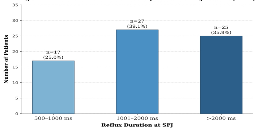
FIGURE. 5 Duration of Reflux at the Saphenofemoral Junction
DISCUSSION:
The present study evaluated the role of duplex color Doppler ultrasonography in the assessment of lower limb venous disease among 85 patients presenting with varicose veins. The findings align with and in several respects, extend the observations of prior comparable studies.
Out of 85 cases in the present study, the majority of patients were in the age group of 41 to 50 years (38.8%), followed by 20.0% in the 51 to 60 year age group, implying that the most commonly affected age group was 41 to 60 years, with a mean age of 46.5 ± 6.2 years. This distribution is consistent with the observation that the peak burden of chronic venous insufficiency and varicose vein disease falls within the fourth and fifth decades of life. The Edinburgh Vein Study, a large community-based cross-sectional survey conducted by Evans et al.1 on 1,566 subjects aged 18 to 64 years, confirmed that the prevalence of all categories of varicose veins and chronic venous insufficiency (CVI) increased progressively with age. The cumulative deterioration of venous valvular competence over time, progressive weakening of the venous wall extracellular matrix, and increasing susceptibility to haemodynamic stress from prolonged orthostatic loading.4
In the present study, a marked male predominance was observed, with 78.8% being males and 21.2% females. A similar gender demographic pattern was observed in the study conducted by Sridhar AS et al.,3 who reported 79% male predominance in their comparable series. The age-adjusted prevalence of trunk varicose veins was actually higher in men (40%) than in women (32%), and that the age-adjusted prevalence of CVI was likewise greater in men (9%) than in women (7%). The male preponderance observed in the present study is further explained by occupational characteristics, with a greater proportion of men in the study population engaged in physically demanding labour requiring prolonged standing — a well-established risk factor for venous hypertension and accelerated valvular degeneration.4
Bilateral lower limb varicosities were most frequently encountered, with an incidence of 41.2%, followed by left lower limb involvement in 30.6% and right lower limb involvement in 28.2% of patients. This bilateral predominance is consistent with Sridhar AS et al.,3 who reported bilateral involvement in 40% of their cohort, with 31% left-sided and 29% right-sided unilateral disease — a distribution that is closely mirrored in the present study. In the study by Irodi A et al.,5 59 patients had left-sided involvement and 41 had right-sided involvement, with no bilateral cases reported. The slight left-sided predominance in unilateral disease, seen both in the present study and in the literature, is anatomically attributable to the compression of the left common iliac vein by the overlying right common iliac artery, resulting in greater venous outflow resistance in the left lower extremity — a mechanism central to the pathophysiology of May-Thurner syndrome.6 The high prevalence of bilateral disease in this cohort highlights the importance of systematic bilateral duplex examination, as limiting assessment to the symptomatic limb alone risks underestimating the full extent of venous pathology and may result in incomplete preoperative mapping.
On analysis of the saphenofemoral junction (SFJ) competency, the present study demonstrated a high prevalence of SFJ incompetence across all patterns of limb involvement. Among the 35 patients with bilateral varicosities, 29 (82.9%) had incompetent SFJ. Of these, Grade I incompetence was most frequent (11 patients, 37.9%), followed by Grade II (10 patients, 34.5%), Grade III (6 patients, 20.7%), and Grade IV (2 patients, 6.9%). Among the 24 patients with right limb involvement, 19 (79.2%) had SFJ incompetence, with Grade I (8/19) and Grade II (9/19) accounting for the majority. Among the 26 patients with left limb disease, 22 (84.6%) had incompetent SFJ, with Grade I (10/22) and Grade II (8/22) predominating. Grade I and Grade II incompetence thus accounted for the majority of SFJ findings across all limb groups, which is consistent with the observations of Sridhar AS et al.,3 who similarly found Grades I and II to be the most frequently observed grades of SFJ incompetence in both bilateral and unilateral disease. In a study by Bashir et al.,7 41.7% of 36 patients had incompetent SFJ, while a higher rate of 92% SFJ incompetence was reported by Irodi A et al.5 in a series where superficial venous reflux in some form was seen in all limbs evaluated.
On analysing the varicosities based on their venous territory, the present study demonstrated that SSV territory involvement was the most prevalent overall (41.2%), particularly in patients with unilateral disease. GSV territory varicosities were present in 32.9% of patients overall, while combined GSV and SSV territory involvement was most commonly observed in patients with bilateral disease, accounting for 51.4% of bilateral cases (18/35). This pattern is consistent with the findings of Sridhar AS et al.,3 who similarly reported SSV territory predominance in unilateral disease and combined GSV-SSV involvement as the most frequent pattern in bilateral cases. In contrast, Azhar et al.10 reported a GSV-predominant distribution (58.5% GSV territory), followed by combined GSV and SSV involvement (39%) and isolated SSV involvement in only 2.1% — a pattern that reflects a different case-mix with a higher proportion of GSV-territory disease in that cohort. Labropoulos N et al.11 demonstrated in a large prospective series that disease was more widespread and involved more venous territories when the SFJ was incompetent, and that varicosities in unilateral disease were most commonly distributed in the calf at or below the anatomical level of GSV incompetence, supporting a descending propagation model of valvular failure. The comprehensive territorial assessment provided by duplex ultrasonography is essential for identifying all contributing sources of reflux, as failure to treat all territories is a well-documented cause of varicose vein recurrence.8
It was observed that many patients in the present study had incompetent perforators at multiple anatomical sites. In patients with unilateral lower limb disease, anterior thigh perforators were the most commonly affected site, whereas posterior mid-calf perforators predominated in patients with bilateral disease (16/35 bilateral cases), followed by medial mid-leg region perforators (10/35 bilateral cases). This anatomical distribution has direct therapeutic relevance, as precise preoperative localisation of incompetent perforating veins — rather than empirical ligation — enables targeted intervention and is associated with improved post-operative outcomes and reduced rates of varicose vein recurrence.6 In an earlier study, Delis KT et al.12 demonstrated predominant involvement of medial leg perforators in patients with CVI. In the study by Maragadha M et al.,13 of 37 participants with perforator incompetence, the majority of incompetent perforators were located medial to the ankle (75%). The anterior accessory GSV as the most common parallel channel to the GSV in the thigh, with reflux within it being frequently responsible for anterior thigh varicose veins — consistent with the predominance of anterior thigh perforator incompetence in unilateral disease observed in the present cohort. Duplex ultrasonography, performed in the standing weight-bearing position, remains the only modality capable of real-time haemodynamic identification of outward perforator flow and precise anatomical localisation for preoperative mapping.
In the present study, reflux duration analysis was performed in 69 patients with confirmed SFJ incompetence. Reflux duration in the 500–1,000 ms range was observed in 17 patients (25.0%), while 27 patients (39.1%) had reflux durations between 1,001 and 2,000 ms, and 25 patients (35.9%) demonstrated reflux exceeding 2,000 ms. Thus, prolonged reflux exceeding 1,000 ms was present in 75.0% of patients with SFJ incompetence, indicating haemodynamically significant, advanced valvular dysfunction in the majority of the cohort. These findings are broadly consistent with Sridhar AS et al.,3 who reported that 74.7% of their patients with SFJ incompetence had reflux durations exceeding 1,001 ms. In the study conducted by Irodi A et al.,5 reflux at the SFJ was continuous in 39/92 patients, between 1,001 and 2,000 ms in 20 patients, and less than 1,000 ms in 10 patients, with comparable proportions distributed across the intermediate and prolonged reflux duration ranges. Labropoulos N et al.11 established that the upper limit of normal reflux duration in superficial veins is 500 ms, and that values exceeding this threshold are indicative of significant incompetence; for femoropopliteal veins the accepted cut-off is >1,000 ms, which in the present study was met by three-quarters of patients with SFJ incompetence. The high prevalence of prolonged reflux in the present cohort — with 35.9% of incompetent limbs exceeding 2,000 ms — is consistent with a tertiary care referral population in whom advanced and symptomatic disease is disproportionately represented, and supports the use of duplex-based reflux duration quantification as a clinically actionable parameter in stratifying disease severity and guiding the extent of surgical or endovascular intervention.
CONCLUSION:
Duplex colour Doppler ultrasonography is a rapid, cost-effective, and non-invasive modality for the comprehensive assessment of lower limb venous insufficiency, enabling simultaneous evaluation of deep, superficial, and perforator valvular incompetence across multiple anatomical levels, with combined multi-site involvement being the most frequently encountered pattern. In The standardised use of physiological manoeuvres during the duplex examination including the Valsalva manoeuvre for SFJ evaluation and weight-bearing positioning for perforator assessment was essential in maximising diagnostic yield. Duplex colour Doppler ultrasonography is therefore the investigation of choice in the preoperative evaluation of lower limb venous disease, enabling precise anatomical and haemodynamic mapping of all incompetent sites, so that intervention is accurately targeted to achieve improved post-operative outcomes and reduced rates of recurrence.
ACKNOWLEDGMENTS
The authors would like to thank all patients who participated in this study and the staff of the Department of Radiodiagnosis for their assistance in data collection.
REFERENCES:
1. Evans CJ, Fowkes FGR, Ruckley CV, Lee AJ. Prevalence of varicose veins and chronic venous insufficiency in men and women in the general population: Edinburgh Vein Study. J Epidemiol Community Health. 1999;53(3):149–153.
2. Cesarone MR, Belcaro G, Nicolaides AN, et al. 'Real' epidemiology of varicose veins and chronic venous diseases: the San Valentino Vascular Screening Project. Angiology. 2002;53(2):119–130.
3. Sridhar AS, Mamtha G, Ravi Shankar M, Rashmi BV, Basti BD. Role of duplex color Doppler ultrasonography for evaluation of lower limb venous disease in patients presenting with varicose veins. European Journal of Cardiovascular Medicine. 2019;9(4):118–123.
4. Robertson L, Evans C, Fowkes FGR. Epidemiology of chronic venous disease. Phlebology. 2008;23(3):103–111.
5. Irodi A, Rajagopalan R, Chacko BR, Mathai J, Rupa V. Duplex colour Doppler assessment of lower limb venous insufficiency. Indian J Radiol Imaging. 2009;19(4):280–287.
6. Labropoulos N, Delis KT, Nicolaides AN, Leon M, Ramaswami G. The role of the distribution and anatomic extent of reflux in the development of signs and symptoms in chronic venous insufficiency. J Vasc Surg. 1996;23(3):504–510.
7. Bashir N, Bashir A, Rather A, et al. Role of colour Doppler ultrasonography in the evaluation of varicose veins of lower limb. Int J Res Med Sci. 2016;4(7):2803–2807.
8. Eberhardt RT, Raffetto JD. Chronic venous insufficiency. Circulation. 2014;130(4):333–346.
9. National Institute for Health and Care Excellence (NICE). Varicose Veins in the Legs: The Diagnosis and Management of Varicose Veins. NICE Clinical Guideline CG168. London: NICE; 2013.
10. Azhar MZ, Akhter W, Javed MA. Colour Doppler imaging in varicose veins. J Pak Med Assoc. 2006;56(11):517–520.
11. Labropoulos N, Tiongson J, Pryor L, et al. Definition of venous reflux in lower-extremity veins. J Vasc Surg. 2003;38(4):793–798.
12. Delis KT, Ibegbuna V, Nicolaides AN, Lauro A, Hafez H. Prevalence and distribution of incompetent perforating veins in chronic venous insufficiency. J Vasc Surg. 1998;28(5):815–825.
13. Maragadha M, Suryanarayana V, Rajini T, Sathyamurthy I. Role of color Doppler ultrasound in assessment of varicose veins of lower limbs. J Evid Based Med Healthc. 2016;3(59):2937–2943.
14. Winokur RS, Khilnani NM, Min RJ. Duplex ultrasound evaluation of patients with chronic venous disease of the lower extremities. AJR Am J Roentgenol. 2014;202(3):633–642.
15. Konoeda H, Yamaki T, Hamahata A, et al. Quantification of superficial venous reflux by duplex ultrasound – role of reflux velocity in the assessment of the clinical stage of chronic venous insufficiency. Phlebology. 2014;29(8):520–528.