Significance of Morphological & Morphometric analysis of adult human dry forearm of Indian origin in forensic science.
- Kavita Modi , Ph.D. Scholar, Dept. of Anatomy, Sardar Patel Medical College, Bikaner, Rajasthan
- Rubina Victor , Associate Professor, Dept. of Anatomy, Gautam Buddha ChikitsaMahavidyalaya, Dehradun, Uttarakhand
- Amarjyoti Chaturvedi , Assistant Professor, Dept. of Anatomy, Ram Krishna Medical College Hospital & Research Centre, Bhopal M.P.
- Mathew Joseph , Assistant Professor, Dept. of Anatomy, Amala Institute of Medical Sciences, Thrissur, Kerala.
Article Information:
Abstract:
Background: Aim: In this study, we will analyse several radius & ulna morphometric & morphological characteristics, search for gender variations in these parameters, and find that the measurements of male and female bones differ considerably. In order to determine whether these characteristics vary between the sexes and it’s significant in forensic sciences. Materials & methods: This study looked at the morphological and morphometric characteristics of dry ulna bones from adult Indians. The research's primary focus was on sexual dimorphism and sideways differences. Hundred of adult human dry forearm bones were obtained from the osteology collections, Ram Krishna Medical College Hospital & Research Center in Bhopal, Madhya Pradesh, Gautam Buddha Chikitsa Mahavidyalaya, Dehradun, Uttarakhand & Amala Institute of Medical Sciences, Thrissur, Kerala. Bones with complete anatomical identity and all of their original structure were included in the collection. With the help of digital Verniercaliper, morphometric parameters of dry forearm recorded. The archeological collections, Ram Krishna Medical College Hospital & Research Center in Bhopal, Madhya Pradesh, Gautam Buddha Chikitsa Mahavidyalaya, Dehradun, Uttarakhand & Amala Institute of Medical Sciences, Thrissur, Kerala, yielded hundred adult human dry forearm bones. Bones with complete anatomical identity and all of their original structure were included in the collection. We measured common morphometric parameters with a digital Verniercaliper and examined morphologically. Results: The radius and ulna bone measurements from both men and women were averaged for this investigation. Maximum length (MLU), olecranon width (OLW), coronoid height (CPH), mid shaft diameter (MSD), and distal end breadth (DEB) were all greater on average in males. In females, the difference between the right and left radii was not statistically significant. In Male, ulna measurements consistently outweigh female values in respect to all parameter and dimensions. At the level of radius, there was a significant difference between the sexes for all parameters, including MLU (259.6 vs. 238.3), OLW (24.55 vs. 22.2), CPH (17.6 vs. 15.4), MSD (9.5 vs. 8.3), and DEB (29.4 vs. 25.55). Conclusion: Results of the study indicate that morphometric & morphological qualities of the radius and ulna are significantly different between both male as well as females. They had somewhat bigger & heavy bones than females. It shows that functional asymmetry was occurring since the right side of the graph had substantially higher values. The results highlight the importance of considering gender and lateral asymmetry in forensics applications. Study shows a lot of sexual dimorphism in adult Indian forearm, with men having much greater values than females. When applied to real-life cases, anatomical studies need to account for differences that are specific to both sexes and sides.
Keywords:
Article :
INTRODUCTION:
Age & Sex determination with the help of forearm skeleton helped forensic science expert. Dominance of the dominant hand, and geographic location are among the various factors that influence small morphometric differences in the human radius. This study revealed that in adult Indian populations, the right radius of males exhibited slightly larger midshaft diameter (MSD), head diameter radius (HDR), neck circumference radius (NCR), mid-length radius (MLR), and distance from the head to the bicipital tuberosity (DEB). The pattern observed consistently indicates that the impact of functional adaptation related to handedness is comparatively minor, despite these differences not reaching statistical significance in earlier studies conducted in other regions.[1-5]
According to literature from both Indian and international,[2-6] the dominant side, which is often the right, was seen to have somewhat bigger dimensions. This might be due to increased mechanical stress and muscle activation.
Similarly, among females of Central Indian descent, MLR, HDR, and DEB showed a small right-sided dominance, but NCR and MSD were marginally more left-sided.[3-8] Earlier research has shown a widespread pattern of dominant-side hypertrophy,[5–10] which is supported by both of these results. Statistical significance was not found in most studies, suggesting a generally symmetrical morphology,[11–13] lending credence to the idea of bilateral anatomical balance despite functional asymmetry. That the morphology is often symmetrical is a key indicator of its significance.
Research shows that there is a strong sexual dimorphism, as men consistently scored better than females on all of the assessed measures.[4-6] Consistent with previous research, which has associated these differences to biomechanical, hormonal, and genetic variables,[3-6] this makes sense. Because it is possible to identify a person by determining their side and sex from isolated bones, these findings have far-reaching consequences for forensic anthropology.[3–7] It is critically important to identify minute side-specific changes in orthopedic operations such as surgical reconstruction and prosthesis design. In this study, we aim to measure the radius's various morphometric properties—its length, breadth, diameters, and indices—to see how these traits differ between the sexes and to find statistically significant.
MATERIALS AND METHODS:
This study looked at the morphological and morphometric characteristics of dry ulna bones from adult Indians. The research's primary focus was on sexual dimorphism and sideways differences. Hundred of adult human dry forearm bones were obtained from the osteology collections, Ram Krishna Medical College Hospital & Research Center in Bhopal, Madhya Pradesh, Gautam Buddha Chikitsa Mahavidyalaya, Dehradun, Uttarakhand & Amala Institute of Medical Sciences, Thrissur, Kerala. Bones with complete anatomical identity and all of their original structure were included in the collection.
With the help of digital Verniercaliper, morphometric parameters of dry forearm recorded. The archeological collections, Ram Krishna Medical College Hospital & Research Center in Bhopal, Madhya Pradesh, Gautam Buddha Chikitsa Mahavidyalaya, Dehradun, Uttarakhand & Amala Institute of Medical Sciences, Thrissur, Kerala, yielded hundred adult human dry forearm bones. Bones with complete anatomical identity and all of their original structure were included in the collection. We measured common morphometric parameters with a digital Verniercaliper and examined morphologically.
The study only considered bones that had all of their anatomical markers and features preserved. Pathological lesions, unknown laterality, or deformities in any of the bones were excluded from the results.
When necessary, we used proven anthropological indicators to verify the sex and side of each bone. To make these observations, we consulted the relevant governmental documents. Digital Vernier callipers with a resolution of 0.01 millimeters were employed to measure a range of conventional morphometric parameters. The measurements taken:
• Maximum Length of Radius (MLR) from the most proximal point of the radial head to the distal styloid process;
• Head Diameter of Radius (HDR) the maximum
transverse diameter of the radial head;
• Neck Circumference of Radius (NCR) measured at the narrowest portion of the radial neck using a flexible measuring tape;
• Midshaft Diameter (MSD) measured at the midpoint of the diaphysis;
• Distance from Head to Bicipital Tuberosity (DEB) measured from the superior articular surface to the most prominent point of the radial tuberosity.
For Ulna bone, parameters are: maximum length (MLU) and olecranon width (OLW), coronoid height (CPH), mid shaft diameter (MSD), and distal end breadth (DEB).
Each measurement was taken three times by two independent observers, and the mean value was used for analysis to reduce inter-observer and intra-observer variability.
Statistical analysis:
The SPSS software version was used to statistically analyze the data. For every parameter, descriptive statistics such as the mean and standard deviation were computed. The paired and unpaired t-tests were used for side-wise and sex-wise comparisons, respectively. P-values below 0.05 were regarded as statistically significant. The results were compared to both domestic and foreign literature, and patterns of asymmetry and dimorphism were interpreted in light of functional and biomechanical considerations.
RESULTS:
In the section that follows, the morphometry of the forearm bones is compared side-wise (right versus left) and sex-wise (male against female), with a special focus on the bone of the radius and the bone of the ulna individually.
Table 1: Radius in males’ side-wise comparison
|
Parameter |
Male Right (n=25) (Mean ± SD) |
Male Left (n=25) (Mean ± SD) |
P value
|
|
Maximum Length (MLR) |
245.9 ± 8.7 |
241.4 ± 9.2
|
T=1.776; df=48; P = 0.081 |
|
Head Diameter (HDR) |
23.1 ± 1.5 |
22.7 ± 1.6 |
T = 0.9119; df =48; P = 0.366 |
|
Neck Circumference (NCR) |
37.8 ± 2.8 |
37.1 ± 3.2 |
T = 0.823; df = 48; P = 0.414 |
|
Midshaft Diameter (MSD) |
9.7 ± 0.8
|
9.5 ± 1.1 |
T = 0.7352; df = 48; P = 0.465 |
|
Distal End Breadth (DEB) |
29.3 ± 2.1 |
28.7 ± 2.3 |
T = 0.963; df = 48; P = 0.340 |
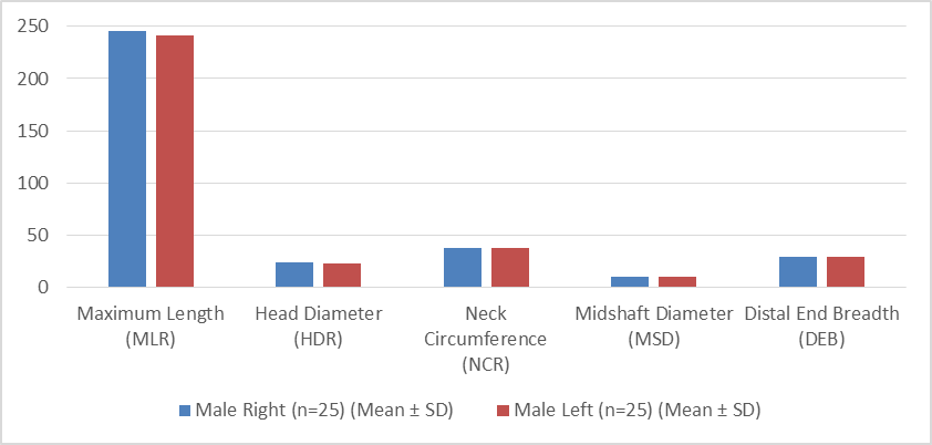
Figure 1: Comparison of the radius in males
Table 2: Radius in females’ side-wise comparison
|
Parameter |
Female Right (n=25) (Mean ± SD) |
Female Left (n=25) (Mean ± SD) |
P value
|
|
Maximum Length (MLR) |
225.1 ± 7.2 |
222.1 ± 8.1 |
T=1.384; df=48; P = 0.172 |
|
Head Diameter (HDR) |
22.9 ± 1.55
|
19.5 ± 1.4 |
T = 0.813; df =48; P = 0.420 |
|
Neck Circumference (NCR) |
37.45 ± 3.0 |
32.5 ± 2.9 |
T = 0.522; df = 48; P = 0.6.3 |
|
Midshaft Diameter (MSD) |
9.6 ± 0.95
|
8.2 ± 0.5 |
T = 0.530; df = 48; P = 0.598 |
|
Distal End Breadth (DEB) |
29 ± 2.2 |
26.3 ± 2.1 |
T = 1.235; df = 48; P = 0.222 |
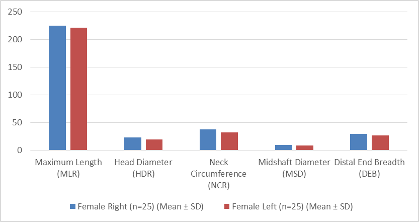
Figure 2: Comparison of the radius in females
Table 3: Radius in both the genders comparison
|
Parameter |
Male (n=25) (Mean ± SD) |
Female (n=25) (Mean ± SD) |
P value
|
|
Maximum Length (MLR) |
243.65 ± 8.7 |
223.6 ± 7.65 |
T=8.221; df=48; P = 0.0001 |
|
Head Diameter (HDR) |
23.1 ± 1.5 |
21.2 ± 1.5 |
T = 4.4783; df =48; P = 0.0001 |
|
Neck Circumference (NCR) |
37.8 ± 2.8 |
35 ± 3.0 |
T = 3.4116; df = 48; P = 0.0013 |
|
Midshaft Diameter (MSD) |
9.7 ± 0.8 |
8.9 ± 0.7 |
T = 3.762; df = 48; P = 0.0005 |
|
Distal End Breadth (DEB) |
29.3 ± 2.1 |
27.6 ± 2.15 |
T = 2.8282; df = 48; P = 0.0068 |
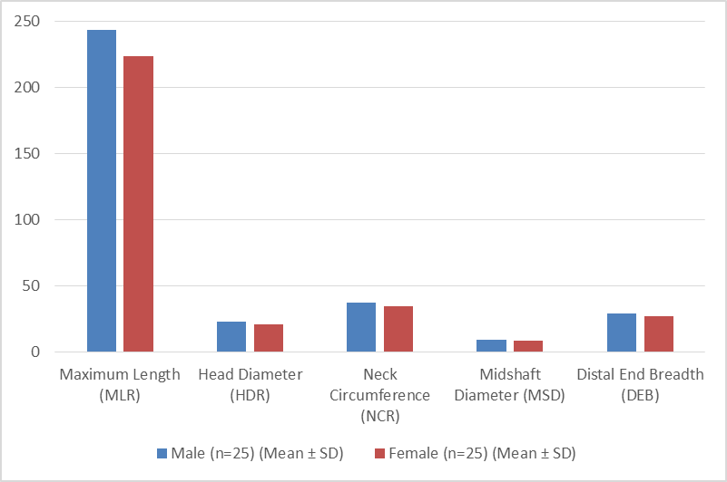
Figure 3: Comparison of radius between genders
Table 4: Ulna in males’ side-wise comparison
|
Parameter |
Male Right (Mean ± SD) |
Male Left (Mean ± SD) |
P value
|
|
Maximum Length (MLU) |
260.3 ± 8.9
|
258.9 ± 9.1 |
T=0.5499; df=48; P = 0.5849 |
|
Olecranon Width (OLW) |
24.9 ± 1.8
|
24.2 ± 1.9 |
T = 1.3373; df =48; P = 0.1874 |
|
Coronoid Height (CPH) |
17.8 ± 1.3 |
17.4 ± 1.5 |
T = 1.0076; df = 48; P = 0.3187 |
|
Midshaft Diameter (MSD) |
9.4 ± 0.9 |
9.6 ± 1.2 |
T = 0.6667; df = 48; P = 0.5082 |
|
Distal End Breadth (DEB) |
29.5 ± 1.9 |
29.3 ± 1.8 |
T = 0.3821; df = 48; P = 0.7041 |
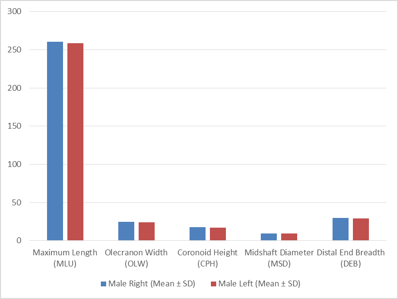
Figure 4: Comparison of the ulna in males
Table 5: Ulna in females’ side-wise comparison
|
Parameter |
Female Right (Mean ± SD) |
Female Left (Mean ± SD) |
P value
|
|
Maximum Length (MLU) |
238.7 ± 7.8 |
237.9 ± 9.9 |
T=0.3174; df=48; P = 0.7523 |
|
Olecranon Width (OLW) |
22.0 ± 1.4
|
22.4 ± 1.6 |
T = 0.9407; df =48; P = 0.3516 |
|
Coronoid Height (CPH) |
15.5 ± 1.1
|
15.3 ± 1.2 |
T = 0.6143; df = 48; P = 0.5419 |
|
Midshaft Diameter (MSD) |
8.2 ± 1.0
|
8.4 ± 0.9 |
T = 0.7433; df = 48; P = 0.4609 |
|
Distal End Breadth (DEB) |
25.7 ± 1.9 |
25.4 ± 1.7 |
T = 0.5883; df = 48; P = 0.5591 |
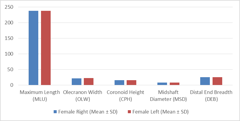
Figure 5: Comparison of the ulna in females
Table 6: Ulna in both the genders comparison
|
Parameter |
Male(Mean ± SD) |
Female (Mean ± SD) |
P value
|
|
Maximum Length (MLU) |
259.6 ± 9.0 |
238.3 ± 8.85 |
T=8.437; df=48; P = 0.0001 |
|
Olecranon Width (OLW) |
24.55 ± 1.85 |
22.2 ± 1.5 |
T = 4.8915; df =48; P = 0.0001 |
|
Coronoid Height (CPH) |
17.6 ± 1.4 |
15.4 ± 1.15 |
T = 6.0714; df = 48; P = 0.0001 |
|
Midshaft Diameter (MSD) |
9.5 ± 1.05 |
8.3 ± 0.95 |
T = 4.2373; df = 48; P = 0.0001 |
|
Distal End Breadth (DEB) |
29.4 ± 1.85 |
25.55 ± 1.8 |
T = 7.4578; df = 48; P = 0.0001 |
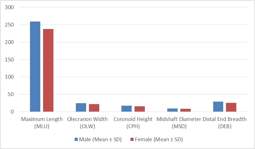
Figure 6: Comparison of ulna between genders
DISCUSSION:
This study show that the following radius morphometric characteristics were significantly higher on the right side of adult male Indians' bodies: Here are the measurements: DEB is 29.3 mm, MSD is 9.7 mm, NCR is 37.8 mm, MLR is 245.9 mm, HDR is 23.1 mm, and HDR is 22.7 mm. The distance from the head to the bicipital tuberosity is also 30.3. Although the pattern does not reach statistical significance, it does suggest a little inclination toward amplification on the dominant side. This pattern may be the product of a handedness-specific functional adaptation in bone morphology. Previous studies have also shown similar variances in negative effects, so our results are consistent with them. For instance, suggesting osseous variation associated to dominance was uncovered in a morphometric study of the radius in male North Indians. There was no statistically significant difference between the right- and left-side values, but the right-side values were somewhat higher.[14] Biomechanical stress effects are likely to blame for this, as was shown by Das et al. (2023),[15]
when they used osteometry to study dry radii and discovered that the dominant side, often the right, had larger dimensions. Literature from various nations lends credence to these conclusions. Hong et al. (2021),[16] used 3D imaging on a Caucasian population and found that right-handed people had considerably greater morphometric values for their right radii. Even if there is functional asymmetry, biological symmetry still persists since the changes did not reach the level of statistical significance (Hong et al., 2021, p. 16). Consistent usage of the upper limb influences radial bone formation, according to a biomechanical research by Sayadizadeh et al. (2024).[17] On the other hand, the changes are still rather little when you look at the two sides side by side.
The little imbalance seen in this study might be explained by differences in the nutrition, lifestyle, and employment habits of Indian males. It is crucial to think about bilateral comparisons within the context of the social and environmental features of the group being researched, based on the findings of Mishra (2016),[18] who claimed that regional and lifestyle differences impact the forearm bone structure in India. Being more dexterous with one side of the body does not always indicate that there are significant anatomical differences between the two in healthy individuals, according to research by Shrestha et al. (2022).[19].
CONCLUSION:
Morphological and morphometric analysis of adult human dry forearm bones—primarily the radius and ulna—has considerable significance in forensic science because skeletal dimensions and anatomical features can provide useful clues for sex estimation, stature estimation, population affinity, and individual identification.
Significance in forensic science
1. Sex estimation
· Measurements such as maximum length, breadth, circumference, and various indices of the radius and ulna can show sexual dimorphism.
· These measurements may assist in estimating sex when only fragmentary upper-limb remains are available.
2. Stature estimation
· The length of long bones, including the radius and ulna, has a relationship with overall body height.
· Population-specific regression equations can therefore be developed to estimate stature from recovered skeletal remains.
3. Population/ancestry assessment
· Morphological characteristics and metric variations of forearm bones may differ among populations.
· Establishing reference values for an Indian population can improve the usefulness of skeletal identification methods in Indian forensic cases.
4. Identification of unknown skeletal remains
· In cases involving mutilated, burned, decomposed, or skeletonized bodies, forearm bones may remain sufficiently preserved for analysis.
· A combination of morphological observations and morphometric measurements can narrow the identification possibilities.
5. Analysis of fragmentary remains
· Complete skeletons are not always recovered from crime scenes or mass disasters.
· Understanding the dimensions and morphology of different portions of the radius and ulna can help derive information even when the bones are incomplete.
6. Development of Indian reference standards
· Skeletal dimensions can vary according to sex, population, geographical region, nutrition, genetics, and secular changes.
· Therefore, data obtained from Indian dry adult forearm bones can contribute to population-specific standards rather than relying exclusively on datasets from Western populations.
7. Forensic anthropology and biological profiling
· Morphometric data can be incorporated into statistical models for constructing a biological profile of unidentified remains.
· When combined with other skeletal elements, these findings can strengthen forensic identification.
8. Medicolegal and anthropological research
· Such studies provide baseline anatomical data useful not only in forensic anthropology but also in anatomy, physical anthropology, and osteological research.
Overall significance
The main value of morphological and morphometric analysis of adult Indian forearm bones is that it can provide objective, measurable skeletal evidence for forensic identification. Establishing reliable measurements and population-specific standards for the Indian population may improve the estimation of sex, stature, and population affinity, particularly when other skeletal parts are unavailable. The Application in Forensic and Anthropological Settings: The results of this investigation will be highly valuable to forensic anthropologists and investigators that focus on medical-legal issues. The incorporation of this morphometric data into standard operating procedures is highly recommended when dealing with forensic skeletal remains. The ability to identify a person's gender from their forearm bones should be a standard component of forensic science training programs. This is particularly crucial information to have when just incomplete remains may be found.
Prompting Additional Investigations: In order to confirm and build upon previous findings, further research is required. Research comparing the population to those of other areas in India might be useful for building more thorough normative statistics. Researching age-related changes and bilateral asymmetry in the forearm bones can provide more precise data on skeletal diversity. In order to create tools that can automatically identify a person's gender, researchers should look at the potential of merging morphometric data with imaging techniques and machine learning algorithms. Fostering Collaboration Among Different Fields: Finally, it is recommended that specialists in biomedical engineering, forensic science, medicine, and anatomy collaborate more closely to develop clinically applicable and therapeutically useful technologies based on anatomical study.
Research results should be used to enhance healthcare service, forensic investigation, and educational opportunities, not just published in academic journals. With the help of interdisciplinary teams, we can achieve this objective.
Conflicts of interest
There are no conflicts of interest.
REFERENCES:
1. Banyeh M, Bani SB, Pathak R, Yakubu DD, Amankwaah E, Ahmed L. Sex estimation from the percutaneous lengths of the femur and the ulna in a Ghanaian population using discriminant function analysis. Canadian Society of Forensic Science Journal. 2022 Jul 3;55(3):142-55. https://doi.org/10.1080/00085030.2022.2028358
2. Bidmos MA, Mazengenya P. Accuracies of discriminant function equations for sex estimation using long bones of upper extremities. International Journal of Legal Medicine. 2021 May;135:1095-102. https://doi.org/10.1007/s43465-021-00540-0
3. Bhattacharyya T, Roy R, Islam M, Agarwal B, Roy SJ. Racial variation of carrying angle of elbow in North-East Indian Population: a clinico-radiological study. Indian Journal of Orthopaedics. 2022 Mar 1:1-7. https://doi.org10.1007/s00276-007-0197-1
4. Alves‐Pimenta S, Soares S, Colaço B, Ginja MM. Effect of elbow angle and weight‐bearing on the evaluation of joint congruence in dogs. The Anatomical Record. 2022 Dec;305(12):3398-409. https://doi.org/10.1007/s00276-022-03372-3
5. SingH A, SingH A. A morphometric study of head of radius and its clinical implication in radial head prosthesis. MH (mm). 2019;8(1.55):5-48. Original Article DOI: 10.7860/IJARS/2019/38115:2452
6. Sridhar S, Jayasree N, Srinivas HL, Shankar DK, Chidambaram S. Morphometry of radial head and neck length in the population of Andhra Pradesh. Int Anat Sci. 2015;6(1):10-5. https://doi.org/10.1016/S0268-0033(01)00075-4
7. Ethiraj S, Jyothi KC, Shetty S. A study of morphology and morphometry of proximal end of dry radius bones with its clinical implications. Int J Anat Res. 2019;7(3):6712-6. DOI: https://dx.doi.org/10.16965/ijar.2019.203
8. Gupta C, Kalthur S, Malsawmzuali J, D'souza A. A morphological and morphometric study of proximal and distal ends of dry radii with its clinical implications. Biomedical journal. 2015 Jul 1;38(4). DOI: 10.4103/23194170.151033
9. Kang SH, Kim DY, Park H, Song HS. Anthropometric study of the proximal radius: does radial head implant fit in all cases?. International orthopaedics. 2015 Aug;39:1553-61. https://link.springer.com/article/10.1007/s00264-015-2773-z
10. Rayna A, FRANCIS Y, GOUTHAMAN P, BEGUM Z, BASKARAN SB, GURUSWAMI K, SANKARAN P, RAGHUNATH G. Morphometric Study of Proximal and Distal End of Radius and its Clinical Significance. Journal of Clinical & Diagnostic Research. 2018 Sep 1;12(9). 10.7860/JCDR/2018/35071.12028
11. Giannicola G, Giulivi A, Polimanti D. Correlations between radial head diameters and humeral articular dimensions: bilateral MRI morphometric analysis on 39 healthy subjects. The Journal of Hand Surgery. 2021 Sep 1;46(9):814-e1. https://doi.org/10.1016/j.jhsa.2020.11.027
12. Deschrijver M, Lamquet S, Planckaert G, Vermue H, De Wilde L, Van Tongel A. Positioning of longest axis of the radial head in neutral forearm rotation. Shoulder & Elbow. 2020 Oct;12(5):362-7. https://doi.org/10.1177/1758573219831285
13. Popovic N, Djekic J, Lemaire R, Gillet P. A comparative study between proximal radial morphology and the floating radial head prosthesis. Journal of shoulder and elbow surgery. 2005 Jul 1;14(4):433-40. https://doi.org/10.1016/j.jse.2004.10.012
14. Gupta C, Kalthur S, Malsawmzuali J, D'souza A. A morphological and morphometric study of proximal and distal ends of dry radii with its clinical implications. Biomedical Journal. 2015 Jul 1;38(4). DOI: 10.4103/2319-4170.151033
15. Das A, Hembram PS, Roy K, Mukherjee S, Bhandari R, Kundu SD. Reconstructions of Length of Radius From its Fragments-A Pilot Study in Eastern Indian Population. Journal of Indian Academy of Forensic Medicine. 2023 Jun;45(2):153-8. https://doi.org/10.48165/jiafm.2023.45.2.14
16. Hong E, Kwak DS, Kim IB. Morphological symmetry of the radius and ulna—can contralateral forearm bones utilize as a reliable template for the opposite side?. PLoS One. 2021 Oct 6;16(10):e0258232.https://doi.org/10.1371/journal.pone.0258232
17. Sayadizadeh M, Daliri M, Rahimi M, Salehipour P, Sadeghi M, Mozafari JK, Moradi A. Grip and pinch strength prediction models based on hand anthropometric parameters: an analytic cross-sectional study. BMC Musculoskeletal Disorders. 2024 Oct 12;25(1):809. https://doi.org/10.1186/s12891-024-07914-z
18. Mishra PK, Nagar M, Gaur SC, Gupta A. Morphometry of distal end radius in the Indian population: A radiological study. Indian Journal of Orthopaedics. 2016 Dec;50:610-5. https://doi.org//10.4103/0019-5413.193482
19. Shrestha S, Mansur DI, Shrestha R, Karki S, Maskey S, Malla M, Basi A, Khadka SK. Radiographic study of distal radius parameters in patients visiting tertiary care hospital. Kathmandu University Medical Journal. 2022 Jun 30;20(2):136-40. https://doi.org/10.3126/kumj.v20i2.50565
20. Rayna A, FRANCIS Y, GOUTHAMAN P, BEGUM Z, BASKARAN SB, GURUSWAMI K, SANKARAN P, RAGHUNATH G. Morphometric Study of Proximal and Distal End of Radius and its Clinical Significance. Journal of Clinical & Diagnostic Research. 2018 Sep 1;12(9). 10.7860/JCDR/2018/35071.12028.