Association Between Vitamin D Levels, Bone Mineral Density and Radiological Severity of Osteoporosis.
- Vishant Gawri , Associate Professor, Department of Orthopaedics Ajay Sangaal Institute Of Medical Sciences & Research And Ayushmaan Hospital, Shamli (U.P) 247773
- Deepak Kumar Arora , Associate Professor, Department of Orthopaedics Ajay Sangaal Institute Of Medical Sciences & Research And Ayushmaan Hospital, Shamli (U.P) 247773
- Amit Garg , Associate Professor, Department of Orthopaedics Ajay Sangaal Institute Of Medical Sciences & Research And Ayushmaan Hospital, Shamli (U.P) 247773
- Sudhir Shandilya , Professor, Department of Orthopaedics Ajay Sangaal Institute Of Medical Sciences & Research And Ayushmaan Hospital, Shamli (U.P) 247773.
- Mukesh Chander Pokhariyal , Professor, Department of Orthopaedics Ajay Sangaal Institute Of Medical Sciences & Research And Ayushmaan Hospital, Shamli (U.P) 247773
- Mukesh Kumar , Professor, Department of Radiology Ajay Sangaal Institute Of Medical Sciences & Research And Ayushmaan Hospital, Shamli (U.P) 247773.
Article Information:
Abstract:
Background: Osteoporosis is a common metabolic skeletal disorder characterized by reduced bone mineral density (BMD), deterioration of bone microarchitecture, and increased risk of fragility fractures. Vitamin D plays a crucial role in calcium homeostasis and bone mineralization; however, deficiency remains prevalent and may contribute to accelerated bone loss. This study was conducted to evaluate the association between serum vitamin D levels, bone mineral density, and radiological severity of osteoporosis. Materials and Methods: A hospital-based cross-sectional observational study was conducted among 90 adult participants undergoing evaluation for skeletal health. Demographic and clinical details were recorded, and serum vitamin D, calcium, phosphate, and parathyroid hormone levels were assessed. Bone mineral density was evaluated using dual-energy X-ray absorptiometry (DXA), and participants were categorized as normal, osteopenia, or osteoporosis according to T-score criteria. The association between vitamin D status, biochemical parameters, BMD, and radiological severity was analyzed using appropriate statistical tests. Results: The mean age of participants was 58.6 ± 12.4 years, with females comprising 57.8% of cases. Vitamin D deficiency was observed in 51.1% of participants, insufficiency in 31.1%, and adequate levels in only 17.8%, with a mean vitamin D level of 21.8 ± 9.6 ng/mL. DXA assessment showed osteoporosis in 44.4%, osteopenia in 40.0%, and normal BMD in 15.6% of participants. Vitamin D deficiency was significantly associated with osteoporosis (p=0.018). Serum vitamin D showed positive correlations with lumbar spine BMD (r=+0.42, p<0.001), femoral neck BMD (r=+0.38, p<0.001), total hip BMD (r=+0.35, p=0.001), and T-score (r=+0.46, p<0.001). Osteoporosis patients had significantly lower vitamin D and calcium levels with higher parathyroid hormone levels. Conclusion: Vitamin D deficiency is significantly associated with reduced bone mineral density and increased severity of osteoporosis. Combined assessment of vitamin D status, biochemical markers, and DXA findings may improve early detection and management of individuals at risk for osteoporotic bone loss.
Keywords:
Article :
INTRODUCTION:
Osteoporosis is a progressive metabolic skeletal disorder characterized by reduced bone mass, deterioration of bone microarchitecture, and increased fragility of bones, resulting in a higher risk of low-trauma fractures.[1] It represents a significant global health concern, particularly among elderly individuals and postmenopausal women, due to its association with increased morbidity, disability, reduced quality of life, and healthcare burden.[2,3] The disease often remains clinically silent until the occurrence of fractures involving commonly affected sites such as the vertebrae, hip, and distal radius. Therefore, early identification of individuals at risk through evaluation of bone health parameters is essential for prevention and appropriate management.
Bone mineral density (BMD) is an important determinant of skeletal strength and is routinely assessed using dual-energy X-ray absorptiometry (DXA), which remains the gold standard for diagnosing osteoporosis. According to the World Health Organization (WHO), osteoporosis is defined by a BMD T-score of ≤ −2.5 standard deviations at the lumbar spine, femoral neck, or total hip.[4] However, BMD alone does not completely represent bone strength, as skeletal quality is also influenced by bone microarchitecture, mineral composition, and metabolic factors involved in bone remodeling.[5,6]
Vitamin D plays a crucial role in maintaining skeletal homeostasis by regulating calcium and phosphate absorption and supporting normal bone mineralization. Serum 25-hydroxyvitamin D [25(OH)D] is considered the most reliable marker for assessing vitamin D status. Vitamin D deficiency results in reduced intestinal calcium absorption, secondary hyperparathyroidism, increased bone turnover, and accelerated loss of bone mass.[7]
Several studies have demonstrated an association between low vitamin D levels, reduced BMD, and increased risk of osteoporotic fractures. However, the strength of this association varies among different populations due to variations in age, nutritional status, sunlight exposure, lifestyle factors, geographical location, and underlying metabolic conditions.[8]
Calcium and phosphate are essential minerals involved in bone formation and remodeling. Alterations in these biochemical parameters, along with vitamin D deficiency, may contribute to impaired mineralization and progression of osteoporosis. Therefore, evaluation of vitamin D status together with related biochemical markers provides a more comprehensive understanding of skeletal health rather than considering vitamin D levels in isolation.[9-11]
Radiological assessment plays an important role in evaluating structural changes associated with osteoporosis. Imaging findings such as cortical thinning, trabecular loss, reduced bone density, and vertebral compression fractures provide valuable information regarding disease severity. Correlating radiological severity with BMD and biochemical parameters may help in better risk stratification and identification of individuals requiring early intervention.[12,13]
Although numerous studies have explored the relationship between vitamin D levels and BMD, findings remain inconsistent, particularly regarding the association between vitamin D status and the radiological severity of osteoporosis. Differences in study populations, measurement techniques, and clinical factors may contribute to these variations.Hence, the present hospital-based cross-sectional study was undertaken to evaluate the association between serum vitamin D levels, bone mineral density, and radiological severity of osteoporosis among adults undergoing skeletal health assessment.
MATERIALS AND METHODS:
The present study was conducted as a hospital-based cross-sectional observational study to evaluate the association between serum vitamin D levels, bone mineral density (BMD), and radiological severity of osteoporosis. The study was carried out in the Department of orthopaedics with relevant clinical departments among adult patients undergoing evaluation for skeletal health.
Study Population
A total of 90 adult participants who underwent evaluation for osteoporosis and bone health assessment were included in the study. The sample size was determined based on the feasibility of patient recruitment and the objectives of assessing the relationship between vitamin D status, BMD measurements, and radiological severity of osteoporosis.
Inclusion Criteria
Participants fulfilling the following criteria were included in the study:
• Adults aged ≥18 years undergoing evaluation for osteoporosis or reduced bone density.
• Patients willing to undergo bone mineral density assessment by dual-energy X-ray absorptiometry (DXA).
• Participants with available serum vitamin D level estimation.
• Patients providing written informed consent for participation in the study.
Exclusion Criteria
Participants were excluded if they had:
• History of major traumatic fractures or fractures due to high-energy trauma.
• Known metabolic bone disorders other than osteoporosis.
• Chronic kidney disease, chronic liver disease, or endocrine disorders affecting bone metabolism.
• History of malignancy involving bone.
• Current treatment with medications significantly affecting bone metabolism, such as long-term corticosteroids or anti-resorptive therapy.
• Incomplete clinical, biochemical, or radiological data.
Clinical and Demographic Assessment
Detailed demographic and clinical information was collected from all participants. Data regarding age, sex, medical history, menopausal status (where applicable), lifestyle factors, history of fractures, and relevant risk factors for osteoporosis were recorded using a structured data collection proforma.
Biochemical Assessment
Venous blood samples were collected from all participants for estimation of biochemical parameters related to bone metabolism. Serum 25-hydroxyvitamin D [25(OH)D] levels were measured using standardized laboratory methods. Serum calcium and phosphate levels were also assessed to evaluate associated mineral metabolism abnormalities.Vitamin D status was categorized according to established clinical criteria into deficient, insufficient, and sufficient groups based on serum 25(OH)D concentration.
Bone Mineral Density Assessment
Bone mineral density was assessed using dual-energy X-ray absorptiometry (DXA). Measurements were obtained at clinically relevant skeletal sites, including the lumbar spine, femoral neck, and total hip. BMD values were expressed as T-scores, and participants were classified according to World Health Organization criteria as:
• Normal bone density (T-score ≥ −1.0)
• Osteopenia (T-score between −1.0 and −2.5)
• Osteoporosis (T-score ≤ −2.5)
Radiological Assessment of Osteoporosis Severity
Radiological evaluation was performed to assess structural changes associated with osteoporosis. Imaging findings were evaluated for features including reduced bone density, cortical thinning, trabecular pattern alterations, vertebral compression changes, and other radiological indicators of skeletal deterioration.
The severity of radiological changes was documented and correlated with BMD measurements and serum vitamin D levels.
Data Collection and Outcome Measures
The primary outcome of the study was to determine the association between serum vitamin D levels and bone mineral density. Secondary outcomes included evaluation of the relationship between vitamin D status, biochemical parameters, and radiological severity of osteoporosis.
Statistical Analysis
Collected data were entered into a Microsoft Excel spreadsheet and analyzed using SPSS.21statistical software. Continuous variables were expressed as mean ± standard deviation or median with interquartile range, depending on data distribution. Categorical variables were presented as frequency and percentage.The association between vitamin D levels, BMD categories, and radiological severity was assessed using appropriate statistical tests. Correlation analysis was performed to determine the relationship between serum vitamin D concentration and BMD values. A p-value of <0.05 was considered statistically significant.
RESULTS:
A total of 90 participants were included in the present study evaluating the association between serum vitamin D levels, bone mineral density (BMD), and radiological severity of osteoporosis. The demographic and clinical characteristics of the study population are summarized in Table 1.
The majority of participants were aged >60 years (42.2%), followed by the 41–60 years age group (37.8%), with a mean age of 58.6 ± 12.4 years. Females constituted the majority of the study population (57.8%), while males accounted for 42.2%. Among female participants, 73.1% were postmenopausal. A history of fragility fracture was present in 26.7% of participants. Regarding physical activity, most participants had a sedentary lifestyle (62.2%) compared with those who were moderately active (37.8%) (Table 1).
Assessment of serum vitamin D status revealed that vitamin D deficiency was common among study participants. Nearly half of the participants had deficient vitamin D levels (51.1%), while 31.1% had insufficient levels and only 17.8% had sufficient vitamin D concentrations. The mean serum vitamin D level among participants was 21.8 ± 9.6 ng/mL (Table 2).
The distribution of vitamin D status among the study population is illustrated in Figure 1, demonstrating a predominance of vitamin D deficiency and insufficiency.Bone mineral density assessment using DXA showed that 44.4% of participants had osteoporosis (T-score ≤ −2.5), whereas 40.0% had osteopenia and 15.6% had normal bone density. The mean lumbar spine BMD was 0.82 ± 0.14 g/cm², while the mean femoral neck BMD was 0.76 ± 0.12 g/cm² (Table 3).
The relationship between vitamin D status and BMD categories demonstrated a significant association between lower vitamin D levels and increased severity of bone loss. Among participants with vitamin D deficiency, 58.7% had osteoporosis, compared with 28.6% among those with insufficient vitamin D and 31.2% among those with sufficient vitamin D levels. This association was statistically significant (p=0.018) (Table 4).
Correlation analysis demonstrated a significant positive relationship between serum vitamin D concentration and various BMD parameters. Serum vitamin D levels showed a positive correlation with lumbar spine BMD (r=+0.42, p<0.001), femoral neck BMD (r=+0.38, p<0.001), total hip BMD (r=+0.35, p=0.001), and overall DXA T-score (r=+0.46, p<0.001) (Table 5).
These findings indicated that higher vitamin D levels were associated with better bone density measurements.Comparison of biochemical parameters between participants with osteoporosis and those with normal bone density/osteopenia showed significant differences. Participants with osteoporosis had significantly lower mean vitamin D levels (17.1 ± 6.7 ng/mL vs 25.6 ± 9.8 ng/mL, p<0.001) and lower serum calcium (8.9 ± 0.7 mg/dL vs 9.3 ± 0.6 mg/dL, p=0.004) and phosphate levels (3.5 ± 0.6 mg/dL vs 3.8 ± 0.5 mg/dL, p=0.018). Conversely, serum parathyroid hormone levels were significantly higher among participants with osteoporosis (62.7 ± 21.5 pg/mL vs 48.2 ± 16.4 pg/mL, p=0.001) (Table 6).
The comparison of biochemical markers between different osteoporosis severity groups is graphically represented in Figure 3.
Table 1: Baseline Demographic and Clinical Characteristics of Study Participants (n=90)
|
Parameter |
Number (n) |
Percentage (%) |
|
Age group (years) |
||
|
18–40 |
18 |
20.0 |
|
41–60 |
34 |
37.8 |
|
>60 |
38 |
42.2 |
|
Mean age (years) |
58.6 ± 12.4 |
|
|
Sex |
||
|
Male |
38 |
42.2 |
|
Female |
52 |
57.8 |
|
Menopausal status (Females, n=52) |
||
|
Premenopausal |
14 |
26.9 |
|
Postmenopausal |
38 |
73.1 |
|
History of fragility fracture |
||
|
Present |
24 |
26.7 |
|
Absent |
66 |
73.3 |
|
Physical activity level |
||
|
Sedentary |
56 |
62.2 |
|
Moderate/Active |
34 |
37.8 |
Table 2: Distribution of Serum Vitamin D Levels Among Study Participants (n=90)
|
Vitamin D Status (25-OH Vitamin D) |
Number (n) |
Percentage (%) |
|
Deficient (<20 ng/mL) |
46 |
51.1 |
|
Insufficient (20–29 ng/mL) |
28 |
31.1 |
|
Sufficient (≥30 ng/mL) |
16 |
17.8 |
|
Mean serum Vitamin D level (ng/mL) |
21.8 ± 9.6 |
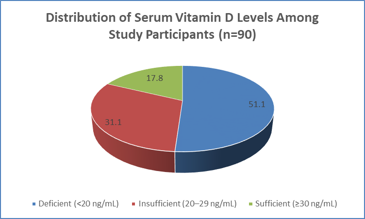
Figure 1 Distribution of Serum Vitamin D Levels Among Study Participants (n=90)
Table 3: Distribution of Bone Mineral Density Status According to DXA Findings (n=90)
|
BMD Category (DXA T-score) |
Number (n) |
Percentage (%) |
|
Normal bone density (T-score ≥ −1.0) |
14 |
15.6 |
|
Osteopenia (T-score −1.0 to −2.5) |
36 |
40.0 |
|
Osteoporosis (T-score ≤ −2.5) |
40 |
44.4 |
|
Mean Lumbar Spine BMD (g/cm²) |
0.82 ± 0.14 |
|
|
Mean Femoral Neck BMD (g/cm²) |
0.76 ± 0.12 |
Table 4: Association Between Vitamin D Status and Bone Mineral Density Category (n=90)
|
Vitamin D Status |
Normal BMD n (%) |
Osteopenia n (%) |
Osteoporosis n (%) |
p-value |
|
Deficient (<20 ng/mL) |
3 (6.5) |
16 (34.8) |
27 (58.7) |
|
|
Insufficient (20–29 ng/mL) |
7 (25.0) |
13 (46.4) |
8 (28.6) |
|
|
Sufficient (≥30 ng/mL) |
4 (25.0) |
7 (43.8) |
5 (31.2) |
|
|
Total |
14 (15.6) |
36 (40.0) |
40 (44.4) |
0.018 |
Table 5: Correlation Between Serum Vitamin D Level and Bone Mineral Density Parameters (n=90)
|
Parameter |
Correlation coefficient (r) |
p-value |
|
Vitamin D level vs Lumbar spine BMD |
+0.42 |
<0.001 |
|
Vitamin D level vs Femoral neck BMD |
+0.38 |
<0.001 |
|
Vitamin D level vs Total hip BMD |
+0.35 |
0.001 |
|
Vitamin D level vs T-score |
+0.46 |
<0.001 |
Statistical test applied: Pearson correlation analysis
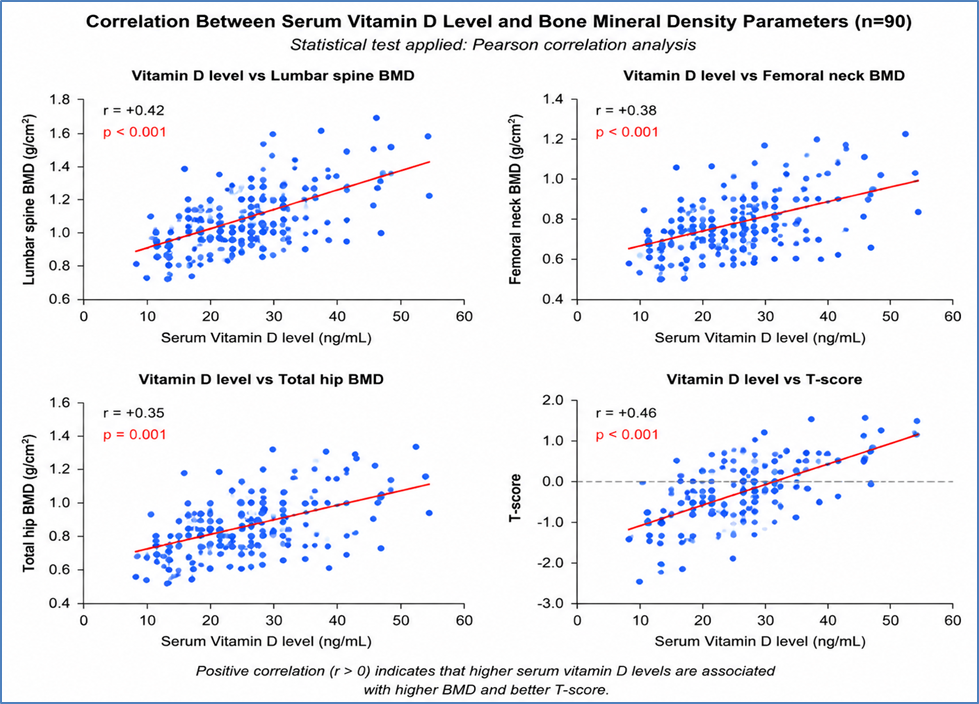
Table 6: Association of Biochemical Parameters with Osteoporosis Severity (n=90)
|
Parameter |
Normal/Osteopenia (n=50) Mean ± SD |
Osteoporosis (n=40) Mean ± SD |
p-value |
|
Serum Vitamin D (ng/mL) |
25.6 ± 9.8 |
17.1 ± 6.7 |
<0.001 |
|
Serum Calcium (mg/dL) |
9.3 ± 0.6 |
8.9 ± 0.7 |
0.004 |
|
Serum Phosphate (mg/dL) |
3.8 ± 0.5 |
3.5 ± 0.6 |
0.018 |
|
Parathyroid hormone (pg/mL) |
48.2 ± 16.4 |
62.7 ± 21.5 |
0.001 |
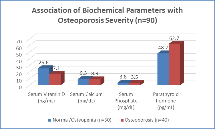
Figure 3 Association of Biochemical Parameters with Osteoporosis Severity (n=90)
DISCUSSION:
Osteoporosis is a common metabolic bone disorder characterized by reduced bone mineral density (BMD) and deterioration of skeletal microarchitecture, leading to increased risk of fragility fractures. Vitamin D deficiency plays an important role in impaired calcium metabolism, increased bone turnover, and accelerated bone loss. The present study evaluated the association between serum vitamin D levels, BMD, and radiological severity of osteoporosis among 90 adult participants and demonstrated a significant relationship between vitamin D deficiency, reduced BMD, and severity of osteoporotic changes.In the present study, the mean age of participants was 58.6 ± 12.4 years, with the majority belonging to the >60 years age group (42.2%). Females constituted 57.8% of cases, and 73.1% of women were postmenopausal.
These findings highlight the increased susceptibility of elderly individuals and postmenopausal women to osteoporosis due to age-related decline in bone formation and estrogen deficiency. Similar observations were reported by Chen et al.[14], who demonstrated a high prevalence of vitamin D deficiency among postmenopausal women with low bone mass and emphasized the influence of aging, hormonal changes, and vitamin D status on skeletal health.Vitamin D deficiency was observed in 51.1% of participants, insufficiency in 31.1%, and adequate levels in only 17.8%, with a mean serum vitamin D level of 21.8 ± 9.6 ng/mL. The high prevalence of suboptimal vitamin D status indicates widespread hypovitaminosis D among individuals at risk for osteoporosis.
Similar findings were reported by Kuchuk et al.[15], who observed inadequate vitamin D levels among postmenopausal women with osteoporosis. Sadat-Ali et al.[16] further demonstrated that vitamin D status significantly influences BMD and emphasized correction of vitamin D deficiency in patients with low bone mass.DXA assessment revealed osteoporosis in 44.4%, osteopenia in 40.0%, and normal bone density in 15.6% of participants. The mean lumbar spine BMD was 0.82 ± 0.14 g/cm², while femoral neck BMD was 0.76 ± 0.12 g/cm². Similar patterns have been reported in studies evaluating individuals undergoing DXA assessment, where a significant proportion showed reduced bone mass. Silambanan et al.[17] highlighted the importance of combined evaluation of BMD and biochemical markers for early detection of osteoporosis risk.
A significant association was observed between vitamin D status and BMD category (p=0.018). Osteoporosis was present in 58.7% of vitamin D-deficient participants compared with 28.6% among those with insufficiency and 31.2% among those with adequate vitamin D levels. Correlation analysis demonstrated significant positive associations between vitamin D levels and lumbar spine BMD (r=+0.42, p<0.001), femoral neck BMD (r=+0.38, p<0.001), total hip BMD (r=+0.35, p=0.001), and T-score (r=+0.46, p<0.001). Similar relationships between vitamin D status and BMD parameters have been reported by Chen et al.[14].Participants with osteoporosis had significantly lower vitamin D (17.1 ± 6.7 vs 25.6 ± 9.8 ng/mL, p<0.001), calcium (8.9 ± 0.7 vs 9.3 ± 0.6 mg/dL, p=0.004), and phosphate levels (3.5 ± 0.6 vs 3.8 ± 0.5 mg/dL, p=0.018), with significantly higher parathyroid hormone levels (62.7 ± 21.5 vs 48.2 ± 16.4 pg/mL, p=0.001). These findings support the physiological mechanism wherein vitamin D deficiency reduces calcium absorption, stimulates secondary hyperparathyroidism, and promotes bone resorption.
CONCLUSION:
The present study demonstrated a significant association between serum vitamin D levels, bone mineral density, and severity of osteoporotic changes among adult participants. Vitamin D deficiency was highly prevalent and was associated with reduced BMD, lower T-scores, and increased proportion of osteoporosis. Participants with osteoporosis showed significantly lower vitamin D, calcium, and phosphate levels along with elevated parathyroid hormone concentrations. Assessment of vitamin D status along with DXA-based BMD evaluation may help in early identification of individuals at increased risk and facilitate appropriate preventive and therapeutic strategies for osteoporosis.
Limitations
The study was conducted with a relatively small sample size of 90 participants and from a single center, which may limit the generalizability of the findings. The cross-sectional study design prevented assessment of the temporal relationship between vitamin D deficiency and progression of osteoporosis. Other influencing factors such as dietary calcium intake, genetic predisposition, sunlight exposure, and lifestyle variations were not evaluated in detail. Long-term prospective studies with larger populations are required to confirm these associations.
REFERENCES:
1. Sunyecz JA. The use of calcium and vitamin D in the management of osteoporosis. Ther Clin Risk Manag. 2008;4(4):827-836.
2. Lanham-New SA. Importance of calcium, vitamin D and vitamin K for osteoporosis prevention and treatment. Proc Nutr Soc. 2008;67(2):163-176.
3. Nieves JW. Osteoporosis: the role of micronutrients. Am J Clin Nutr. 2005;81(5):1232S-1239S.
4. Vidal M, Lane NE. Osteoporosis: definition, diagnosis, and considerations prior to starting treatment. Explor Endocr Metab Dis. 2026;3:101456.
5. Hejazi J, Davoodi A, Khosravi M. Nutrition and osteoporosis prevention and treatment. Biomed Res Ther. 2020;7:3709-3720.
6. Yeum KJ, Ju S, Choe U. Strategies for preventing bone loss in populations with insufficient calcium and vitamin D intake. Nutr Res Pract. 2025;19(2):155-169.
7. Jiang M, Li G, Yang K, Tao L. Role of vitamins in the development and treatment of osteoporosis (review). Int J Mol Med. 2025;56:109.
8. Wang D, Yang Y. The relationship between serum 25-hydroxyvitamin D levels and osteoporosis in postmenopausal women. Clin Interv Aging. 2023;18:619-627.
9. Chen X, Shen L, Gao C. Vitamin D status and its associations with bone mineral density, bone turnover markers, and parathyroid hormone in Chinese postmenopausal women with osteopenia and osteoporosis. Front Nutr. 2023;10:1307896.
10. Dhanwal DK, Kochupillai N, Gupta N, Cooper C, Dennison EM. Hypovitaminosis D and bone mineral metabolism and bone density in hyperthyroidism. J Clin Densitom. 2010;13(4):462-466.
11. Chongsrisawat V, Ruttanamongkol P, Chaiwatanarat T, Chandrakamol B, Poovorawan Y. Bone density and 25-hydroxyvitamin D level in extrahepatic biliary atresia. Pediatr Surg Int. 2001;17(8):604-608.
12. Al-Khakani MF, Radhi SW, Almohanna AM. Assessment of serum calcium, phosphorus, magnesium, iron, and zinc in osteoporosis postmenopausal women. Iraq Med J. 2018;2:51-56.
13. Liu L, Luo P, Wen P, Xu P. The role of magnesium in the pathogenesis of osteoporosis. Front Endocrinol (Lausanne). 2024;15:1406248.
14. Chen X, Shen L, Gao C, Weng R, Fan Y, Xu S, et al. Vitamin D status and its associations with bone mineral density, bone turnover markers, and parathyroid hormone in Chinese postmenopausal women with osteopenia and osteoporosis. Front Nutr. 2024;10:1307896.
15. Kuchuk N, van Schoor NM, Pluijm SMF, Chines A, Lips P. Vitamin D status, parathyroid function, bone turnover, and bone mineral density in postmenopausal women with osteoporosis: global perspective. J Bone Miner Res. 2009;24(4):693-701.
16. Sadat-Ali M, Al Elq AH, Al-Turki HA, Al-Mulhim FA, Al-Ali AK. Influence of vitamin D levels on bone mineral density and osteoporosis. Ann Saudi Med. 2011;31(6):602-608.
17. Modagan, Silambanan S, Menon PG, Arunalatha P. Comparison of bone mineral density with biochemical parameters and prevalence of osteopenia and osteoporosis in South Indian population. Biomed Pharmacol J. 2018;11(4):2209-2214.