Accuracy of bedside physical examination in distinguishing Different Types of Shock.
- Mansee Bhagat , Assistant professor, Dept of Medicine, Government Medical College, Nagpur, Maharashtra, INDIA.
- Vishakha Jain , Additional Professor, Department of Medicine, AIIMS Bibinagar, Hyderabad, INDIA.
- Siddharth Rao , Additional Professor, Department of Surgery, AIIMS Bibinagar, Hyderabad, INDIA.
- Saurabh Chahande , Consultant Rheumatologist, Nagpur, Maharashtra, INDIA.
Article Information:
Abstract:
Background: Early recognition and timely management of shock are crucial for improving outcomes, particularly in resource-limited intensive care settings. This diagnostic accuracy study evaluated the ability of structured bedside physical examination to differentiate shock categories using an adjudicated reference standard. Methods: A total of 298 patients aged >14 years with sustained non-surgical hypotension admitted to the Medicine ICU/CICU were included. The study is reported in accordance with STARD 2015 principles. Index tests included capillary refill time (CRT), skin temperature, pulse character, jugular venous pressure (JVP), S3 gallop, and lung crackles. Residents and attending physicians assessed clinical signs independently. Final shock type was adjudicated by a senior physician using predefined clinical, laboratory, electrocardiographic, echocardiographic, and treatment-response criteria while blinded to the prospectively recorded index-test findings. Results: Septic shock was most common (44.97%), followed by cardiogenic shock (29.19%), hypovolemic shock (12.08%), and unspecified shock (13.76%). No obstructive shock cases were identified as a separate final category. Overall mortality was 68.12%. Fast CRT showed high sensitivity but low specificity for septic shock, whereas warm skin and bounding pulse demonstrated high diagnostic performance with accuracies of 96.97% and 95.97%, respectively. Raised JVP showed 90.60% accuracy for cardiogenic shock; S3 gallop was highly specific but insensitive. Slow CRT, cold skin, and weak pulse demonstrated >90% accuracy for low-output shock. Conclusion: Structured bedside physical examination remains a rapid, inexpensive, and clinically useful first-line tool for differentiating shock types, especially where echocardiography or advanced monitoring is delayed. These findings require multicentric validation.
Keywords:
Article :
INTRODUCTION:
Shock is a serious medical emergency characterized by rapid progression and high risk of morbidity and mortality, leading to significant healthcare burden. It is a common condition in critical care settings, affecting nearly one-third of patients admitted to intensive care units (ICUs).¹ Shock is broadly classified into four types: distributive, cardiogenic, hypovolemic, and obstructive. However, these categories are not mutually exclusive, and many patients present with overlapping or multifactorial shock. The condition arises due to underlying pathophysiological mechanisms such as reduced circulating volume, impaired cardiac function, mechanical obstruction to blood flow, or abnormal distribution of blood due to vasodilation.² According to the European Society of Intensive Care Medicine (ESICM) consensus statement 2014, circulatory shock is defined as a life-threatening, generalized form of acute circulatory failure associated with inadequate cellular oxygen utilization.³
A diagnosis of shock is based on clinical, hemodynamic, and biochemical signs, which can broadly be summarized into three components.¹ Among these, bedside physical examination remains a fundamental component as it is rapid, repeatable, non-invasive, cost-effective, and readily available.⁴ Despite these advantages, its role has been increasingly underutilized in modern clinical practice, potentially contributing to diagnostic delays and inaccuracies.⁵ Early recognition and prompt hemodynamic management are essential to prevent progression to organ dysfunction and reduce mortality.¹
Although physical examination is routinely used in clinical settings, there is limited evidence evaluating its diagnostic accuracy in differentiating the types of shock, particularly in critically ill patients. Previous studies have explored its utility in specific conditions such as sepsis, cardiovascular disorders, and assessment of fluid responsiveness, but comprehensive evaluation in the context of shock classification remains inadequate.⁶⁻⁸ Furthermore, the predictive value of structured bedside examination algorithms has not been extensively studied. In this context, the present study was conducted to determine the diagnostic accuracy of bedside physical examination in distinguishing various types of shock using a final adjudicated diagnosis as the gold standard.
MATERIALS AND METHODS:
After obtaining approval from the Institutional Ethics Committee and written informed consent from all participants or their relatives, the study was conducted in the Medicine Intensive Care Unit (ICU) of a rural tertiary care teaching hospital in central India over a two-year period, from October 2016 to September 2018. During the study period, approximately 2,184 patients developed shock. Of these, 298 patients aged more than 14 years who were admitted to the ICU and CICU and had sustained hypotension (defined as systolic blood pressure <90 mmHg for more than 30 minutes due to non-surgical causes) were enrolled in the study. Patients with obvious surgical causes of shock, such as hemorrhagic shock, those with transient hypotension that resolved within 30 minutes, patients who died within 24 hours of admission and those who, or whose relatives, did not provide consent to participate were excluded.
All participants underwent detailed evaluation, including history, clinical examination, and relevant laboratory investigations. A structured proforma was used to collect demographic details, comorbidities, vital parameters, pulse pressure, Glasgow Coma Scale, capillary refill time, skin temperature, pulse character, jugular venous pressure (JVP), S3 gallop, lung crackles, neurological status, oxygen requirement, mechanical ventilation, PaO₂/FiO₂ ratio, urine output, biochemical parameters, ECG, and two-dimensional echocardiography. Severity of illness was assessed using APACHE II and SOFA scores. Patients were initially evaluated by a trainee resident and independently reassessed by the attending physician, with findings documented separately. The timing of index-test assessment was at first ICU/CICU evaluation after sustained hypotension was identified and before final diagnostic adjudication. Reporting follows STARD 2015 guidelines for diagnostic accuracy studies. After discharge or death, all available clinical, laboratory, ECG, echocardiographic, imaging, treatment-response, and outcome data were reviewed by a senior physician for final classification. The adjudicator was blinded to patient identity and to the prospectively recorded bedside index-test results (CRT, skin temperature, pulse character, JVP, S3, and crackles) used for diagnostic accuracy calculations. The final adjudicated diagnosis was considered the reference standard.
During the study period, 2,184 ICU/CICU patients developed shock. Patients were screened for eligibility. Exclusions included surgical causes of shock, transient hypotension resolving within 30 minutes, death within 24 hours, age ≤14 years, and lack of consent. A total of 298 eligible patients were included in the diagnostic accuracy analysis. No enrolled patient was excluded because of missing index-test or reference-standard data.
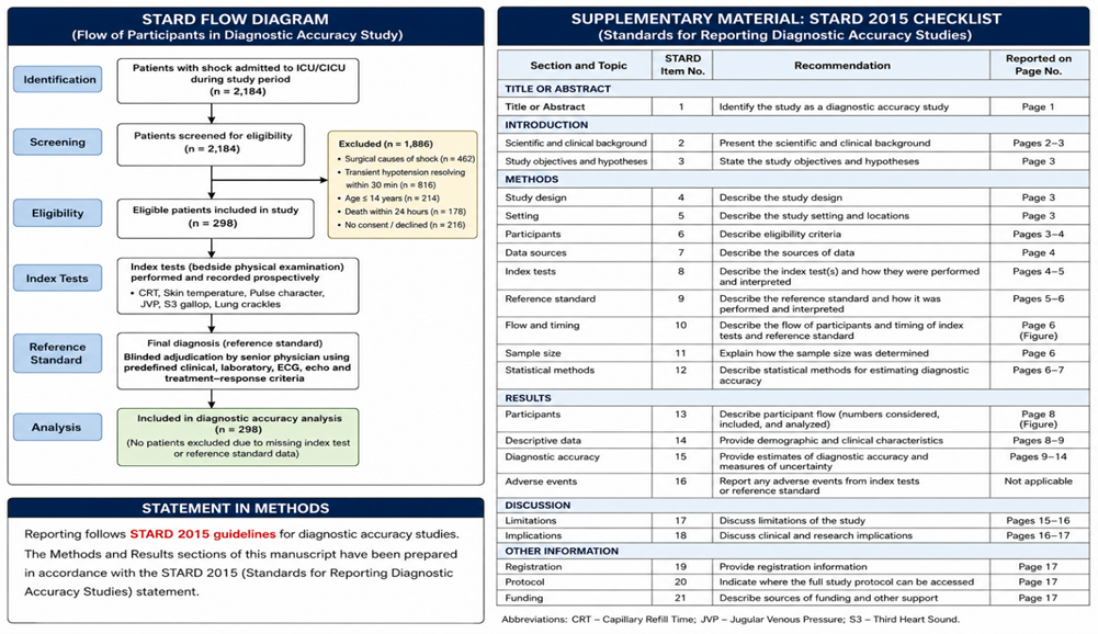
Table A: Predefined reference-standard criteria used for shock classification
|
Shock category |
Operational criteria for final adjudication |
|
Septic/distributive shock |
Suspected or documented infection with sustained hypotension requiring resuscitation, compatible systemic inflammatory/organ dysfunction features, raised inflammatory markers where available, and no primary cardiogenic or hypovolemic explanation. Sepsis-3 concepts were applied where feasible considering the 2016-2018 study period. |
|
Cardiogenic shock |
Hypotension with clinical or ECG evidence of acute coronary syndrome, arrhythmia, valvular disease, cardiomyopathy, congenital heart disease, or echocardiographic evidence of cardiac dysfunction, with supportive findings such as raised JVP, pulmonary crackles, S3, elevated troponin, low LVEF, or regional/global wall-motion abnormality. |
|
Hypovolemic shock |
Hypotension attributed to volume depletion or reduced effective circulating volume due to non-surgical causes such as gastrointestinal fluid loss, anemia, non-surgical hemorrhage, or adrenal crisis, supported by history, examination, laboratory profile, collapsible IVC where available, and response to fluid/steroid/blood-product therapy. |
|
Obstructive/unspecified shock |
Obstructive shock was to be diagnosed when tamponade, pulmonary embolism, tension pneumothorax, or other mechanical obstruction was the dominant mechanism. In this dataset, no case fulfilled criteria for obstructive shock as a separate final category. Cases with insufficient evidence for a single dominant mechanism were classified as unspecified. |
STARD participant flow: During the study period, 2,184 ICU/CICU patients developed shock. Patients were screened for eligibility; exclusions included surgical causes of shock, transient hypotension resolving within 30 minutes, death within 24 hours of admission, age <=14 years, and non-consent. A total of 298 eligible patients were included in the diagnostic accuracy analysis. No enrolled patient was excluded from final analysis due to missing index-test or reference-standard data.
Statistical analysis:
Data was entered into Microsoft Excel and analyzed using STATA version 14. Diagnostic accuracy of bedside physical examination was assessed by calculating sensitivity, specificity, positive likelihood ratio (LR+), and negative likelihood ratio (LR–), along with 95% confidence intervals. Additional measures including positive predictive value (PPV), negative predictive value (NPV), and overall accuracy were also calculated using standard formulas.
RESULTS:
In the present study, the majority of patients (38.92%) were in the geriatric age group (>60 years), with a mean age of 53.20±18.23 years and a male predominance (61.07%). The mean hospital and ICU stay were 6.02±7.27 days and 5.1±5.75 days, respectively, with most patients staying less than 5 days. Most patients were non-smokers (89.60%) and about two- thirds were non- or occasional alcohol consumers, while comorbidities were common, particularly coronary artery disease/heart failure (29.19%), hypertension (28.18%), and diabetes (17.11%). ECG commonly showed sinus tachycardia (47.65%), while echocardiography was normal in 56.38% cases, with the rest showing abnormalities such as regional wall motion defects and global hypokinesia; the mean LVEF was 43.62±12.43%, and most patients had a collapsible IVC (61.4%), (Table 1).
Table 1: Baseline characteristics of study population
|
Baseline characteristics |
No. of patients |
Percentage (%) |
|
|
Age group (years) |
≤20 |
13 |
4.36 |
|
21–30 |
30 |
10.07 |
|
|
31–40 |
38 |
12.75 |
|
|
41–50 |
50 |
16.78 |
|
|
51–60 |
51 |
17.11 |
|
|
61–70 |
67 |
22.48 |
|
|
71–80 |
35 |
11.74 |
|
|
81–90 |
11 |
3.69 |
|
|
91–100 |
03 |
1.01 |
|
|
Gender |
Male |
182 |
61.07 |
|
Female |
116 |
38.93 |
|
|
Duration of ICU stay (days) |
1 to 5 |
216 |
72.48% |
|
6 to 10 |
49 |
16.44% |
|
|
11 to 15 |
17 |
5.70% |
|
|
16 to 20 |
06 |
2.01% |
|
|
>20 |
10 |
3.36% |
|
|
Duration of hospital stay (days) |
1 to 5 |
199 |
66.78% |
|
6 to 10 |
55 |
18.46% |
|
|
11 to 15 |
21 |
7.05% |
|
|
16 to 20 |
08 |
2.68% |
|
|
>20 |
15 |
5.03% |
|
|
H/O smoking |
No |
267 |
89.60% |
|
Yes |
31 |
10.40% |
|
|
H/O alcohol |
No |
200 |
67.11% |
|
Yes |
98 |
32.89% |
|
|
Comorbidities |
HTN |
84 |
28.18% |
|
DM |
51 |
17.11% |
|
|
COPD |
06 |
2.01% |
|
|
CAD/Heart failure |
87 |
29.19% |
|
|
Chronic liver disease/hepatic encephalopathy |
30 |
10.06% |
|
|
H/O UGI bleed |
16 |
5.36% |
|
|
Chronic kidney disease |
31 |
10.40% |
|
|
H/O metastatic malignancy |
06 |
2.01% |
|
|
H/O AIDS |
06 |
2.01% |
|
|
ECG diagnosis |
Normal |
54 |
18.12 |
|
Sinus tachycardia |
142 |
47.65 |
|
|
Sinus bradycardia |
07 |
2.35 |
|
|
STEMI |
35 |
11.74 |
|
|
NSTEMI |
26 |
8.72 |
|
|
Arrhythmias |
14 |
4.70 |
|
|
LVH |
02 |
0.67 |
|
|
Tall T wave |
03 |
1.01 |
|
|
P pulmonale |
02 |
0.67 |
|
|
P mitrale |
01 |
0.34 |
|
|
Poor R-wave progression |
11 |
3.69 |
|
|
LBBB |
05 |
1.68 |
|
|
RBBB |
03 |
1.01 |
|
|
AV dissociation |
02 |
0.67 |
|
|
Low-voltage complexes |
02 |
0.67 |
|
|
ECHO findings |
Normal |
168 |
56.38 |
|
RWMA |
49 |
16.44 |
|
|
Valvular lesion |
32 |
10.74 |
|
|
Cardiomyopathy |
06 |
2.01 |
|
|
Septal defect |
04 |
1.34 |
|
|
LVH |
08 |
2.68 |
|
|
Pericardial effusion |
02 |
0.67 |
|
|
Clot |
03 |
1.01 |
|
|
LV dysfunction |
02 |
0.67 |
|
|
Pulmonary HTN |
14 |
4.70 |
|
|
Global hypokinesia |
49 |
16.44 |
The study population showed marked hemodynamic instability with a mean heart rate of 109 ± 27.08 bpm, temperature of 37.52 ± 0.77°C, systolic BP of 71.1 ± 10.97 mmHg, diastolic BP of 43.22 ± 12.78 mmHg, and pulse pressure of 25.51 ± 10.38 mmHg. The mean GCS, PaO₂/FiO₂ ratio and urine output were 8.18 ± 3.65, 2.64 ± 1.31 and 628 ± 287.9, respectively. Biochemical parameters showed mean BUN 37.71 ± 33.06, blood urea 80.93 ± 70.84, creatinine 2.98 ± 4.22, RBS 132.13 ± 70.9, bilirubin 1.91 ± 3.82, lactate 3.19 ± 3.5, and troponin T 0.24 ± 0.96. Hematological values included hemoglobin 10.26 ± 3.07 g/dL, hematocrit 31.45 ± 10.62%, WBC 14.33 ± 9.44 × 10³/µL, platelets 171.27 ± 110.78 × 10³/µL, neutrophils 77.77 ± 13.59%, and lymphocytes 18.2%. Electrolyte and ABG analysis showed potassium 4.5 ± 0.93 mEq/L, sodium 132.76 ± 8.16 mEq/L, pH 7.28 ± 0.18, and bicarbonate 16.71 ± 9.48 mmol/L.
Out of 298 patients, the most common diagnosis was high-output septic shock in 134 (44.97%). Among low-output shock, cardiogenic shock was seen in 87 (29.19%), predominantly due to acute myocardial infarction (69), followed by valvular heart disease (7), cardiomyopathy (4), rhythm disturbances (3), and congenital heart disease (2). Hypovolemic shock occurred in 36 patients (12.08%), mainly due to haemorrhage (19), anemia (15), and adrenal crisis (2), while 41 patients (13.76%) had unspecified shock (Figure 1).
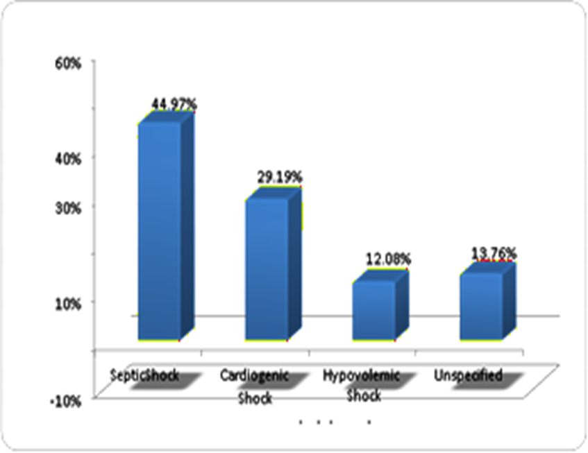
Figure 1: Distribution of patients according to final diagnosis categorized
Out of 298 patients, overall mortality was 68.12% (203 deaths) with 31.88% discharges (95patients). Septicshock(44.97%) had 66.42%mortality, cardiogenic shock(29.19%) had 62.07%, hypovolemic shock (12.08%) had 77.78%, and unspecified shock (13.76%) had 78.05% mortality. The difference in outcomes among shock types was not statistically significant (p = 0.160), (Table 2).
Table2: Distribution of final diagnosis categorized according to outcome
|
Final Diagnosis |
No of patients |
Discharge |
Death |
P value |
|
Highoutput SepticShock |
134(44.97%) |
45(33.58%) |
89(66.42%) |
0.160 |
|
LowoutputCardiogenic Shock |
87(29.19%) |
33(37.93%) |
54(62.07%) |
|
|
Hypovolemicshock |
36(12.08%) |
8(22.22%) |
28(77.78%) |
|
|
Unspecified |
41(13.76%) |
9(21.95%) |
32(78.05%) |
|
|
Total |
298(100%) |
95(31.88%) |
203(68.12%) |
|
The mean APACHE II score was 21.37 ± 6.60 (range 4–42), with 60.06% of patients having a score >20, which was associated with higher mortality; most patients had scores between 20–24. The mean SOFA score was 9.96 ± 3.3 (range 2–19), with nearly 66% of patients in the 7–12 range and the highest frequency between 10–12. The mean APACHE II and SOFA scores among patients who died were 23.48 ± 5.59 and 10.81 ±3.04, respectively. Both scores showed a significant correlation with patient outcomes, (p=0.0001).
Bothwarmskintemperatureandboundingpulsewerestatisticallysignificantpredictors of septic shock (p=0.0001), whereas fast CRT was not statistically significant (p=0.47) as shown in Table 3.
Table 3: Predictive characteristics of bedside physical examination for septic shock
|
|
FastCRT |
Warm Skin Temperature |
Bounding Pulse |
|
Sensitivity |
100% (95.12-100%) |
98.99% (94.17-99.41%) |
94.12% (87.64-97.81%) |
|
Specificity |
40.51% (23.42-55.45%) |
97.45% (94.15-99.17%) |
96.94% (93.46-98.87%) |
|
PPV |
54.34% (35.21-56.31%) |
95.15% (89.03-98.41%) |
94.12% (87.64-97.81%) |
|
NPV |
100% (95.12-100%) |
99.48% (90.26-99.92%) |
96.94% (93.46-98.87%) |
|
Accuracy |
44.56% |
96.97% |
95.97% |
|
LikelihoodRatio |
1.005 |
38.80 |
30.75 |
|
p-value |
0.47, NS |
0.0001, S |
0.0001, S |
The area under the ROC curve (AUC) for fast CRT in detecting septic shock was 0.708, indicating fair discrimination rather than chance-level performance (Figure 2).
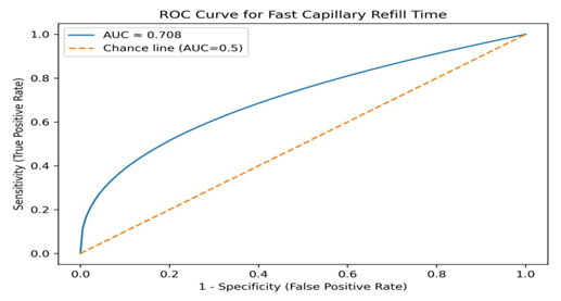
Figure 2: ROC curve for Fast CRT
The AUC for warm skin temperature in detecting septic shock was 0.732, indicating fair-to-moderate discrimination (Figure 3).
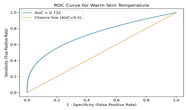
Figure 3: ROC for warm skin temperature
The AUC for bounding pulse in detecting septic shock was close to 1.0, indicating excellent discrimination in this dataset (Figure 4).
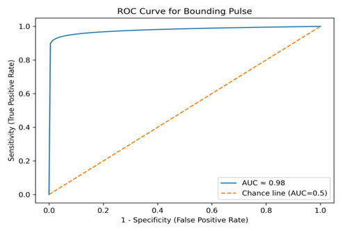
Figure 4: ROC for bounding pulse
Combination of bedside findings showed high diagnostic accuracy for septic shock, with Fast CRT plus warm skin temperature having the highest accuracy (98.64%). Other combinations also demonstrated high accuracy (95.97–96.64%). All combinations were statistically significant predictors of septic shock (p=0.0001), (Table 4).
Predictive characteristics of serum lactate >2 for the final diagnosis of septic shock showed low sensitivity (23.53%) and specificity (57.65%), with PPV of 22.43%, NPV of 59.16%,andanoverallaccuracyof45.97%.Despiteitslimiteddiagnosticperformance,itwas statistically significant (p=0.0014).
Table 4: Predictive characteristics of combination of bedside physical examinations for septic shock
|
|
Fast CRT+ Warm Skin Temperature |
FastCRT+ Bounding Pulse |
Warm Skin Temperature+ Bounding Pulse |
Fast CRT+ Warm SkinTemperature+ Bounding Pulse |
|
Sensitivity |
96.08% |
94.12% |
94.12% |
96.08% (90.26-98.92%) |
|
Specificity |
97.45% |
96.94% |
97.45% |
96.94% (93.46-98.87%) |
|
PPV |
95.15% |
94.12% |
95.05% |
94.23% (87.87-97.85%) |
|
NPV |
97.95% |
96.94% |
96.95% |
97.94% (94.81-99.44%) |
|
Accuracy |
98.64% |
95.97% |
96.30% |
96.64% |
|
LikelihoodRatio |
37.66 |
30.75 |
36.89 |
31.39 |
|
p-value |
0.0001, S |
0.0001, S |
0.0001, S |
0.0001, S |
Raised JVP showed high sensitivity (86.79%) and specificity (92.71%) with 90.60% accuracy. S3gallophad100%specificity but low sensitivity(19.81%), while crackles showed good sensitivity (80.19%) and specificity (84.38%). All findings were statistically significant predictors of cardiogenic shock (p=0.0001), (Table 5).
Table 5: Predictive characteristics of bedside physical examination for cardiogenic shock
|
|
↑ JVP |
S3 Gallop |
Cracklesto1/3rdof lung |
|
Sensitivity |
86.79% (82.26-98.92%) |
19.81% (17.21-22.21%) |
80.19% (79.21-92.15%) |
|
Specificity |
92.71% (87.64-95.81%) |
100% (92.21-100%) |
84.38% (78.52-86.31%) |
|
PPV |
86.79% (82.26-98.92%) |
100% (92.21-100%) |
73.91% (65.19-77.12%) |
|
NPV |
92.71% (87.64-95.81%) |
67.61% (59.26-77.92%) |
88.52% (81.26-92.51%) |
|
Accuracy |
90.60% |
69.12% |
82.88% |
|
LikelihoodRatio |
11.90 |
∞ |
5.13 |
|
p-value |
0.0001, S |
0.0001, S |
0.0001, S |
Combinationsofbedsidefindingsshowedhighspecificityforcardiogenicshock.↑JVP with crackles had the best performance (sensitivity 72.64%, specificity- 97.92%, accuracy 88.92%),
whileothercombinationshad100%specificitybutlowsensitivity.Allcombinations were statistically significant (p=0.0001), (Table 6).
Table 6: Predictive characteristics of combinations of bedside physical examinations for cardiogenic shock
|
|
↑JVP+ S3 Gallop |
↑ JVP+ Crackles to 1/3rdof lung |
S3Gallop+ Cracklesto 1/3rdof lung |
↑JVP+S3 Gallop+ Cracklesto1/3rdof lung |
|
Sensitivity |
11.32% (7.51-15.21%) |
72.64% (62.59- 76.15%) |
11.32% (7.51-15.21%) |
66.37 % (62.47-69.81%) |
|
Specificity |
100% (92.12-100%) |
97.92% (90.21- 99.52%) |
100% (92.12-100%) |
88.69 % (84.27-91.57%) |
|
PPV |
100% (92.12-100%) |
95.06% (85.62- 97.16%) |
100% (92.12-100%) |
79.34% (76.37-86.1%) |
|
NPV |
67.13% (56.51- 69.21%) |
86.64% (78.51- 89.21%) |
67.13% (56.51-69.21%) |
74.39 % (71.84-79.94%) |
|
Accuracy |
68.45% |
88.92% |
68.45% |
80.86 % |
|
Likelihood Ratio |
∞ |
34.87 |
∞ |
7.98 |
|
P-value |
0.0001, S |
0.0001, S |
0.0001, S |
0.0001, S |
Collapsible IVC showed moderate sensitivity (64.15%) but low specificity (40.10%), while low LVEF had low sensitivity (37.74%) and moderate specificity (61.46%). Both had low accuracy and were not statistically significant predictors of hypovolemic shock (p=0.47 and p=0.89), (Table 7).
Table 7: Predictive characteristics of collapsible IVC and low LVEF for hypovolemic shock
|
|
Collapsible IVC |
LowLVejection fraction |
|
Sensitivity |
64.15% |
37.74% |
|
|
(62.19-72.21%) |
(31.51-39.21%) |
|
Specificity |
40.10% (32.56-45.12%) |
61.46% (58.62-65.26%) |
|
PPV |
37.16% (32.12-45.26%) |
35.09% (31.12-39.56%) |
|
NPV |
66.96% (61.52-69.21%) |
64.13% (56.52-69.31%) |
|
Accuracy |
48.65% |
53.02% |
|
LikelihoodRatio |
1.07 |
0.97 |
|
p-value |
0.47, NS |
0.89, NS |
All the three physical signs i.e. slow CRT, cold skin and weak pulse have >90% sensitivity, specificity and accuracy in detection of low output shock (cardiogenic and hypovolemic shock), (Table 8).
Table 8: Predictive characteristics of bedside physical examination for low cardiac output i.e. cardiogenic and hypovolemic shock
|
|
Slow CRT |
ColdSkin |
Weak Pulse |
|
Sensitivity |
95.92% (92.34-97.4%) |
93.37% (89.27-96.52%) |
95.41% (91.37-98.54%) |
|
Specificity |
95.10% (93.14-96.3%) |
96.08% (91.57-99.37%) |
95.10% (89.93-96.45%) |
|
PPV |
97.41% (93.43-98.19%) |
97.86% (94.33-99.37%) |
97.40% (96.37-98.37%) |
|
NPV |
92.38% (87.52-96.57%) |
88.29% (84.55-92.34%) |
91.51% (88.49-93.46%) |
|
Accuracy |
95.63% |
94.29% |
95.30% |
|
LikelihoodRatio |
19.57% |
23.81 |
19.46 |
|
p-value |
0.0001, S |
0.0001, S |
0.0001, S |
Slow CRT, cold skin and weak pulse have >90% sensitivity, specificity and accuracy indetection of hypo volemic shock and all the three physical signs have significant co-relation, (Table 9).
Table 9: Predictive characteristics of combinations of bed side physical examinations for hypovolemic shock
|
|
SlowCRT+ Cold Skin |
SlowCRT+ Weak Pulse |
ColdSkin+ Weak Pulse |
|
Sensitivity |
93.37% |
95.41% |
95.92% |
|
Specificity |
96.08% |
96.08% |
96.08% |
|
PPV |
97.86% |
97.91% |
97.92% |
|
NPV |
88.29% |
91.59% |
92.45% |
|
Accuracy |
94.29% |
95.63% |
95.97% |
|
LikelihoodRatio |
23.81 |
24.33 |
24.46 |
|
p-value |
0.0001, S |
0.0001, S |
0.0001, S |
Table10showstheinterobservercorrelationbetween0.75-0.9forallthephysicalsigns like CRT, hand skin temperature, pulse pressure, JVP and crackles in lung with minimal variance which suggests that these signs are reproduced fairly good in 2 different observers.
Table10: Interobserver Reliability for clinical examinations
|
Clinical Examinations |
ICC |
Variance |
p-value |
|
CapillaryRefill Time(sec) |
0.836 |
0.004 |
0.0001, S |
|
Handskintemperature |
0.917 |
0.000 |
0.0001, S |
|
Pulse Pressure |
0.862 |
0.012 |
0.0001, S |
|
JVP |
0.915 |
0.000 |
0.0001, S |
|
Cracklesinlung |
0.830 |
0.001 |
0.0001, S |
Kappa coefficient for fast CRT was 0.789, showing moderate agreement between two observers. Kappa coefficient was 0.804 for warm skin temperature, suggesting strong agreement between two observers. Kappa for bounding pulse was 0.765 which showed moderate interobserver agreement. K value for raised JVP was 0.298 suggesting minimal interobserver agreement. Crackles to 1/3rd of lungs have kappa coefficient of 0.801 which showed strong interobserver agreement, (Table 11).
Table11: Kappa Statistics for clinical parameters
|
|
Kappa |
p-value |
Interpretation |
|
FastCRT |
0.789 |
0.0001, S |
Moderate |
|
WarmSkinTemperature |
0.804 |
0.0001, S |
Strong |
|
Bounding Pulse |
0.765 |
0.0001, S |
Moderate |
|
Increased JVP |
0.298 |
0.563, NS |
Minimal |
|
Crackles-1/3rdoflung |
0.801 |
0.0001, S |
Strong |
Out of all the patients with shock included in our study, shock due to acute myocardial infarction was the leading cause, constituting 23.15% of the study population. The next leading cause of shock was sepsis due to respiratory cause in 17.79% of the patients. Septic shock due to not specified cause was found in 15.1% of the patients. In around 13.76% of the patients, the cause of shock could not be ascertained and was categorized as unspecified cause of shock (Table 12).
Table 12: Distribution of patients according to final diagnosis
|
Shock Category |
Final Diagnosis |
No. of Patients |
Percentage (%) |
|
High-output shock |
Sepsis due to respiratory cause |
53 |
17.79 |
|
Sepsis due to GIT cause |
12 |
4.03 |
|
|
Sepsis due to CNS cause |
5 |
1.68 |
|
|
Sepsis due to UTI |
5 |
1.68 |
|
|
Sepsis due to bone |
1 |
0.34 |
|
|
Sepsis due to skin |
13 |
4.36 |
|
|
Sepsis due to unspecified cause |
45 |
15.10 |
|
|
Low-output shock |
Cardiogenic shock – AMI |
69 |
23.15 |
|
Cardiogenic shock – Cardiomyopathy |
4 |
1.34 |
|
|
Cardiogenic shock – Rhythm disturbances |
3 |
1.01 |
|
|
Cardiogenic shock – Valvular heart disease |
7 |
2.35 |
|
|
Cardiogenic shock – Congenital heart disease |
2 |
0.67 |
|
|
Cardiogenic shock – Tamponade |
2 |
0.67 |
|
|
Hypovolemic shock – Haemorrhage |
19 |
6.38 |
|
|
Hypovolemic shock – Diarrhoea |
0 |
0.00 |
|
|
Hypovolemic shock – Adrenal crisis |
2 |
0.67 |
|
|
Hypovolemic shock – Anemia |
15 |
5.03 |
|
|
Unspecified category / unknown cause of shock |
— |
41 |
13.76 |
|
Total |
— |
298 |
100.00 |
DISCUSSION:
In the present study, the maximum 44.96% of patients had high output, i.e. septic shock, followed by cardiogenic shock in 29.19% patients. Hypovolemic shock was less common and was found in 12.08% of patients. 13.75% of patients were found to have an unspecified cause of shock in whom the cause of shock could not be ascertained. These findings are comparable with the studies done by Vincent JL et al.¹ and Chen JT et al.⁹
Fast CRT had maximum (100%) sensitivity in detecting septic shock but was less specific (40.51%), having 54.34% PPV, 100% NPV, and 44.56% accuracy in detecting septic shock, which is comparable with the study done by Vazquez R et al.⁸ Similarly, Fleming et al.¹⁰ reported slow CRT with 84.6% sensitivity and 92.3% specificity for hypovolemic shock, while our study demonstrated higher sensitivity (95.92%) and specificity (95.1%). Warm skin temperature and bounding pulse had >90% sensitivity and specificity in detecting septic shock, with accuracy being around 95%. Both of these parameters had a significant correlation with septic shock. All three parameters taken together increased the sensitivity of detecting septic shock to 96.08%, specificity to 96.94%, with an accuracy of 96.94%. All these three parameters of clinical examination in combination had a significant correlation with septic shock. These findings are correlated with the study done by Vazquez R et al.⁸
Serum lactate >2 showed low sensitivity (23.53%) and specificity (57.65%), with an accuracy of 45.97% for detecting septic shock, although it had a significant correlation. While lactate is a useful marker for shock, it lacks specificity as it can be elevated in conditions such as metformin toxicity, diabetic ketoacidosis, and alcoholism. However, higher lactate levels, especially >4 mmol/L, are strongly associated with increased mortality, particularly in septic shock.¹¹ Increased JVP had maximum sensitivity (86.79%), with specificity of 92.71% and accuracy of 90.60% in detecting cardiogenic shock, which is comparable with the studies done by Sakka SG et al.⁵ and Butman et al.¹²
S3 gallop was less sensitive (19.81%) but 100% specific, with an accuracy of 69.12% in detecting cardiogenic shock, which is correlated with the study done by Scott HF et al.⁷ Crackles to one-third of the lung were 80.19% sensitive and 84.38% specific and had an accuracy of 82.88% in the detection of cardiogenic shock. The combination of all these three physical signs had a sensitivity of 66.37%, specificity of 88.69%, and accuracy of 80.86%. All three parameters individually and in combination showed a significant correlation with cardiogenic shock. Further, raised JVP had 82% sensitivity, 79% specificity, and 80% accuracy in detecting cardiogenic shock, which is comparable with the findings of Butman et al.¹² and Stevenson LW et al.¹³ Crackles to one-third of the lung had 55% sensitivity, 71% specificity, and 64% accuracy for the detection of cardiogenic shock. When these two physical signs were combined, sensitivity was 55%, specificity increased to 100%, and accuracy was 80% in the detection of cardiogenic shock.
In our study, collapsible IVC had 64.15% sensitivity, 40.1% specificity, and 48.65% accuracy in the detection of hypovolemic shock. All three physical signs, i.e. slow CRT, cold skin, and weak pulse, individually had >90% sensitivity, specificity, and accuracy in the detection of low-output shock (cardiogenic and hypovolemic shock), and all three physical signs had a significant correlation with hypovolemic shock. Whereas low LV ejection fraction had 37.74% sensitivity, 61.46% specificity, and 53.02% accuracy. Schriger DL et al.¹⁴ reported that capillary refill time >2 seconds had low sensitivity (59%) for diagnosing hypovolemia, though it increased to 77% of patients with hypovolemic shock. McGee et al.¹⁵ also suggested that capillary refill is less reliable, with postural vital changes being more accurate, and found that raised JVP (>7 cm H₂O) is a better predictor of cardiogenic shock than pulmonary crackles.
We also studied the interobserver variability by calculating Cohen’s kappa statistic (κ). Kappa’s coefficient for fast CRT was 0.789, showing moderate agreement between two observers. The kappa coefficient was 0.804 for warm skin temperature, suggesting strong agreement between two observers. Kappa for bounding pulse was 0.765, which showed moderate interobserver agreement. The κ value for raised JVP was 0.298, suggesting minimal interobserver agreement. Crackles to one-third of the lungs had a kappa coefficient of 0.801, which showed strong interobserver agreement. Except for raised JVP, all four physical signs had significant interobserver correlation with minimal variance, which suggests that these signs are fairly reproducible between two different observers.
In the present study, mortality was 66.42% in patients with septic shock, 62.07% in cardiogenic shock, and 77.78% in hypovolemic shock. The mortality rates are around 17.9% for sepsis, 28.6% for severe sepsis, and higher than 40% for septic shock. Similar findings have been reported in previous studies.¹⁶–¹⁸ The higher mortality in the present cohort should be interpreted in the context of a rural tertiary-care ICU, delayed referrals, high baseline severity scores, pre-COVID care pathways, and the high proportion of patients with cardiovascular comorbidity and organ dysfunction.
Recent evidence supports the value of point-of-care ultrasound (POCUS), including RUSH-type protocols, for improving diagnostic confidence in undifferentiated shock; however, ultrasound availability, operator skill, and time to imaging may vary across resource-limited settings. Therefore, the findings of the present study should not be interpreted as replacing echocardiography or POCUS. Rather, they support structured bedside signs as a rapid first step to guide initial resuscitation while laboratory tests and imaging are being arranged. Training residents to assess CRT, skin temperature, pulse character, JVP, S3 gallop, and lung crackles in a standardized way may improve early triage and treatment decisions when advanced monitoring is delayed.²⁰˒²¹
This study is strengthened by its clinically relevant diagnostic accuracy design, consecutive enrollment, relatively large sample size compared with the earlier pilot study, independent assessment of bedside signs by two observers, and evaluation of combinations of signs rather than isolated parameters alone. However, several limitations must be acknowledged. First, the study was conducted at a single rural tertiary-care center, limiting generalizability to high-resource ICUs and different case-mix settings. Second, the reference standard was based on single-expert adjudication; multiple independent adjudicators were not feasible in the study setting, and this may have introduced classification bias. Third, because the adjudicator reviewed routine clinical data also used in real-world bedside assessment, incorporation bias cannot be completely excluded, although prospectively recorded index-test findings were not used for final adjudication. Fourth, obstructive shock was not identified as a separate final category, and 13.76% of cases remained unspecified, reflecting overlap and diagnostic uncertainty in critically ill patients. Fifth, no a priori sample-size calculation was performed; however, the final sample was substantially larger than previous pilot data and provided adequate precision for key sensitivity and specificity estimates. Finally, data collection was completed in 2018; sepsis definitions and shock-management pathways have evolved since then. The results should therefore be interpreted as real-world evidence from a resource-constrained pre-COVID ICU setting and require external multicentric validation.
CONCLUSION:
The present study demonstrates that structured bedside physical examination can help differentiate major categories of shock with clinically useful diagnostic accuracy. Fast CRT was highly sensitive but poorly specific for septic shock, whereas warm skin and bounding pulse showed high accuracy for septic/distributive shock. Raised JVP and lung crackles supported cardiogenic shock, while S3 gallop was highly specific but insensitive. Slow CRT, cold skin, and weak pulse were useful markers of low-output shock. These findings support renewed emphasis on standardized bedside clinical assessment as an immediate, low-cost, first-line approach, particularly in resource-limited settings where echocardiography, POCUS, laboratory tests, or invasive monitoring may be delayed. Bedside signs should be used in combination and interpreted within the clinical context rather than as replacements for definitive investigations. Multicentric, STARD-compliant validation with multiple adjudicators and integrated POCUS comparison is recommended before routine protocol adoption.
REFERENCES:
1. Vincent JL, De Backer D. Circulatory shock. N Engl J Med. 2013 Oct 31;369(18):1726-34.
2. Haseer Koya H, Paul M. Shock. [Updated 2023 Jul 24]. In: StatPearls [Internet]. Treasure Island (FL): StatPearls Publishing; 2026 Jan-. Available from: https://www.ncbi.nlm.nih.gov/books/NBK531492/
3. Cecconi M, De Backer D, Antonelli M, et al. Consensus on circulatory shock and hemodynamic monitoring. Task force of the European Society of Intensive Care Medicine. Intensive Care Med. 2014;40:1795–815.
4. Sakr Y, Reinhart K, Vincent JL, Sprung CL, Moreno R, Ranieri VM, et al. Does dopamine administration in shock influence outcome? Results of the Sepsis Occurrence in Acutely Ill Patients (SOAP) Study. Crit Care Med. 2006;34:589–597.
5. Sakka SG, Ruhl CC, Pfeiffer UJ, Beale R, McLuckie A, Reinhart K, et al. Assessment of cardiac preload and extravascular lung water by single transpulmonary thermodilution. Intensive Care Med. 2000;26:180–7.
6. Goldberg RJ, Spencer FA, Gore JM, Lessard D, Yarzebski J. Thirty-year trends (1975 to 2005) in the magnitude of, management of, and hospital death rates associated with cardiogenic shock in patients with acute myocardial infarction: a population-based perspective. Circulation. 2009 Mar 10;119(9):1211-9.
7. Scott HF, Donoghue AJ, Gaieski DF, et al. Effectiveness of physical exam signs for early detection of critical illness in pediatric systemic inflammatory response syndrome. BMC Emerg Med. 2014;14:24.
8. Vazquez R, Gheorghe C, Kaufman D, Manthous CA. Accuracy of bedside physical examination in distinguishing categories of shock: a pilot study. J Hosp Med. 2010 Oct;5(8):471-4.
9. Chen JT, Roberts R, Fazzari MJ, Kashani K, Qadir N, Cairns CB, et al. Variation in fluid and vasopressor use in shock with and without physiologic assessment: a multicenter observational study. Crit Care Med. 2020 Oct;48(10):1436-1444.
10. Fleming S, Gill P, Jones C, Taylor JA, Van den Bruel A, Heneghan C, et al. The diagnostic value of capillary refill time for detecting serious illness in children: a systematic review and meta-analysis. PLoS One. 2015 Sep 16;10(9):e0138155.
11. Freeman R, Lirofonis V, Farquhar WB, Risk M. Limb venous compliance in patients with idiopathic orthostatic intolerance and postural tachycardia. J Appl Physiol (1985). 2002 Aug;93(2):636-44.
12. Butman SM, Ewy GA, Standen JR, Kern KB, Hahn E. Bedside cardiovascular examination in patients with severe chronic heart failure: importance of rest or inducible jugular venous distension. J Am Coll Cardiol. 1993 Oct;22(4):968-74.
13. Stevenson LW, Perloff JK. The limited reliability of physical signs for estimating hemodynamics in chronic heart failure. JAMA. 1989 Feb 10;261(6):884-8.
14. Schriger DL, Baraff LJ. Capillary refill—is it a useful predictor of hypovolemic states? Ann Emerg Med. 1991 Jun;20(6):601-5.
15. McGee SR. Physical examination of venous pressure: a critical review. Am Heart J. 1998 Jul;136(1):10-8.
16. Bone RC, Balk RA, Cerra FB, Dellinger RP, Fein AM, Knaus WA, Schein RM, Sibbald WJ. Definitions for sepsis and organ failure and guidelines for the use of innovative therapies in sepsis. The ACCP/SCCM Consensus Conference Committee. American College of Chest Physicians/Society of Critical Care Medicine. Chest. 1992 Jun;101(6):1644-55.
17. Martin GS, Mannino DM, Eaton S, Moss M. The epidemiology of sepsis in the United States from 1979 through 2000. N Engl J Med. 2003 Apr 17;348(16):1546-54.
18. Jawad I, Luksic I, Rafnsson SB. Assessing available information on the burden of sepsis: global estimates of incidence, prevalence and mortality. J Glob Health. 2012;2:010404.