Role Of Fetal Doppler Parameters In Predicting Adverse Neonatal Outcomes In Pregnancies Complicated By Fetal Growth Restriction
- Gurmit Singh , MBBS,MD Assistant Professor, Department of Pediatrics, Ajay Sangaal Institute Of Medical Sciences & Research And Ayushmaan Hospital, Shamli (U.P) 247773.
- Patwinder Bedi , MBBS, DNB , MNAMS, Assistant Professor, Department of Radiodiagnosis, Ajay Sangaal Institute Of Medical Sciences & Research And Ayushmaan Hospital, Shamli (U.P) 247773.
- Shamma Raj Rani Kapoor , MBBS, MS, Assistant Professor, Department of Obstetrics and Gynaecology, Ajay Sangaal Institute Of Medical Sciences & Research And Ayushmaan Hospital, Shamli (U.P) 247773.
Article Information:
Abstract:
Background: Fetal growth restriction (FGR) is an important cause of perinatal morbidity and mortality, predominantly resulting from placental insufficiency and chronic fetal hypoxemia. Fetal Doppler assessment provides information regarding placental resistance and fetal circulatory adaptation and may facilitate early identification of fetuses at risk of adverse neonatal outcomes. Methods: This prospective observational study included 90 singleton pregnancies complicated by FGR. Doppler assessment included umbilical artery pulsatility index (UA-PI), middle cerebral artery pulsatility index (MCA-PI), cerebroplacental ratio (CPR), and ductus venosus (DV) Doppler. Participants were followed until delivery, and neonatal outcomes were recorded. The association and predictive performance of Doppler abnormalities for adverse neonatal outcomes were evaluated using appropriate statistical tests and receiver operating characteristic analysis. Results: At least one abnormal Doppler parameter was identified in 55.6% of pregnancies, and 51.1% of neonates experienced an adverse outcome. Abnormal UA-PI, MCA-PI, reduced CPR, and abnormal DV were observed in 34.4%, 38.9%, 45.6%, and 11.1%, respectively. NICU admission occurred in 43.3%, respiratory distress in 26.7%, and neonatal mortality in 4.4%. Adverse outcomes occurred in 80.6% of pregnancies with abnormal UA-PI, 77.1% with abnormal MCA-PI, 80.5% with reduced CPR, and 90.0% with abnormal DV. Reduced CPR demonstrated the best overall predictive performance (AUC 0.82; sensitivity 71.7%; specificity 81.8%), while abnormal DV showed the highest specificity (97.7%). Conclusion: Fetal Doppler parameters were useful predictors of adverse neonatal outcomes in FGR. A multivessel Doppler approach may improve antenatal risk stratification and facilitate timely obstetric intervention.
Keywords:
Article :
INTRODUCTION:
Fetal growth restriction (FGR) is a major obstetric complication characterized by failure of the fetus to achieve its genetically determined growth potential. It affects approximately 3–10% of pregnancies and is associated with increased risks of stillbirth, preterm birth, neonatal intensive care unit (NICU) admission, and neonatal mortality.[1] Growth-restricted infants are also at increased risk of long-term neurodevelopmental, cardiovascular, metabolic, and endocrine complications. [2] Accurate diagnosis and appropriate antenatal surveillance are therefore essential to improve perinatal outcomes. FGR should be differentiated from a constitutionally small but otherwise healthy fetus. Although an estimated fetal weight (EFW) or abdominal circumference below the 10th percentile is commonly used to identify small fetuses, fetal size alone cannot reliably establish pathological growth restriction.
Assessment of fetal growth velocity, amniotic fluid volume, placental function, and Doppler parameters can improve differentiation between FGR and constitutionally small-for-gestational-age fetuses.[3] Placental insufficiency represents one of the principal mechanisms underlying FGR. Abnormal trophoblastic invasion, inadequate spiral artery remodeling, maternal vascular malperfusion, and placental infarction may increase uteroplacental resistance and compromise oxygen and nutrient transfer to the fetus.[4] Persistent placental insufficiency produces adaptive redistribution of fetal circulation to maintain perfusion of vital organs. With progressive deterioration, these compensatory mechanisms may become inadequate, resulting in fetal hypoxemia, acidemia, cardiovascular compromise, and adverse perinatal outcomes.[5] FGR is commonly categorized as early-onset (<32 weeks) or late-onset (≥32 weeks).
Early-onset FGR is generally associated with more severe placental dysfunction and recognizable Doppler abnormalities, whereas late-onset FGR may involve subtle placental dysfunction despite apparently normal umbilical artery findings.[6] Consequently, identifying fetuses at greatest risk, particularly in late-onset disease, remains challenging. Doppler ultrasonography is an important non-invasive method for assessing uteroplacental and fetoplacental circulation and fetal cardiovascular adaptation to chronic hypoxemia.[7] Umbilical artery (UA) Doppler reflects placental vascular resistance. Increased UA pulsatility index (PI) and absent or reversed end-diastolic flow indicate progressively severe placental dysfunction and are associated with increased perinatal morbidity and mortality.[8] However, UA Doppler alone may have limited sensitivity in late-onset FGR. The middle cerebral artery (MCA) Doppler provides information regarding fetal cerebral redistribution. Chronic hypoxemia results in cerebral vasodilatation and reduced MCA-PI, representing the characteristic brain-sparing response.[9]
The cerebroplacental ratio (CPR), calculated from MCA-PI and UA-PI, simultaneously reflects cerebral and placental circulations. Reduced CPR has been associated with fetal distress, emergency cesarean delivery, low Apgar scores, metabolic acidosis, and NICU admission and may identify compromised fetuses even when isolated UA findings remain normal.[10] In advanced FGR, ductus venosus (DV) Doppler provides information regarding fetal cardiac function and central venous pressure. Absent or reversed flow during atrial contraction represents advanced cardiovascular compromise and is particularly relevant in severe early-onset FGR when determining the timing of delivery.[11] Other Doppler indices, including the umbilicocerebral ratio, cerebroplacentouterine ratio, uterine artery, aortic isthmus, and renal artery Doppler, have also been investigated for improving fetal risk assessment.[12] As individual Doppler parameters reflect different components of fetal hemodynamic adaptation, a multivessel assessment may provide better prognostic information than any single parameter. Therefore, the present study was designed to evaluate the role of fetal Doppler parameters in predicting adverse neonatal outcomes in pregnancies complicated by FGR and to identify the Doppler indices with the greatest predictive value for neonatal morbidity and mortality.
MATERIALS AND METHODS:
This hospital-based prospective observational study was conducted a tertiary care teaching hospital. Pregnant women diagnosed with fetal growth restriction (FGR) who fulfilled the predefined eligibility criteria were consecutively enrolled.
Study Population
The study included pregnant women with singleton pregnancies complicated by FGR who underwent antenatal fetal Doppler assessment and subsequently delivered at the study institution. FGR was diagnosed on the basis of ultrasonographic fetal biometry, with an estimated fetal weight (EFW) and/or abdominal circumference below the 10th percentile for gestational age, along with clinical and Doppler findings suggestive of pathological growth restriction where applicable.A total of 90 pregnant women with FGR fulfilling the eligibility criteria were included in the study. Participants were enrolled consecutively until the required sample size was achieved.
Inclusion Criteria
Pregnant women were included if they had a singleton pregnancy, gestational age of ≥28 weeks, a live fetus, and ultrasonographically diagnosed FGR with EFW and/or abdominal circumference below the 10th percentile for gestational age. Only women who provided written informed consent and for whom complete antenatal Doppler and neonatal outcome data were available were included.
Exclusion Criteria
Pregnancies with major fetal structural or chromosomal abnormalities, multiple gestations, intrauterine fetal demise at initial assessment, and cases with incomplete Doppler or neonatal outcome data were excluded. Women who delivered outside the study institution and for whom reliable neonatal follow-up information was unavailable were also excluded.
Data Collection and Clinical Assessment
After enrollment, detailed demographic, obstetric, and clinical information was recorded using a predefined study proforma. Maternal variables included age, parity, gestational age at enrollment, relevant medical and obstetric history, and associated pregnancy complications. Gestational age was determined from the first day of the last menstrual period and was confirmed by first-trimester ultrasonography wherever available.
All participants underwent routine antenatal examination, including assessment of maternal vital signs, abdominal examination, fetal heart rate, and other investigations as clinically indicated. The pregnancy was subsequently monitored according to institutional protocols, and the mode and timing of delivery were determined by the treating obstetric team based on maternal and fetal condition.
Ultrasonographic Assessment
Ultrasonography was performed using a standard ultrasound system equipped with obstetric and pulsed-wave Doppler facilities. Fetal biometry included measurement of the biparietal diameter, head circumference, abdominal circumference, and femur length. Estimated fetal weight was calculated using the ultrasound machine's incorporated fetal weight estimation algorithm. Amniotic fluid volume and placental characteristics were also assessed. Fetal growth restriction was categorized according to gestational age at diagnosis, where appropriate, as early-onset FGR (<32 weeks) or late-onset FGR (≥32 weeks).
Doppler Velocimetry
Color and pulsed-wave Doppler examinations were performed with the mother in a semi-recumbent position and during periods of fetal quiescence. The insonation angle was kept as low as technically possible, and measurements were obtained from at least three consecutive uniform waveforms. Doppler indices were interpreted according to gestational age-specific reference ranges.
The principal Doppler parameters evaluated were the umbilical artery pulsatility index (UA-PI), middle cerebral artery pulsatility index (MCA-PI), cerebroplacental ratio (CPR), and ductus venosus (DV) waveform, where technically feasible and clinically indicated.
Umbilical Artery Doppler
Umbilical artery Doppler waveforms were obtained from a free-floating loop of the umbilical cord. The UA-PI and end-diastolic flow pattern were recorded. Increased UA resistance was considered abnormal when the PI exceeded the gestational age-specific reference limit. The presence of absent end-diastolic flow (AEDF) or reversed end-diastolic flow (REDF) was separately documented as an advanced Doppler abnormality.
Middle Cerebral Artery Doppler
The MCA was visualized using color Doppler at the level of the circle of Willis. The Doppler sample volume was positioned in the proximal portion of the MCA near its origin from the internal carotid artery. MCA-PI was recorded, and a reduction below the gestational age-specific reference range was considered suggestive of cerebral vasodilatation or fetal brain-sparing.
Cerebroplacental Ratio
The CPR was calculated as:
CPR = MCA-PI / UA-PI
A reduced CPR according to the gestational age-specific reference range was considered abnormal and indicative of redistribution of fetal circulation.
Ductus Venosus Doppler
Ductus venosus Doppler was assessed particularly in fetuses with severe or early-onset FGR and/or abnormal arterial Doppler findings. The DV waveform was evaluated with particular attention to the a-wave. Increased DV pulsatility and absent or reversed a-wave were considered abnormal and indicative of advanced fetal cardiovascular compromise.
Classification of Doppler Findings
For analysis, participants were categorized according to the presence or absence of abnormal Doppler findings. Individual abnormalities in UA, MCA, CPR, and DV were documented separately. The number and combination of abnormal Doppler parameters were also recorded to evaluate whether multiple Doppler abnormalities were associated with a greater risk of adverse neonatal outcomes.
Obstetric Management and Delivery
Participants were followed until delivery. Antenatal surveillance was performed using fetal growth assessment, Doppler velocimetry, cardiotocography, and/or biophysical profile according to clinical requirements. The decision regarding expectant management or delivery was made by the treating obstetric team based on gestational age, severity of FGR, Doppler findings, fetal surveillance results, maternal condition, and established institutional protocols.
Gestational age at delivery, onset of labor, indication for delivery, and mode of delivery were recorded. Cesarean deliveries performed because of non-reassuring fetal status or fetal compromise were documented separately.
Neonatal Assessment
Immediately after delivery, neonatal characteristics and outcomes were recorded. These included gestational age at birth, sex, birth weight, Apgar scores at 1 and 5 minutes, requirement for neonatal resuscitation, and need for admission to the NICU.
Neonates were followed during their hospital stay for the occurrence of clinically important complications, including respiratory distress, need for oxygen or respiratory support, hypoglycemia, metabolic acidosis, sepsis, feeding difficulty, and other significant neonatal morbidity. Duration of NICU stay and neonatal survival until discharge were also recorded.
Outcome Measures
The primary outcome of the study was the occurrence of an adverse neonatal outcome among pregnancies complicated by FGR.
Adverse neonatal outcome was defined as the presence of one or more of the following: low Apgar score, requirement for active neonatal resuscitation, NICU admission, respiratory distress, need for continuous positive airway pressure or mechanical ventilation, metabolic acidosis, hypoglycemia, neonatal sepsis, prolonged NICU stay, or neonatal death.
The secondary outcomes included the association of individual UA, MCA, CPR, and DV abnormalities with specific neonatal outcomes and comparison of the predictive performance of different Doppler parameters for identifying neonates at risk of adverse outcomes.
Statistical Analysis
Data were entered into a Microsoft Excel spreadsheet and analyzed using SPSS. 25 statistical software. Continuous variables were summarized as mean ± standard deviation (SD) for normally distributed data and median with interquartile range (IQR) for non-normally distributed data. Categorical variables were expressed as frequencies and percentages.
Continuous variables between groups with and without adverse neonatal outcomes were compared using the independent-samples Student's t-test or Mann–Whitney U test, as appropriate. Categorical variables were compared using the Chi-square test or Fisher's exact test when expected cell frequencies were small.
The association between abnormal fetal Doppler parameters and adverse neonatal outcomes was evaluated. Univariate and multivariable logistic regression analyses were performed, where appropriate, to identify independent predictors of adverse neonatal outcomes, and the results were expressed as odds ratios (ORs) with 95% confidence intervals (CIs).
The predictive performance of UA-PI, MCA-PI, CPR, and other continuous Doppler parameters was assessed using receiver operating characteristic (ROC) curve analysis. The area under the ROC curve (AUC), optimal cut-off value, sensitivity, specificity, positive predictive value, and negative predictive value were calculated where applicable. The optimal cut-off was determined using the Youden index. A two-sided p-value <0.05 was considered statistically significant.
RESULTS:
A total of 90 pregnant women with fetal growth restriction (FGR) were included in the study. The mean maternal age was 28.7 ± 4.3 years, and the mean gestational age at Doppler assessment was 34.2 ± 2.8 weeks. Late-onset FGR (≥32 weeks) was observed in 66 (73.3%) pregnancies, whereas 24 (26.7%) had early-onset FGR. The baseline maternal and obstetric characteristics are presented in (Table 1, Figure 1).
Table 1. Baseline Maternal and Obstetric Characteristics of the Study Population (N = 90)
|
Characteristic |
n (%) / Mean ± SD |
|
Maternal age (years) |
28.7 ± 4.3 |
|
Age <25 years |
16 (17.8) |
|
Age 25–29 years |
35 (38.9) |
|
Age 30–34 years |
27 (30.0) |
|
Age ≥35 years |
12 (13.3) |
|
Primigravida |
42 (46.7) |
|
Multigravida |
48 (53.3) |
|
Gestational age at Doppler (weeks) |
34.2 ± 2.8 |
|
Early-onset FGR (<32 weeks) |
24 (26.7) |
|
Late-onset FGR (≥32 weeks) |
66 (73.3) |
|
Hypertensive disorder of pregnancy |
29 (32.2) |
|
Oligohydramnios |
24 (26.7) |
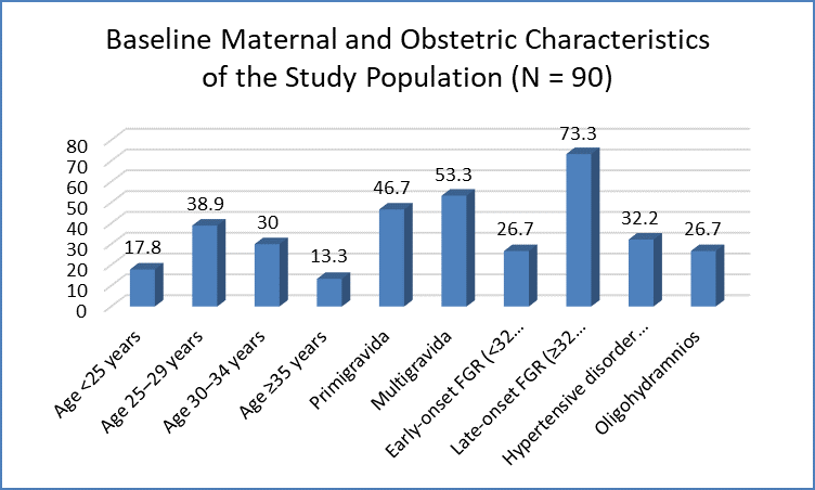
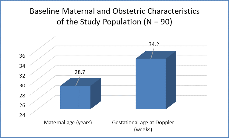
Figure 1 Baseline Maternal and Obstetric Characteristics of the Study Population (N = 90)
At least one abnormal fetal Doppler parameter was identified in 50 (55.6%) pregnancies. Abnormal UA Doppler was observed in 31 (34.4%), while abnormal MCA-PI indicating cerebral redistribution was present in 35 (38.9%) cases. A reduced CPR was the most frequently observed Doppler abnormality, occurring in 41 (45.6%) pregnancies. Abnormal DV Doppler was identified in 10 (11.1%) cases. Among the 31 pregnancies with abnormal UA Doppler, AEDF and REDF were observed in 8 (8.9%) and 3 (3.3%) of the total study population, respectively (Table 2, Figure 2).
Table 2. Distribution of Fetal Doppler Findings (N = 90)
|
Doppler finding |
n (%) |
|
Any abnormal Doppler parameter |
50 (55.6) |
|
Abnormal UA-PI |
31 (34.4) |
|
Abnormal MCA-PI |
35 (38.9) |
|
Reduced CPR |
41 (45.6) |
|
Abnormal DV Doppler |
10 (11.1) |
|
UA absent end-diastolic flow |
8 (8.9) |
|
UA reversed end-diastolic flow |
3 (3.3) |
|
≥2 abnormal Doppler parameters |
29 (32.2) |
|
No Doppler abnormality |
40 (44.4) |
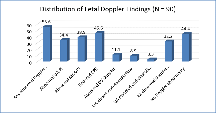
Figure 2 Distribution of Fetal Doppler Findings (N = 90)
The mean gestational age at delivery was 35.6 ± 2.5 weeks, and the mean neonatal birth weight was 1895 ± 436 g. Preterm delivery before 37 weeks occurred in 52 (57.8%) pregnancies. Cesarean delivery was performed in 58 (64.4%) cases, including 31 (34.4%) deliveries performed because of fetal compromise or a non-reassuring fetal status. A birth weight below 2000 g was observed in 49 (54.4%) neonates (Table 3).
Table 3. Delivery and Neonatal Characteristics (N = 90)
|
Characteristic |
n (%) / Mean ± SD |
|
Gestational age at delivery (weeks) |
35.6 ± 2.5 |
|
Preterm delivery (<37 weeks) |
52 (57.8) |
|
Vaginal delivery |
32 (35.6) |
|
Cesarean delivery |
58 (64.4) |
|
Cesarean for fetal compromise |
31 (34.4) |
|
Birth weight (g) |
1895 ± 436 |
|
Birth weight <2000 g |
49 (54.4) |
|
Male neonate |
49 (54.4) |
|
Female neonate |
41 (45.6) |
Overall, 46 (51.1%) neonates experienced at least one adverse neonatal outcome. NICU admission was required in 39 (43.3%) neonates, while 24 (26.7%) developed respiratory distress. A 5-minute Apgar score <7 was observed in 14 (15.6%), and 21 (23.3%) neonates required active resuscitation at birth. Respiratory support was required in 20 (22.2%) neonates. Hypoglycemia and neonatal sepsis occurred in 16 (17.8%) and 11 (12.2%) neonates, respectively. There were 4 (4.4%) neonatal deaths during hospitalization (Table 4).
Table 4. Distribution of Adverse Neonatal Outcomes (N = 90)
|
Neonatal outcome |
n (%) |
|
Any adverse neonatal outcome |
46 (51.1) |
|
NICU admission |
39 (43.3) |
|
5-minute Apgar score <7 |
14 (15.6) |
|
Need for active resuscitation |
21 (23.3) |
|
Respiratory distress |
24 (26.7) |
|
Need for respiratory support |
20 (22.2) |
|
Hypoglycemia |
16 (17.8) |
|
Metabolic acidosis |
12 (13.3) |
|
Neonatal sepsis |
11 (12.2) |
|
NICU stay ≥5 days |
25 (27.8) |
|
Neonatal mortality |
4 (4.4) |
Adverse neonatal outcomes occurred significantly more frequently among pregnancies with abnormal fetal Doppler findings. Among the 31 pregnancies with abnormal UA Doppler, 25 (80.6%) had an adverse neonatal outcome compared with 21 of 59 (35.6%) pregnancies with normal UA Doppler (p<0.001). Similarly, adverse outcomes were observed in 27 of 35 (77.1%) pregnancies with abnormal MCA-PI compared with 19 of 55 (34.5%) with normal MCA-PI (p<0.001).
A reduced CPR demonstrated a particularly strong association with adverse neonatal outcomes, with 33 of 41 (80.5%) pregnancies with abnormal CPR experiencing an adverse outcome compared with 13 of 49 (26.5%) pregnancies with normal CPR (p<0.001). Adverse neonatal outcomes were also observed in 9 of 10 (90.0%) pregnancies with abnormal DV Doppler compared with 37 of 80 (46.3%) pregnancies with normal DV findings (p=0.009). The presence of two or more abnormal Doppler parameters was associated with an adverse outcome in 26 of 29 (89.7%) cases (Table 5).
Table 5. Association of Fetal Doppler Findings With Adverse Neonatal Outcomes
|
Doppler parameter |
Abnormal Doppler n/N (%) |
Normal Doppler n/N (%) |
p-value |
|
UA-PI |
25/31 (80.6) |
21/59 (35.6) |
<0.001 |
|
MCA-PI |
27/35 (77.1) |
19/55 (34.5) |
<0.001 |
|
CPR |
33/41 (80.5) |
13/49 (26.5) |
<0.001 |
|
DV Doppler |
9/10 (90.0) |
37/80 (46.3) |
0.009 |
|
≥2 abnormal Doppler parameters |
26/29 (89.7) |
20/61 (32.8) |
<0.001 |
Receiver operating characteristic (ROC) analysis was performed to assess the ability of different Doppler parameters to predict adverse neonatal outcomes. CPR showed the highest overall discriminatory performance among the individual arterial Doppler parameters, with an AUC of 0.82 (95% CI: 0.73–0.91), sensitivity of 71.7%, and specificity of 81.8%. UA Doppler demonstrated an AUC of 0.74, with sensitivity and specificity of 54.3% and 86.4%, respectively. MCA Doppler demonstrated an AUC of 0.76, with sensitivity of 58.7% and specificity of 81.8%.
Although abnormal DV Doppler demonstrated high specificity (97.7%), its sensitivity was low (19.6%), reflecting its occurrence predominantly in advanced fetal compromise. The presence of ≥2 abnormal Doppler parameters demonstrated an AUC of 0.78, sensitivity of 56.5%, and specificity of 93.2% (Table 6, Figure 3).
Table 6. Predictive Performance of Doppler Parameters for Adverse Neonatal Outcomes
|
Doppler parameter |
AUC (95% CI) |
Sensitivity (%) |
Specificity (%) |
PPV (%) |
NPV (%) |
p-value |
|
Abnormal UA-PI |
0.74 (0.64–0.84) |
54.3 |
86.4 |
80.6 |
64.4 |
<0.001 |
|
Abnormal MCA-PI |
0.76 (0.66–0.86) |
58.7 |
81.8 |
77.1 |
65.5 |
<0.001 |
|
Reduced CPR |
0.82 (0.73–0.91) |
71.7 |
81.8 |
80.5 |
73.5 |
<0.001 |
|
Abnormal DV Doppler |
0.59 (0.51–0.68) |
19.6 |
97.7 |
90.0 |
53.8 |
0.009 |
|
≥2 abnormal Doppler parameters |
0.78 (0.69–0.87) |
56.5 |
93.2 |
89.7 |
67.2 |
<0.001 |
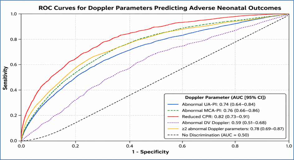
Figure 3 Predictive Performance of Doppler Parameters for Adverse Neonatal Outcomes
DISCUSSION:
The present study evaluated the predictive role of fetal Doppler parameters for adverse neonatal outcomes in 90 pregnancies complicated by fetal growth restriction (FGR). At least one abnormal Doppler parameter was present in 55.6% of pregnancies, while 51.1% of neonates developed at least one adverse outcome. Abnormal UA-PI, MCA-PI, CPR, and ductus venosus Doppler were all significantly associated with adverse neonatal outcomes. Among the evaluated parameters, reduced CPR showed the best overall predictive performance (AUC 0.82, sensitivity 71.7%, specificity 81.8%), whereas abnormal DV Doppler had the highest specificity (97.7%). The mean maternal age was 28.7 ± 4.3 years, and the mean gestational age at Doppler assessment was 34.2 ± 2.8 weeks. Early-onset FGR accounted for 26.7%, while 73.3% had late-onset FGR. Hypertensive disorders and oligohydramnios were present in 32.2% and 26.7%, respectively. Comparable evidence from the multicenter PORTO study involving 1,116 FGR pregnancies showed that 28% of neonates required NICU admission and 5.2% experienced a composite adverse perinatal outcome, particularly in association with severe growth restriction and abnormal Doppler findings. In the present study, abnormal UA-PI was observed in 34.4%, abnormal MCA-PI in 38.9%, reduced CPR in 45.6%, and abnormal DV Doppler in 11.1%.
AEDF and REDF were present in 8.9% and 3.3%, respectively. The higher frequency of abnormal CPR and MCA findings compared with severe UA and DV abnormalities may be related to the predominance of late-onset FGR. Previous guidance has reported that approximately 20% of term small-for-gestational-age fetuses with normal UA Doppler may still demonstrate reduced MCA-PI, indicating cerebral redistribution despite apparently preserved placental Doppler. The mean gestational age at delivery was 35.6 ± 2.5 weeks, with 57.8% delivered preterm. Mean birth weight was 1895 ± 436 g, and 64.4% underwent cesarean delivery. Overall, 43.3% required NICU admission, 26.7% developed respiratory distress, 23.3% required active resuscitation, 15.6% had a 5-minute Apgar score <7, and neonatal mortality was 4.4%. In the PORTO cohort, NICU admission occurred in approximately 28%, suggesting a greater burden of neonatal morbidity in the present study. Abnormal UA Doppler was strongly associated with adverse neonatal outcome, occurring in 80.6% of pregnancies with abnormal UA-PI compared with 35.6% with normal UA findings (p<0.001).
UA Doppler showed an AUC of 0.74, sensitivity of 54.3%, and specificity of 86.4%. Similarly, the PORTO study reported adverse outcomes in 11.5% of fetuses with abnormal UA Doppler compared with only 1.3% with normal UA Doppler. Abnormal MCA-PI was associated with adverse outcomes in 77.1% compared with 34.5% among those with normal MCA Doppler (p<0.001). MCA Doppler demonstrated an AUC of 0.76. A systematic review involving 128 studies and 47,748 women also demonstrated that MCA and CPR abnormalities were associated with adverse perinatal outcomes, with CPR generally performing better than MCA alone for composite outcomes. Reduced CPR showed the strongest arterial predictive value in the present study. Adverse neonatal outcomes occurred in 80.5% of pregnancies with reduced CPR compared with 26.5% with normal CPR (p<0.001). Nassr et al. [13] in a meta-analysis of 1,428 fetuses, reported that abnormal CPR was associated with increased odds of cesarean delivery for fetal distress (OR 4.49), low Apgar score (OR 4.01), NICU admission (OR 9.65), and neonatal complications (OR 11.00).
Conde-Agudelo et al. [14] analyzing 4,301 pregnancies, reported a summary AUC of 0.83, sensitivity of 93%, and specificity of 76% for CPR in predicting perinatal death, closely comparable to the present AUC of 0.82. Abnormal DV Doppler was uncommon (11.1%) but was associated with adverse outcomes in 90.0% of cases and demonstrated 97.7% specificity, supporting its role as a marker of advanced fetal compromise. This is consistent with the TRUFFLE trial of 503 very preterm FGR pregnancies, where DV-based monitoring was associated with improved survival without neurodevelopmental impairment. Overall, the findings suggest that no single Doppler parameter is sufficient in isolation. Reduced CPR provided the best overall discrimination, while abnormal DV and multiple Doppler abnormalities identified particularly high-risk fetuses. A multivessel Doppler approach integrating UA, MCA, CPR, and DV with gestational age and clinical surveillance may therefore improve risk stratification and guide timely delivery in pregnancies complicated by FGR.
CONCLUSION:
Fetal Doppler assessment was found to be a valuable tool for predicting adverse neonatal outcomes in pregnancies complicated by FGR. Abnormal UA, MCA, CPR, and DV findings were significantly associated with increased neonatal morbidity. Among these, reduced CPR showed the best overall predictive performance, while abnormal DV Doppler demonstrated the highest specificity for severe fetal compromise. A multivessel Doppler approach may therefore improve antenatal risk stratification, guide timely delivery, and help reduce adverse neonatal outcomes.
LIMITATIONS
The study was limited by its relatively small sample size and single-center design, which may restrict the generalizability of the findings. The inclusion of both early- and late-onset FGR may have introduced heterogeneity in Doppler patterns and neonatal outcomes. Long-term neurodevelopmental outcomes were not assessed, and larger multicenter prospective studies are required to validate the predictive performance of individual and combined Doppler parameters.
REFERENCES:
1. Nardozza LM, Caetano AC, Zamarian AC, Mazzola JB, Silva CP, Marçal VM, et al. Fetal growth restriction: current knowledge. Arch Gynecol Obstet. 2017;295:1061-77.
2. Placental lesions associated with maternal underperfusion are more frequent in early-onset than in late-onset preeclampsia. J Perinat Med. 2011;39:641-52.
3. Egbor M, Ansari T, Morris N, Green CJ, Sibbons PD. Morphometric placental villous and vascular abnormalities in early- and late-onset pre-eclampsia with and without fetal growth restriction. BJOG. 2006;113:580-9.
4. Parra-Saavedra M, Crovetto F, Triunfo S, Savchev S, Parra G, Sanz M, et al. Added value of umbilical vein flow as a predictor of perinatal outcome in term small-for-gestational-age fetuses. Ultrasound Obstet Gynecol. 2013;42:189-95.
5. Murray E, Fernandes M, Fazel M, Kennedy SH, Villar J, Stein A, et al. Differential effect of intrauterine growth restriction on childhood neurodevelopment: a systematic review. BJOG. 2015;122:1062-72.
6. Caradeux J, Martinez-Portilla RJ, Peguero A, Sotiriadis A, Figueras F. Diagnostic performance of third-trimester ultrasound for the prediction of late-onset fetal growth restriction: a systematic review and meta-analysis. Am J Obstet Gynecol. 2019;220:449-59.
7. Figueras F, Caradeux J, Crispi F, Eixarch E, Peguero A, Gratacos E, et al. Diagnosis and surveillance of late-onset fetal growth restriction. Am J Obstet Gynecol. 2018;218(2S):S790-S802.e1.
8. Oros D, Figueras F, Cruz-Martinez R, Padilla N, Meler E, Hernandez-Andrade E, et al. Middle versus anterior cerebral artery Doppler for the prediction of perinatal outcome and neonatal neurobehavior in term small-for-gestational-age fetuses with normal umbilical artery Doppler. Ultrasound Obstet Gynecol. 2010;35:456-61.
9. Haugen G, Godfrey K, Crozier S, Hanson M. Doppler blood flow velocity waveforms in the fetal renal arteries: variability at proximal and distal sites in the right and left arteries. Ultrasound Obstet Gynecol. 2004;23:590-3.
10. Salomon LJ, Alfirevic Z, Da Silva Costa F, Deter RL, Figueras F, Ghi T, et al. ISUOG Practice Guidelines: ultrasound assessment of fetal biometry and growth. Ultrasound Obstet Gynecol. 2019;53:715-23.
11. Hadlock FP, Harrist RB, Sharman RS, Deter RL, Park SK. Estimation of fetal weight with the use of head, body, and femur measurements: a prospective study. Am J Obstet Gynecol. 1985;151:333-7.
12. Rizzo G, Mappa I, Bitsadze V, Słodki M, Khizroeva J, Makatsariya A, et al. Role of Doppler ultrasound at time of diagnosis of late-onset fetal growth restriction in predicting adverse perinatal outcome: prospective cohort study. Ultrasound Obstet Gynecol. 2020;55:793-8.
13. Nassr AA, Abdelmagied AM, Shazly SA. Fetal cerebro-placental ratio and adverse perinatal outcome: systematic review and meta-analysis of the association and diagnostic performance. J Perinat Med. 2016;44(2):249-56.
14. Conde-Agudelo A, Villar J, Kennedy SH, Papageorghiou AT. Predictive accuracy of cerebroplacental ratio for adverse perinatal and neurodevelopmental outcomes in suspected fetal growth restriction: systematic review and meta-analysis. Ultrasound Obstet Gynecol. 2018;52(4):430-41.