Evaluation of Intraocular Pressure Changes Across Different Stages Of Chronic Kidney Disease And Their Clinical Correlation: A Hospital-Based Cross-Sectional Observational Study.
- Anushree M , Assistant professor, Department of Ophthalmology, Basaveshwara Medical College and Hospital, Chitradurga, Karnataka, India.
- Ashwini K G , Assistant professor, Department of Ophthalmology, Basaveshwara Medical College and Hospital, Chitradurga, Karnataka, India.
- Gagan Kumar H A , Postgraduate Student, Department of Ophthalmology, Basaveshwara Medical College and Hospital, Chitradurga, Karnataka, India.
Article Information:
Abstract:
Background: Chronic kidney disease (CKD) is a progressive systemic disorder associated with multiple vascular and metabolic abnormalities that can affect various organs, including the eye. Alterations in body fluid balance, plasma osmolarity, vascular endothelial function, and ocular perfusion may influence intraocular pressure (IOP), thereby increasing the risk of glaucoma and other optic nerve disorders. Although several studies have investigated ocular manifestations in CKD, limited Indian data are available regarding the relationship between CKD severity and intraocular pressure. Objectives: 1. To evaluate intraocular pressure in patients with chronic kidney disease. 2. To assess the association between intraocular pressure and different stages of chronic kidney disease. 3. To correlate intraocular pressure with renal function parameters, dialysis status, and associated systemic comorbidities. Materials and Methods: A hospital-based cross-sectional observational study was conducted among 50 patients diagnosed with chronic kidney disease attending the Departments of Ophthalmology and Nephrology of a tertiary care teaching hospital. After obtaining informed consent, demographic details, clinical history, CKD stage, duration of disease, dialysis status, serum creatinine, estimated glomerular filtration rate (eGFR), and associated comorbidities were recorded. Comprehensive ophthalmic examination, including Goldmann applanation tonometry for intraocular pressure measurement, was performed in all patients. Data were analyzed using IBM SPSS Statistics version 26.0. Results: The mean age of the study population was 53.8 ± 11.6 years, and males constituted 64% of the participants. Stage 5 CKD was observed in 50% of patients, while hypertension and diabetes mellitus were present in 70% and 56%, respectively. Mean intraocular pressure increased significantly with advancing CKD stage, measuring 15.2 ± 1.8 mmHg in Stage 3, 16.5 ± 2.1 mmHg in Stage 4, and 18.4 ± 2.7 mmHg in Stage 5 (ANOVA, p = 0.001). Serum creatinine demonstrated a significant positive correlation with intraocular pressure (r = 0.52, p < 0.001), whereas eGFR showed a significant negative correlation (r = –0.48, p = 0.001). Patients receiving maintenance hemodialysis had significantly higher mean intraocular pressure than non-dialysis patients (18.1 ± 2.4 mmHg vs. 16.2 ± 2.0 mmHg, p = 0.003). Conclusion: Advancing chronic kidney disease is associated with progressive elevation of intraocular pressure, particularly among patients undergoing maintenance hemodialysis. Significant correlations between intraocular pressure and renal function parameters suggest that worsening kidney function adversely influences ocular physiology. Routine ophthalmic screening, including intraocular pressure assessment and glaucoma evaluation, should be incorporated into the comprehensive management of patients with chronic kidney disease to facilitate early detection of ocular complications and prevent irreversible vision loss.
Keywords:
Article :
INTRODUCTION:
Chronic kidney disease (CKD) is a progressive and irreversible disorder characterized by structural or functional abnormalities of the kidneys persisting for more than three months, resulting in a gradual decline in glomerular filtration rate (GFR) and impairment of metabolic, endocrine, and excretory renal functions. ¹ CKD has emerged as a major global public health challenge because of its increasing prevalence, high morbidity, mortality, and economic burden. According to the Global Burden of Disease (GBD) Study, CKD is among the fastest-growing causes of death worldwide, affecting approximately 10–15% of the adult population, with diabetes mellitus and hypertension accounting for nearly two-thirds of all cases. ¹˒² The increasing life expectancy and rising prevalence of metabolic disorders have further contributed to the escalating burden of CKD across both developed and developing countries. ²
The kidney and the eye share several embryological, anatomical, physiological, and microvascular characteristics. Both organs possess specialized vascular networks with autoregulatory mechanisms that maintain tissue perfusion despite fluctuations in systemic blood pressure. Consequently, systemic diseases affecting the renal microvasculature often manifest as ocular abnormalities involving the retina, optic nerve, and anterior chamber structures. ³ Similar pathogenic mechanisms such as endothelial dysfunction, oxidative stress, chronic inflammation, activation of the renin–angiotensin–aldosterone system, and vascular calcification contribute to damage in both renal and ocular tissues. ³˒⁴
Intraocular pressure (IOP) represents the pressure exerted by the aqueous humour within the globe and is determined by the dynamic balance between aqueous humour production by the ciliary body and its drainage through the trabecular meshwork and uveoscleral pathways.⁵ Maintenance of normal IOP is essential for preserving optic nerve function and retinal perfusion. Although elevated IOP is the most important modifiable risk factor for glaucoma, variations in systemic hydration, plasma osmolarity, blood pressure, renal function, and fluid balance may also influence IOP. ⁵ These physiological interactions become particularly relevant in patients with CKD, who frequently experience alterations in body fluid volume, electrolyte balance, and vascular homeostasis.
Patients with CKD undergo multiple metabolic and hemodynamic changes capable of influencing ocular physiology. Fluid overload, hypoalbuminemia, increased venous pressure, anaemia, electrolyte disturbances, secondary hyperparathyroidism, and repeated haemodialysis sessions can alter aqueous humour dynamics and ocular perfusion pressure. ⁴˒⁶ Haemodialysis-induced osmotic shifts may result in transient increases or decreases in intraocular pressure depending on plasma osmolarity, ultrafiltration volume, and anterior chamber anatomy. Several studies have demonstrated variable changes in IOP before and after dialysis, indicating that renal replacement therapy itself may influence ocular biomechanics and optic nerve health. ⁶
Recent epidemiological evidence has highlighted an important association between CKD and glaucoma. A systematic review and meta-analysis involving nearly two million participants demonstrated that individuals with CKD have significantly higher odds of developing glaucoma, while patients with glaucoma also exhibit an increased long-term risk of developing CKD, suggesting a bidirectional relationship between the two conditions.⁷ Shared mechanisms include vascular dysregulation, oxidative stress, endothelial injury, chronic inflammation, genetic susceptibility, and abnormalities in the renin–angiotensin system.⁷ These findings emphasize the importance of ophthalmic evaluation in patients with chronic renal disease. Globally, CKD affects approximately one in ten adults and continues to increase because of the growing burden of diabetes, hypertension, obesity, and aging populations.¹˒² In India, CKD has become a significant public health concern, with recent community-based meta-analytic evidence estimating an overall prevalence of approximately 13.2%, with even higher prevalence reported in rural populations and southern India.² The coexistence of CKD with diabetes mellitus and hypertension, both established risk factors for glaucoma, further increases the likelihood of ocular complications among Indian patients. Despite this, routine ophthalmological assessment in CKD patients remains underutilized, and available Indian literature evaluating intraocular pressure changes across different stages of CKD is limited.²
Understanding the relationship between CKD and intraocular pressure has important clinical implications. Early identification of abnormal IOP may facilitate timely diagnosis of glaucoma, enable appropriate monitoring during dialysis, and help prevent irreversible optic nerve damage. Furthermore, correlating IOP changes with renal function parameters, CKD stage, duration of disease, dialysis status, and associated systemic illnesses may provide valuable insights into the pathophysiological interactions between renal dysfunction and ocular health. Therefore, the present study was undertaken to evaluate intraocular pressure changes in patients with chronic kidney disease and to determine their clinical correlation in a tertiary care hospital setting. ³˒⁷
Aim
To evaluate intraocular pressure changes in patients with chronic kidney disease and determine their clinical correlation with the stage of CKD, renal function parameters, and associated systemic comorbidities.
Objectives
Primary Objective
1. To evaluate intraocular pressure (IOP) in patients with chronic kidney disease attending a tertiary care hospital.
Secondary Objectives
2. To assess the association between intraocular pressure and the severity (stage) of chronic kidney disease.
3. To correlate intraocular pressure with renal function parameters (serum creatinine and estimated glomerular filtration rate), dialysis status, and associated systemic comorbidities such as diabetes mellitus and hypertension.
MATERIALS AND METHODS:
Study Design
A Hospital-based cross-sectional observational study.
Study Setting
The study will be conducted in the Departments of Ophthalmology and Nephrology.
Sample Size
A total of 50 patients with chronic kidney disease will be included in the study.
Sample Size Calculation
The sample size was calculated using the formula for estimating a proportion:
Where:
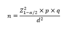
n = Required sample size, Z = Standard normal variate at 95% confidence interval = 1.96
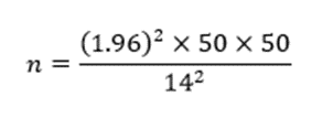
p = Expected prevalence of ocular manifestations in CKD = 50% (chosen to obtain the maximum sample size in the absence of precise local prevalence), q = 100 − p = 50
d = Absolute precision = 14%
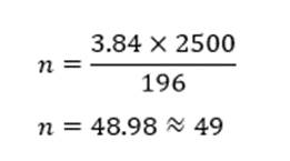
After rounding off and accounting for possible incomplete data, the final sample size will be 50 participants.
Inclusion Criteria
• Patients aged 18 years and above.
• Patients diagnosed with chronic kidney disease according to KDIGO guidelines.
• Patients willing to provide written informed consent.
• Patients with CKD irrespective of dialysis status.
Exclusion Criteria
• Known primary glaucoma or ocular hypertension.
• History of ocular trauma or intraocular surgery within the previous six months.
• Corneal disorders affecting accurate Goldmann applanation tonometry.
• Active ocular infection or inflammation.
• Patients receiving topical or systemic corticosteroids.
• Patients with congenital ocular anomalies.
• Patients unwilling to participate.
Methodology
After obtaining approval from the Institutional Ethics Committee and written informed consent, eligible patients fulfilling the inclusion criteria will be enrolled consecutively until the desired sample size of 50 is achieved.
A detailed clinical history including age, sex, duration of chronic kidney disease, history of diabetes mellitus, hypertension, dialysis status, and current medications will be recorded using a structured proforma.
General physical examination and systemic examination will be performed.
Renal disease severity will be assessed using:
• Serum creatinine
• Blood urea
• Estimated glomerular filtration rate (eGFR)
• CKD stage according to KDIGO classification
All patients will undergo a comprehensive ophthalmic examination including:
• Best corrected visual acuity (BCVA)
• Slit-lamp biomicroscopy
• Goldmann applanation tonometry for intraocular pressure measurement
• Gonioscopy (when indicated)
• Fundus examination using indirect ophthalmoscopy and slit-lamp biomicroscopy with a 90D lens
For patients undergoing maintenance haemodialysis, intraocular pressure will be measured immediately before and within one hour after completion of dialysis whenever feasible.
The mean intraocular pressure of both eyes will be recorded for statistical analysis.
Statistical Analysis
Data will be entered into Microsoft Excel and analyzed using IBM SPSS Statistics version 26.0. Continuous variables will be expressed as mean ± standard deviation (SD). Categorical variables will be expressed as frequency and percentage. Independent Student's t-test or Mann–Whitney U test will be used to compare continuous variables between two groups. One-way ANOVA or Kruskal–Wallis test will be used for comparison among different CKD stages. Chi-square test or Fisher's exact test will be used for categorical variables. Pearson's or Spearman's correlation coefficient will be used to assess the relationship between intraocular pressure and renal function parameters. A p-value <0.05 will be considered statistically significant.
RESULTS:
Table 1. Demographic Characteristics and Clinical Profile of Patients with Chronic Kidney Disease (n = 50)
|
Variable |
Frequency (n) |
Percentage (%) |
|
Age Group (years) |
||
|
18–30 |
4 |
8.0 |
|
31–45 |
12 |
24.0 |
|
46–60 |
21 |
42.0 |
|
>60 |
13 |
26.0 |
|
Gender |
||
|
Male |
32 |
64.0 |
|
Female |
18 |
36.0 |
|
Mean age (years) |
53.8 ± 11.6 |
|
|
Duration of CKD |
||
|
<2 years |
11 |
22.0 |
|
2–5 years |
20 |
40.0 |
|
>5 years |
19 |
38.0 |
|
Hemodialysis |
||
|
Yes |
24 |
48.0 |
|
No |
26 |
52.0 |
Interpretation: The mean age of the study participants was 53.8 ± 11.6 years, with most patients (42%) belonging to the 46–60 years age group. Males constituted 64% of the study population. Nearly half of the participants (48%) were receiving maintenance haemodialysis.
Table 2. Distribution of Chronic Kidney Disease Stages and Associated Comorbidities (n = 50)
|
Variable |
Frequency (n) |
Percentage (%) |
|
CKD Stage |
||
|
Stage 3 |
8 |
16.0 |
|
Stage 4 |
17 |
34.0 |
|
Stage 5 |
25 |
50.0 |
|
Diabetes Mellitus |
28 |
56.0 |
|
Hypertension |
35 |
70.0 |
|
Both DM and HTN |
21 |
42.0 |
Interpretation: Half of the patients had Stage 5 CKD, indicating advanced renal disease. Hypertension (70%) was the most common comorbidity, followed by diabetes mellitus (56%).
Table 3. Comparison of Mean Intraocular Pressure Across Different Stages of Chronic Kidney Disease
|
CKD Stage |
n |
Mean IOP (mmHg) ± SD |
p-value |
|
Stage 3 |
8 |
15.2 ± 1.8 |
0.001 |
|
Stage 4 |
17 |
16.5 ± 2.1 |
|
|
Stage 5 |
25 |
18.4 ± 2.7 |
Interpretation: The mean intraocular pressure increased progressively with advancing CKD stage. Patients with Stage 5 CKD had significantly higher mean IOP compared with those in Stages 3 and 4 (p = 0.001).
Table 4. Correlation Between Intraocular Pressure and Renal Function Parameters
|
Variable |
Correlation Coefficient (r) |
p-value |
|
Serum Creatinine |
0.52 |
<0.001 |
|
eGFR |
–0.48 |
0.001 |
|
Duration of CKD |
0.39 |
0.005 |
Interpretation: A moderate positive correlation was observed between intraocular pressure and serum creatinine (r = 0.52), while eGFR showed a moderate negative correlation (r = –0.48). These findings suggest that worsening renal function is associated with increasing intraocular pressure.
Table 5. Comparison of Intraocular Pressure According to Haemodialysis Status
|
Hemodialysis Status |
n |
Mean IOP (mmHg) ± SD |
p-value |
|
On Hemodialysis |
24 |
18.1 ± 2.4 |
0.003 |
|
Not on Hemodialysis |
26 |
16.2 ± 2.0 |
Interpretation: Patients undergoing maintenance hemodialysis demonstrated a significantly higher mean intraocular pressure than those not receiving dialysis (18.1 ± 2.4 mmHg vs. 16.2 ± 2.0 mmHg; p = 0.003). This finding suggests that fluid and osmotic changes associated with dialysis may influence intraocular pressure.
DISCUSSION:
The present hospital-based cross-sectional observational study evaluated intraocular pressure and its clinical correlation among 50 patients with chronic kidney disease. The principal findings were a progressive rise in mean IOP with advancing CKD stage, a positive correlation of IOP with serum creatinine and duration of CKD, a negative correlation with eGFR, and significantly higher IOP among patients receiving maintenance haemodialysis. Together, these observations suggest that worsening renal dysfunction and dialysis-related physiological changes may influence aqueous humour dynamics and ocular pressure.
The mean age of the study population was 53.8 ± 11.6 years, and the largest proportion of patients belonged to the 46–60-year age group. Males constituted 64% of the participants. This age distribution was expected because CKD commonly becomes clinically apparent during middle and older age, particularly in individuals with longstanding diabetes mellitus, hypertension, or both. In the Singapore Malay Eye Study, Nongpiur et al. evaluated the relationship between CKD, IOP and glaucoma in a population-based sample and reported a mean IOP of approximately 15.4 ± 3.7 mmHg. CKD remained independently associated with higher IOP even after adjustment for age, diabetes and glaucoma status.⁸ This supports the possibility that renal impairment itself may influence IOP independently of traditional ocular risk factors.
In the present study, hypertension was observed in 70%, diabetes mellitus in 56%, and coexistence of diabetes and hypertension in 42% of patients. These findings indicate that vascular and metabolic disorders constituted the major associated conditions in the study population. Both hypertension and diabetes can affect renal and ocular microcirculation through endothelial dysfunction, oxidative stress, basement-membrane changes and altered autoregulation. Therefore, a proportion of the observed IOP variation may reflect the combined influence of CKD severity and associated systemic vascular disease. Nevertheless, the association of CKD with higher IOP has been demonstrated even after adjustment for diabetes and other confounders. ⁸
Half of the participants had Stage 5 CKD, while 34% had Stage 4 and 16% had Stage 3 disease. The mean IOP increased progressively from 15.2 ± 1.8 mmHg in Stage 3 to 16.5 ± 2.1 mmHg in Stage 4 and 18.4 ± 2.7 mmHg in Stage 5. This difference was statistically significant by one-way ANOVA (F = 8.96, p = 0.001). The progressive elevation suggests that increasing renal dysfunction may be accompanied by alterations in fluid volume, plasma osmolarity, venous pressure and aqueous humour outflow.
The findings are clinically important because even IOP values falling within the conventionally accepted normal range may carry additional risk when accompanied by repeated pressure fluctuations or reduced ocular perfusion pressure. Hu et al. studied IOP and ocular perfusion during haemodialysis and demonstrated a statistically significant increase in IOP together with a reduction in systolic, diastolic and mean ocular perfusion pressures. The authors observed that a substantial proportion of eyes reached perfusion-pressure levels associated with glaucoma development or progression.⁹ These findings indicate that the ocular consequences of renal failure may depend not only on the absolute IOP value but also on its interaction with systemic blood pressure and optic-nerve perfusion.
In the present study, serum creatinine showed a moderate positive correlation with IOP (r = 0.52, p < 0.001), while eGFR demonstrated a moderate negative correlation (r = −0.48, p = 0.001). IOP also increased with longer CKD duration (r = 0.39, p = 0.005). These correlations reinforce the stage-wise analysis and suggest that declining renal filtration capacity is associated with increasing ocular pressure. Accumulation of uraemic solutes, extracellular fluid expansion, altered colloid osmotic pressure and vascular dysregulation may contribute to this relationship.
However, published findings regarding IOP changes in renal failure and during haemodialysis have been heterogeneous. Doshiro et al. reported an overall reduction in IOP during haemodialysis and suggested that increasing plasma colloid osmotic pressure promoted movement of fluid from the eye into the circulation. They also observed that patients undergoing dialysis for longer periods were more likely to demonstrate a post-dialysis rise in IOP.¹⁰ This variation indicates that the direction of IOP change may depend on the relative magnitude of plasma osmolarity reduction, colloid osmotic pressure elevation, ultrafiltration and aqueous outflow resistance. Wang et al. evaluated IOP changes according to anterior-chamber angle configuration and found that patients with narrow angles were more susceptible to dialysis-related IOP elevation than those with wider angles.¹¹ This is relevant because rapid reduction in plasma osmolarity during dialysis may create an osmotic gradient between the plasma and intraocular fluids, encouraging movement of water into the aqueous compartment. When aqueous outflow is adequate, this additional fluid can be drained; in eyes with compromised or narrow angles, however, IOP may rise substantially.
A meta-analysis by Chen et al. confirmed that the effect of haemodialysis on IOP varied according to study period, dialysis technique, baseline angle status and timing of pressure measurement.¹² Earlier dialysis protocols were more often associated with IOP elevation, whereas some studies using modern slower dialysis techniques reported stable or decreased IOP. This inconsistency explains why individual patient characteristics and standardized measurement timing are essential when evaluating IOP in dialysis populations.
The present study found that patients receiving maintenance haemodialysis had a significantly higher mean IOP than non-dialysis patients—18.1 ± 2.4 mmHg versus 16.2 ± 2.0 mmHg, respectively (t = 3.08, p = 0.003). This difference may partly reflect the greater CKD severity of the dialysis group, as most haemodialysis patients are likely to have Stage 5 disease. Therefore, dialysis status and CKD stage may exert overlapping effects and should ideally be evaluated using multivariable analysis in larger studies.
Kilavuzoglu et al. examined patients before and after haemodialysis and demonstrated significant changes in ocular and systemic parameters during treatment.¹³ Their observations highlighted that ultrafiltration, changes in body weight, blood pressure and serum osmolarity could alter IOP during a dialysis session. Similarly, Chelala et al. assessed visual acuity, macular thickness and IOP in patients with CKD undergoing haemodialysis and reported measurable ocular changes after dialysis, emphasizing that renal replacement therapy can influence both anterior- and posterior-segment parameters.¹⁴
Samsudin et al. also investigated the effect of haemodialysis on IOP and reported that pressure responses were not uniform across all patients.¹⁵ The variability across studies may be related to differences in dialysis duration, ultrafiltration volume, dialysate composition, systemic blood pressure, hydration status, anterior-chamber anatomy, timing of tonometry and presence of glaucoma. Consequently, a single pre- or post-dialysis IOP measurement may fail to identify clinically relevant intradialytic pressure peaks. Although the current study focused on IOP rather than confirmed glaucoma, the findings have implications for glaucoma risk.
Hsu et al., in a nationwide propensity-matched cohort involving 723,216 patients with CKD and an equal number of controls, reported a higher risk of newly diagnosed glaucoma in patients with CKD (adjusted hazard ratio 1.29; 95% CI 1.26–1.32). Haemodialysis patients had an adjusted hazard ratio of approximately 1.98 for glaucoma compared with non-CKD individuals.¹⁶ These large-scale data support regular ophthalmic surveillance in advanced CKD and dialysis patients. Ro et al. similarly reported that CKD was a significant risk factor for open-angle glaucoma and that the association became stronger with increasing CKD severity.¹⁷ Such findings are consistent with the progressive rise in mean IOP across CKD stages observed in the present study. Nevertheless, IOP alone does not establish glaucoma; optic-disc evaluation, retinal nerve-fibre-layer imaging and visual-field assessment remain necessary for diagnosing glaucomatous optic neuropathy.
Levy et al., in a review of IOP during haemodialysis, concluded that published studies had demonstrated increases, decreases and unchanged IOP values during dialysis.¹⁸ They emphasized that patients with glaucoma, narrow angles or impaired aqueous drainage may be at greater risk of clinically significant pressure elevation. This review supports an individualized approach rather than assuming a uniform dialysis-related IOP response.
The higher IOP observed among haemodialysis patients in the present study should therefore be interpreted as the combined effect of advanced renal disease, fluid and osmotic changes and possible variation in aqueous drainage. Repeated fluctuations may be particularly harmful when accompanied by dialysis-related hypotension, because the resulting reduction in ocular perfusion pressure could compromise blood flow to the optic nerve even when IOP is not markedly elevated.
CONCLUSION:
The present study demonstrated that intraocular pressure tends to increase with advancing stages of chronic kidney disease. A significant positive correlation was observed between intraocular pressure and serum creatinine, while an inverse correlation was found with estimated glomerular filtration rate, indicating that worsening renal function is associated with higher intraocular pressure. Patients undergoing maintenance haemodialysis exhibited significantly higher intraocular pressure than those not receiving dialysis, suggesting that dialysis-related fluid and osmotic changes may influence ocular physiology. Hypertension and diabetes mellitus were the predominant comorbidities among patients with CKD and may further contribute to ocular vascular alterations. These findings emphasize the importance of routine ophthalmic evaluation, including intraocular pressure measurement and glaucoma screening, in patients with chronic kidney disease, particularly those with advanced disease and those undergoing haemodialysis. Early identification and appropriate management of ocular changes may help prevent irreversible visual impairment and improve the overall quality of life in this high-risk population.
REFERENCES:
1. Kidney Disease: Improving Global Outcomes (KDIGO) CKD Work Group. KDIGO 2024 Clinical Practice Guideline for the Evaluation and Management of Chronic Kidney Disease. Kidney Int Suppl. 2024;14(Suppl 1):S1-S150.
2. Talukdar R, Ajayan R, Gupta S, Biswas S, Parveen M, Sadhukhan D, et al. Chronic Kidney Disease Prevalence in India: A Systematic Review and Meta-Analysis From Community-Based Representative Evidence Between 2011 to 2023. Nephrology (Carlton). 2025;30:e14420. doi:10.1111/nep.14420.
3. Cheung N, Wong TY. Ocular manifestations of systemic vascular diseases. Lancet. 2014;383:1410-1421.
4. Liu W, Guo R, Huang D, Ji J, Gansevoort RT, Snieder H, et al. Co-occurrence of chronic kidney disease and glaucoma: Epidemiology and etiological mechanisms. Surv Ophthalmol. 2023;68(1):1-16. doi:10.1016/j.survophthal.2022.09.001.
5. Weinreb RN, Aung T, Medeiros FA. The pathophysiology and treatment of glaucoma. JAMA. 2014;311(18):1901-1911. doi:10.1001/jama.2014.3192
6. Tawara A, Kobata H, Fujisawa K, Abe T, Ohnishi Y. Effects of hemodialysis on intraocular pressure and ocular perfusion. J Glaucoma. 1998;7(6):396-401.
7. Ng FYC, Song HJJD, Tan BKJ, Teo CB, Cheng CY, et al. Bidirectional association between glaucoma and chronic kidney disease: A systematic review and meta-analysis. eClinicalMedicine. 2022;49:101498. doi:10.1016/j.eclinm.2022.101498.
8. Nongpiur ME, Wong TY, Sabanayagam C, Lim SC, Tai ES, Aung T. Chronic kidney disease and intraocular pressure: the Singapore Malay Eye Study. Ophthalmology. 2010;117(3):477–483. doi:10.1016/j.ophtha.2009.07.029.
9. Hu J, Bui KM, Patel KH, Kim H, Arruda JA, Wilensky JT, et al. Effect of hemodialysis on intraocular pressure and ocular perfusion pressure. JAMA Ophthalmol. 2013;131(12):1525–1531. doi:10.1001/jamaophthalmol.2013.5599.
10. Doshiro A, Ban Y, Kobayashi L, Yoshida Y, Uchiyama H. Intraocular pressure change during hemodialysis. Am J Ophthalmol. 2006;142(2):337–339. doi:10.1016/j.ajo.2006.03.017.
11. Wang F, Wang L, Yu Z, Chen N, Wang D. Effects of hemodialysis on intraocular pressure and ocular biological parameters in different angle structures. Dis Markers. 2022;2022:9261653. doi:10.1155/2022/9261653.
12. Chen SH, Lu DW, Ku WC, Chuang LH, Ferng SH, Chen YJ, et al. Changes in intraocular pressure during hemodialysis: a meta-analysis. J Glaucoma. 2021;30(9):866–873. doi:10.1097/IJG.0000000000001842.
13. Kilavuzoglu AEB, Yurteri G, Guven N, Marsap S, Celebi ARC, Cosar CB. The effect of hemodialysis on intraocular pressure. Adv Clin Exp Med. 2018;27(1):105–110. doi:10.17219/acem/68234.
14. Chelala E, Dirani A, Fadlallah A, Slim E, Abdelmassih Y, Fakhoury H, et al. Effect of hemodialysis on visual acuity, intraocular pressure, and macular thickness in patients with chronic kidney disease. Clin Ophthalmol. 2015;9:109–114. doi:10.2147/OPTH.S74481.
15. Samsudin A, Mimiwati Z, Soong T, Fauzi MS, Zabri K. Effect of haemodialysis on intraocular pressure. Eye (Lond). 2010;24(1):70–73. doi:10.1038/eye.2009.33.
16. Hsu AY, Shao YC, Lin CJ, Chen HS, Lin CL, Cho DY, et al. Evaluating the risk of new-onset glaucoma in chronic kidney disease patients: a nationwide cohort study. BMC Nephrol. 2024;25(1):263. doi:10.1186/s12882-024-03698-5.
17. Ro JS, Lee CJ, Shin JH, Lee YH, Chung HS, Kim JA. Association between chronic kidney disease and open-angle glaucoma in South Korea: a 12-year nationwide retrospective cohort study. Sci Rep. 2022;12:2198. doi:10.1038/s41598-022-07190-8.
18. Levy J, Tovbin D, Lifshitz T, Zlotnik M, Tessler Z. Intraocular pressure during haemodialysis: a review. Eye (Lond). 2005;19(12):1249–1256. doi:10.1038/sj.eye.6701755.