A Prospective Observational Study Of Various Anatomical Positions Of Appendix In Cases Of Acute Appendicitis In A Tertiary Care Hospital.
- Asutosh Swain , Senior Resident, MS (General Surgery), Department of General Surgery, M. R. Bangur Super Speciality Hospital, 249, Deshapran Sasmal Road, Tollygunge, Kolkata – 700033.
- Ritam Mukhopadhyay , Assistant Professor, MS (General Surgery), Department of General Surgery, Jhargram Government Medical College and Hospital, Vidyasagar Pally, Jhargram, District – Jhargram, West Bengal – 721507.
- Abhishek Roy , Senior Resident, MS (General Surgery), Department of General Surgery, College of Medicine and Sagore Dutta Hospital, B. T. Road, Kamarhati, Kolkata - 700058, West Bengal.
Article Information:
Abstract:
Introduction: Acute appendicitis is one of the most common abdominal surgical emergencies. The anatomical position of the vermiform appendix varies considerably among individuals and may influence clinical presentation, diagnostic accuracy, operative difficulty, and postoperative outcomes. Variations such as retrocecal, pelvic, subcecal, pre-ileal, and post-ileal positions can produce atypical symptoms and may contribute to delayed diagnosis or surgical challenges. Aims and objectives: To study various anatomical positions of the appendix in acute appendicitis and correlate these positions with clinical features, symptoms, and presentation patterns for improving diagnosis and management. Materials and Methods: A hospital-based prospective observational study was conducted in the Department of General Surgery, Calcutta National Medical College and Hospital, Kolkata, over 18 months. A total of 115 patients with acute appendicitis undergoing emergency appendectomy were enrolled and evaluated using a structured proforma after ethical approval. Results: Among 115 patients, acute appendicitis was most common in the 10–20 years age group (48, 41.80%) with female predominance (59, 51.30%). Appendix length was mainly 7–12 cm (71, 61.80%). Anorexia (85, 74%) was the commonest symptom. Pelvic appendix was predominant, and appendix position significantly influenced clinical presentation (p<0.0001) but not complications (p=0.2457). Conclusion: Understanding anatomical variations of the appendix is essential for accurate diagnosis and effective surgical management of acute appendicitis. Recognition of these variations may help surgeons anticipate operative challenges, improve clinical decision-making, and reduce postoperative complications.
Keywords:
Article :
INTRODUCTION:
The vermiform appendix is a narrow, worm-like structure present in humans and certain primates. It is considered a vestigial organ; however, it contains abundant lymphoid tissue and may contribute to immune function. The appendix is one of the most variable organs in the human body, showing significant variations in length, position, peritoneal attachment, and relation with surrounding structures.
It originates from the posteromedial wall of the cecum, approximately 2 cm below the ileocecal valve, at the site of convergence of the three taenia coli. The length of the appendix varies from 2 cm to 20 cm, with an average length of approximately 9 cm. The base of the appendix remains relatively constant, whereas the position of its tip varies considerably within the abdominal cavity [1].
Acute appendicitis is one of the most common surgical emergencies and remains the leading cause of acute abdomen requiring operative management, particularly among young individuals. Although the diagnosis is primarily based on clinical examination and evaluation, anatomical variations in the position of the appendix often make diagnosis difficult.
The classical clinical presentation includes periumbilical pain migrating to the right lower quadrant, anorexia, nausea, vomiting, fever, and localized tenderness. However, atypical appendix positions may produce variable clinical signs and symptoms, leading to delayed diagnosis and increased risk of complications [2].
The appendix can occupy different anatomical positions, including retrocecal, pelvic, subcecal, pre-ileal, retroileal, and ectopic locations [3]. These variations influence the clinical manifestations of acute appendicitis and may mimic other abdominal or pelvic disorders. A pelvic appendix may present with urinary symptoms or resemble pelvic inflammatory disease, ovarian pathology, torsion of ovarian cyst, or ruptured ectopic pregnancy. A retroileal appendix may mimic ureteric colic, whereas a subhepatic appendix may resemble hepatobiliary disease [4].
Therefore, knowledge of the anatomical position of the appendix is essential for accurate diagnosis and appropriate management. The first comprehensive anatomical evaluation of appendix position demonstrated considerable variation among individuals. Earlier studies based on anatomical dissections and operative observations have reported differences in the frequency of various appendix positions.
The most common position of the appendix remains controversial, with studies reporting different predominant locations among different populations. Variations related to age, sex, ethnicity, geographic region, and dietary factors have been suggested to influence appendix position [3]. Acute appendicitis occurring in an unusual anatomical position may present with atypical symptoms and may be misdiagnosed, resulting in delayed treatment and increased incidence of perforation and complications [4].
The clinical course of appendicitis in relation to appendix position remains debated. Some studies have suggested that retrocecal appendices may have a different clinical presentation, while others have reported no significant difference in disease progression or complication rates [5]. These variations highlight the importance of understanding the relationship between appendix anatomy and clinical outcomes.
The position of the appendix is also important from a surgical perspective. Anatomical variations may increase operative difficulty, prolong operative time, require modification of surgical approach, and influence postoperative recovery. A misplaced or unusually located appendix may require additional dissection during appendectomy and may increase the risk of complications. Awareness of these anatomical variations is therefore important for preoperative planning, especially during laparoscopic procedures [6].
To study various anatomical positions of the appendix in acute appendicitis and correlate these positions with clinical features, symptoms, and presentation patterns for improving diagnosis and management.
MATERIALS AND METHODS:
Study design- Hospital-based, prospective observational study
Place of study- Case selection and research analysis was done in The Department of General Surgery in Calcutta National Medical College and Hospital, Kolkata.
Study period- 18 months’ duration, starting after getting clearance from hospital ethical committee and acceptance from the West Bengal University of Health Sciences.
Study population- All patients admitted at surgery emergency with features of acute appendicitis planned for emergency appendectomy.
Sample size- A total of 115 patients was included in the study. Then patient was evaluated according to the proforma attached.
Inclusion Criteria:
• Age group-10-65 year
• All patients presenting to emergency with RIF pain and clinically diagnosed as acute appendicitis after ruling out other causes of RIF pain.
• Radiologically diagnosed acute appendicitis or its sequelae.
Exclusion Criteria:
• All patients with RIF pain other than appendicitis.
• All patients with well localized appendicular lump.
• All patients with appendicular abscess.
• All Patients unfit for General anesthesia or Spinal anesthesia.
Study Variable:
• Age and Gender
• Appendix Position and Length
• Clinical Symptoms and Signs
• Ultrasonography and Intraoperative Findings
• Complicated and Uncomplicated Appendicitis
Statistical Analysis: Data were entered into Microsoft Excel and analyzed using SPSS version 27.0 and GraphPad Prism version 5. Continuous variables were expressed as mean ± standard deviation, while categorical variables were presented as frequency and percentage.
Independent and paired t-tests were used for comparison of numerical variables. One-way ANOVA was applied for comparison among multiple groups. Categorical variables were analyzed using Chi-square test or Fisher’s exact test. Non-parametric data were analyzed using Mann–Whitney U test.
Pearson correlation analysis was used to assess relationships between variables, and logistic regression was performed to identify associated risk factors. A p-value ≤0.05 was considered statistically significant.
RESULTS:
Table 1: Age and Gender-wise Distribution of Study Participants (N = 115)
|
|
Number of Cases (n) |
Percentage of Cases (%) |
|
|
Age Group (Years) |
10–20 |
48 |
41.80% |
|
21–30 |
33 |
28.70% |
|
|
31–40 |
16 |
14.00% |
|
|
41–50 |
8 |
6.90% |
|
|
51–60 |
8 |
6.90% |
|
|
61–65 |
2 |
1.70% |
|
|
Gender |
Male |
56 |
48.70% |
|
Female |
59 |
51.30% |
|
Table 2: Distribution of Appendix Length, Symptoms, and Clinical Signs Among Study Participants (N = 115)
|
|
Number of Cases (n) |
Percentage of Cases (%) |
|
|
Appendix Length (cm) |
<7 cm |
18 |
15.60% |
|
7–12 cm |
71 |
61.80% |
|
|
>12 cm |
26 |
22.60% |
|
|
Symptoms |
Fever |
69 |
60.00% |
|
Anorexia |
85 |
74.00% |
|
|
Nausea |
47 |
41.00% |
|
|
Vomiting |
35 |
30.00% |
|
|
Clinical Signs |
Rovsing Sign |
41 |
35.60% |
|
Psoas Test |
23 |
20.00% |
|
|
Obturator Test |
26 |
22.60% |
|
Table 3: Distribution of Ultrasonography Findings and Atypical Symptoms Among Study Participants (N = 115)
|
|
Frequency (n) |
Percentage (%) |
|
|
USG Findings |
Acute Appendicitis |
36 |
31.30% |
|
Subacute Appendicitis |
34 |
29.50% |
|
|
Probe Tenderness |
10 |
8.60% |
|
|
Not Detected |
35 |
30.40% |
|
|
Atypical Symptoms |
Urinary Symptoms |
16 |
13.90% |
|
Gynecological Symptoms |
6 |
5.20% |
|
|
Bowel Disturbances |
5 |
4.30% |
|
Table 4: Distribution of Site of Maximum Pain, Peritoneal Relation, and Intraoperative Findings Among Study Participants (N = 115)
|
|
Frequency (n) |
Percentage (%) |
|
|
Site of Maximum Pain |
Right Iliac Fossa (RIF) |
97 |
84.30% |
|
Right Flank (RF) |
11 |
9.50% |
|
|
Right Lumbar Region (RL) |
5 |
4.30% |
|
|
Diffuse Abdomen (D) |
2 |
1.70% |
|
|
Relation with Peritoneal Cavity |
Intraperitoneal |
101 |
87.80% |
|
Extraperitoneal |
14 |
12.10% |
|
|
Intraoperative Findings |
Inflamed Appendix |
110 |
95.60% |
|
Adhesions |
34 |
29.50% |
|
|
Distended Appendix |
7 |
6.10% |
|
|
Fixed Appendix |
15 |
13.00% |
|
|
Gangrenous Appendix |
15 |
13.00% |
|
|
Perforation |
13 |
11.30% |
|
Table 5: Association Between Appendix Position and Typical/Atypical Clinical Presentation (N = 115)
|
Appendix Position |
Typical (n) |
Atypical (n) |
p-value |
|
Pelvic |
29 |
12 |
<0.0001 |
|
Retrocaecal |
15 |
14 |
|
|
Pre-Ileal |
18 |
0 |
|
|
Post-Ileal |
3 |
10 |
|
|
Paracaecal |
8 |
0 |
|
|
Subcaecal |
4 |
0 |
|
|
Subhepatic |
0 |
2 |
|
|
Left-sided |
0 |
1 |
Table 6: Association between Appendix Position and Complicated/Uncomplicated Appendicitis (N = 115)
|
Appendix Position |
Complicated (n) |
Uncomplicated (n) |
p-value |
|
Pelvic |
13 |
28 |
0.2457 |
|
Retrocaecal |
10 |
19 |
|
|
Pre-Ileal |
3 |
15 |
|
|
Post-Ileal |
2 |
11 |
|
|
Paracaecal |
1 |
7 |
|
|
Subcaecal |
0 |
4 |
|
|
Subhepatic |
0 |
2 |
|
|
Left-sided |
1 |
0 |
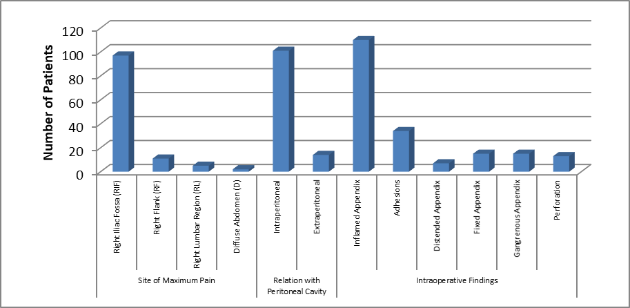
Figure 1: Distribution of Site of Maximum Pain, Peritoneal Relation, and Intraoperative Findings Among Study Participants
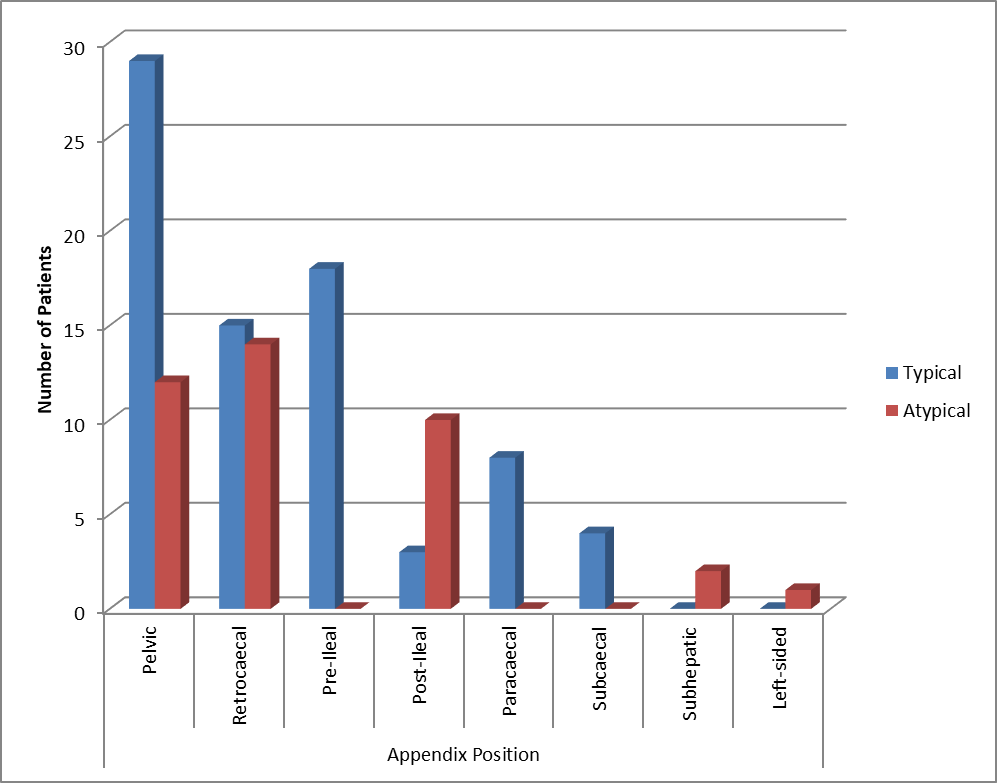
Figure 2: Association between Appendix Position and Typical/Atypical Clinical Presentation
Age and Gender-wise Distribution of Study Participants
Results:
Among the 115 study participants, the highest number of cases was observed in the 10–20 years age group (48 cases, 41.80%), followed by 21–30 years (33 cases, 28.70%). The distribution of cases decreased progressively with increasing age, with 16 cases (14.00%) in the 31–40 years group, 8 cases each (6.90%) in the 41–50 years and 51–60 years groups, and 2 cases (1.70%) in the 61–65 years age group. Regarding gender distribution, female patients constituted 59 cases (51.30%), whereas male patients accounted for 56 cases (48.70%).
Interpretation:
Acute appendicitis was predominantly observed among younger age groups, with more than two-thirds of cases occurring between 10–30 years. A slight female predominance was noted, with females representing 51.30% of the study population compared with 48.70% males.
Distribution of Appendix Length, Symptoms, and Clinical Signs Among Study Participants
Results:
The appendix length was most commonly 7–12 cm, observed in 71 cases (61.80%), followed by >12 cm in 26 cases (22.60%) and <7 cm in 18 cases (15.60%). Among presenting symptoms, anorexia was the most frequent symptom reported in 85 cases (74.00%), followed by fever in 69 cases (60.00%), nausea in 47 cases (41.00%), and vomiting in 35 cases (30.00%). Among clinical signs, Rovsing sign was positive in 41 cases (35.60%), followed by obturator test positivity in 26 cases (22.60%) and psoas test positivity in 23 cases (20.00%).
Interpretation:
The majority of patients had an appendix length between 7–12 cm. Classical symptoms of acute appendicitis, particularly anorexia (74.00%) and fever (60.00%), were commonly observed. Clinical signs were present in a smaller proportion of patients, indicating variability in physical examination findings.
Distribution of Ultrasonography Findings and Atypical Symptoms Among Study Participants
Results:
Ultrasonography findings showed acute appendicitis in 36 cases (31.30%), subacute appendicitis in 34 cases (29.50%), and probe tenderness in 10 cases (8.60%). In 35 cases (30.40%), ultrasonography did not detect definitive findings. Among atypical symptoms, urinary symptoms were present in 16 cases (13.90%), followed by gynecological symptoms in 6 cases (5.20%) and bowel disturbances in 5 cases (4.30%).
Interpretation:
Ultrasonography identified acute or subacute appendicitis in approximately 60% of cases, while nearly one-third of patients had non-diagnostic findings. Atypical symptoms were less frequent but clinically important due to their potential to mimic other abdominal or pelvic disorders.
Distribution of Site of Maximum Pain, Peritoneal Relation, and Intraoperative Findings Among Study Participants
Results:
The most common site of maximum pain was the right iliac fossa (97 cases, 84.30%), followed by right flank (11 cases, 9.50%), right lumbar region (5 cases, 4.30%), and diffuse abdominal pain (2 cases, 1.70%). Regarding appendix relation with the peritoneal cavity, 101 cases (87.80%) were intraperitoneal, while 14 cases (12.10%) were extraperitoneal. Intraoperative findings revealed inflamed appendix in 110 cases (95.60%), adhesions in 34 cases (29.50%), fixed appendix in 15 cases (13.00%), gangrenous appendix in 15 cases (13.00%), perforation in 13 cases (11.30%), and distended appendix in 7 cases (6.10%).
Interpretation:
Most patients presented with classical right iliac fossa pain (84.30%) and had an intraperitoneal appendix (87.80%). Intraoperative examination showed inflammation as the predominant finding (95.60%), while advanced pathological changes such as gangrene and perforation were observed in a smaller proportion.
Association Between Appendix Position and Typical/Atypical Clinical Presentation
Results:
A statistically significant association was observed between appendix position and typical or atypical clinical presentation (p < 0.0001). The pelvic appendix was the most common position, accounting for 41 cases, of which 29 cases (70.70%) presented typically and 12 cases (29.30%) presented atypically.
The retrocaecal appendix was observed in 29 cases, with 15 typical presentations (51.70%) and 14 atypical presentations (48.30%). The pre-ileal appendix showed only typical presentations (18 cases, 100%). The post-ileal appendix demonstrated predominantly atypical presentations (10 cases, 76.90%), with only 3 typical cases (23.10%). Subhepatic and left-sided appendix positions were associated exclusively with atypical presentations (2 cases and 1 case, respectively).
Interpretation:
The findings demonstrate that appendix position significantly affects clinical presentation. Unusual positions such as post-ileal, subhepatic, and left-sided appendix were more commonly associated with atypical symptoms, which may contribute to delayed diagnosis.
Association Between Appendix Position and Complicated/Uncomplicated Appendicitis
Results:
The association between appendix position and complicated appendicitis was statistically non-significant (p = 0.2457). Pelvic appendix had 13 complicated cases (31.70%) and 28 uncomplicated cases (68.30%) among 41 cases. Retrocaecal appendix showed 10 complicated cases (34.50%) and 19 uncomplicated cases (65.50%) among 29 cases. Pre-ileal appendix had 3 complicated cases (16.70%) and 15 uncomplicated cases (83.30%).
Post-ileal appendix showed 2 complicated cases (15.40%) and 11 uncomplicated cases (84.60%). Paracaecal appendix had 1 complicated case (12.50%) and 7 uncomplicated cases (87.50%), while subcaecal and subhepatic positions showed no complicated cases. The left-sided appendix had 1 complicated case (100%), but the sample size was very small.
Interpretation:
Although complication rates varied among different appendix positions, the association was not statistically significant (p = 0.2457). Therefore, appendix position alone does not appear to be an independent determinant of complications in acute appendicitis. Other factors such as duration of symptoms, severity of inflammation, and delayed presentation may influence disease outcome.
DISCUSSION:
Acute appendicitis remains one of the most common causes of acute abdomen requiring emergency surgical intervention. In the present prospective observational study involving 115 patients, acute appendicitis was predominantly observed among younger individuals, with the highest incidence in the 10–20 years age group (48 cases, 41.80%), followed by 21–30 years (33 cases, 28.70%).
A slight female predominance was observed, with 59 females (51.30%) and 56 males (48.70%). Similar demographic patterns have been reported in recent studies, where acute appendicitis showed a higher incidence among adolescents and young adults. Bhangu et al. emphasized that appendicitis remains a major surgical emergency worldwide, with peak incidence occurring in younger age groups [7]. Ferris et al. also demonstrated that age-related variation significantly influences the epidemiology and clinical presentation of appendicitis [8].
The vermiform appendix is considered one of the most variable organs of the gastrointestinal tract, showing considerable variation in length, position, and relationship with surrounding structures. In the present study, appendix length was most commonly between 7–12 cm (71 cases, 61.80%), followed by >12 cm (26 cases, 22.60%) and <7 cm (18 cases, 15.60%). Recent anatomical studies have highlighted that variations in appendix morphology and position may significantly influence clinical manifestations and surgical approach.
Vieira ED et al. reported that appendiceal location is highly variable and that anatomical differences may contribute to atypical clinical presentations and diagnostic challenges [9]. In the present study, anorexia was the most frequent symptom (85 cases, 74.00%), followed by fever (69 cases, 60.00%), nausea (47 cases, 41.00%), and vomiting (35 cases, 30.00%). Classical symptoms of appendicitis are usually related to inflammatory progression from visceral irritation to localized peritoneal involvement.
However, anatomical variations may modify symptom localization and severity. Echevarria et al. reported that atypical presentations of appendicitis are associated with delayed diagnosis and increased diagnostic uncertainty, particularly in patients with abnormal appendix locations [10].
Ultrasonography findings in the current study showed acute appendicitis in 36 cases (31.30%), subacute appendicitis in 34 cases (29.50%), probe tenderness in 10 cases (8.60%), while 35 cases (30.40%) had no definitive ultrasonographic detection. These findings demonstrate the diagnostic limitations of ultrasonography in patients with variable appendix anatomy. Di Saverio et al. highlighted that although imaging plays an important role in appendicitis diagnosis, anatomical variations and operator dependency may influence diagnostic accuracy [11].
The present study observed that the majority of patients presented with classical right iliac fossa pain (97 cases, 84.30%), while atypical pain locations including right flank (11 cases, 9.50%) and right lumbar region (5 cases, 4.30%) were also documented. Most appendices were intraperitoneal (101 cases, 87.80%), whereas 14 cases (12.10%) were extraperitoneal. Variations in appendix position may alter the site of inflammation and produce symptoms mimicking other abdominal, urinary, or gynecological disorders. Echevarria et al. emphasized that atypical appendix locations can result in unusual clinical presentations and may delay appropriate management [10].
Intraoperative evaluation in the present study demonstrated that inflamed appendix was the most common finding (110 cases, 95.60%), followed by adhesions (34 cases, 29.50%), fixed appendix (15 cases, 13.00%), gangrenous appendix (15 cases, 13.00%), perforation (13 cases, 11.30%), and distension (7 cases, 6.10%). These findings indicate the importance of early diagnosis and intervention to prevent progression to complicated appendicitis. Di Saverio et al. reported that delayed diagnosis and advanced inflammation are major contributors to perforation and postoperative complications [11].
A major finding of the present study was the statistically significant association between appendix position and typical/atypical clinical presentation (p <0.0001). The pelvic appendix was the most common position, accounting for 41 cases, among which 29 cases (70.70%) showed typical presentation and 12 cases (29.30%) showed atypical presentation. Retrocaecal appendix demonstrated 15 typical cases (51.70%) and 14 atypical cases (48.30%). The post-ileal appendix was predominantly associated with atypical presentation (10 cases, 76.90%). Subhepatic and left-sided appendix positions also showed atypical presentations.
Similar findings were reported by Gomes et al., who demonstrated that appendiceal position influences clinical manifestations and may create diagnostic difficulty [12]. Ferrie et al. also described that retrocaecal and unusual appendix positions may mimic other abdominal conditions and delay diagnosis [13]. The present study further evaluated the relationship between appendix position and complicated appendicitis. Although pelvic and retrocaecal positions showed higher absolute numbers of complications (13 cases and 10 cases respectively), the association was statistically non-significant (p = 0.2457).
This suggests that appendix position alone may not determine disease severity. Sakpal et al. reported that complications of appendicitis are multifactorial and are influenced by duration of symptoms, inflammatory severity, and delay in treatment rather than anatomical position alone [14]. Recent surgical literature has also emphasized that anatomical variations of the appendix are important during laparoscopic appendectomy.
Sabry AA et al. reported that unusual appendix positions may increase operative complexity and require modification of surgical technique [15]. Similarly, Kaur et al. highlighted that atypical appendix locations can mimic other abdominal emergencies and require increased clinical awareness for appropriate diagnosis and surgical planning [16].
CONCLUSION:
The present prospective observational study concludes that anatomical variations in the position of the appendix significantly influence the clinical presentation of acute appendicitis. Pelvic and retrocaecal positions were the most frequently observed, while atypical appendix locations were associated with unusual symptoms and diagnostic difficulty.
A significant association was found between appendix position and typical or atypical clinical presentation, highlighting the importance of anatomical knowledge during evaluation. However, appendix position alone was not significantly associated with complicated appendicitis. Understanding these variations can assist clinicians in improving diagnostic accuracy, reducing delays in treatment, and planning appropriate surgical management for acute appendicitis.
REFERENCES:
1. Sabiston DC, Townsend CM, Beauchamp R, Evers BM, Mattox KL. Sabiston textbook of surgery: the biological basis of modern surgical practice. Philadelphia: Wb Saunders; 2001.
2. Schwartz SI, editor. Principles of surgery: Pretest self-assessment and review. McGraw-Hill, Health Professions Division; 1999.
3. Denjalic A, Delic J, Delic-Custendil S, Muminagic S. Variations in position and place of formation of appendix vermiformis found in the course of open appendectomy. Medical Archives. 2009 Mar 1;63(2):100.
4. Ramsden WH, Mannion RA, Simpkins KC, DeDombal FT. Is the appendix where you think it is—and if not does it matter?. Clinical radiology. 1993 Feb 1;47(2):100-3.
5. Chaudhari ML, Kanani S. Anatomical variations of vermiform appendix in Gujarat. Int J Anat Res. 2018;6(1.1):4815-8.
6. Grunditz T, Ryden CI, Janzon L. Does the retrocecal position influence the course of acute appendicitis.Actachirurgicascandinavica. 1983 Jan 1;149(7):707-10.
7. Bhangu A, Søreide K, Di Saverio S, Assarsson JH, Drake FT. Acute appendicitis: modern understanding of pathogenesis, diagnosis, and management. The Lancet. 2015 Sep 26;386(10000):1278.
8. Ferris M, Quan S, Kaplan BS, Molodecky N, Ball CG, Chernoff GW, Bhala N, Ghosh S, Dixon E, Ng S, Kaplan GG. The global incidence of appendicitis: a systematic review of population-based studies. Annals of surgery. 2017 Aug 1;266(2):237-41.
9. Vieira ED, Bonato LM, da Silva GG, Gurgel JL. Congenital abnormalities and anatomical variations of the vermiform appendix and mesoappendix. Journal of Coloproctology. 2019 Sep;39(03):279-87.
10. Echevarria S, Rauf F, Hussain N, Zaka H, Ahsan N, Broomfield A, Akbar A, Khawaja UA, Farwa UE. Typical and atypical presentations of appendicitis and their implications for diagnosis and treatment: a literature review. Cureus. 2023 Apr 2;15(4).
11. Di Saverio S, Podda M, De Simone B, Ceresoli M, Augustin G, Gori A, Boermeester M, Sartelli M, Coccolini F, Tarasconi A, De’Angelis N. Diagnosis and treatment of acute appendicitis: 2020 update of the WSES Jerusalem guidelines. World journal of emergency surgery. 2020 Apr 15;15(1):27.
12. Gomes CA, Junior CS, de Peixoto RO, Netto JM, Gomes CC, Gomes FC. Appendiceal stump closure by metal endoclip in the management of complicated acute appendicitis. World Journal of Emergency Surgery. 2013 Sep 18;8(1):35.
13. Ferrie A, Barghash M, Mansour M. A retrocaecal appendix presenting with recurrent right upper quadrant pain (RUQ): a rare presentation of a rather common surgical pathology. Cureus. 2025 Oct 4;17(10).
14. Sakpal SV, Bindra SS, Chamberlain RS. Laparoscopic appendectomy conversion rates two decades later: an analysis of surgeon and patient-specific factors resulting in open conversion. Journal of Surgical Research. 2012 Jul 1;176(1):42-9.
15. Sabry AA, Menesy M, Kassem M, Elkeleny MR. The outcomes of laparoscopic versus open appendectomies for complicated appendicitis. The Egyptian Journal of Surgery. 2023 Oct 6;42(3).