Optic Nerve Sheath Diameter as a Non-Invasive Predictor of Perioperative Intracranial Pressure Changes.
- Syeda Maryam Quadri , Senior Resident Department of Anaesthesiology ESIC Medical College, Kalaburagi, Karnataka
- Veereshkumar Gobburkar , Senior Resident Department of Anesthesiology ESIC Medical College, Gulbarga, Karnataka
- J Vennela , Senior Resident Department of Anesthesiology ESIC Medical College, Gulbarga, Karnataka.
Article Information:
Abstract:
Background: Intracranial pressure (ICP) monitoring is essential in neurosurgical patients; however, invasive monitoring techniques are associated with limitations and risks. Ultrasonographic measurement of optic nerve sheath diameter (ONSD) has emerged as a promising non-invasive surrogate marker for detecting intracranial pressure variations. The present study evaluated the utility of ONSD as a predictor of perioperative ICP changes. Methods: This prospective observational study included 120 patients undergoing elective neurosurgical procedures under general anaesthesia. Bilateral ONSD measurements were performed using ultrasonography at predefined perioperative time points: preoperatively, after induction of anaesthesia, after endotracheal intubation, intraoperatively, and postoperatively. Associations between ONSD changes and clinical, radiological, and perioperative variables were analysed. Results: The mean baseline bilateral ONSD was 5.10 ± 0.40 mm, which increased significantly after induction (5.26 ± 0.43 mm) and reached maximum values after intubation (5.44 ± 0.46 mm; p<0.001). Higher ONSD values were significantly associated with hydrocephalus, midline shift, tumour size, and elevated end-tidal carbon dioxide levels. ROC analysis demonstrated that an ONSD cut-off value of ≥5.6 mm predicted significant ICP changes with 82.5% sensitivity, 78.4% specificity, and AUC of 0.86 (95% CI: 0.79–0.92). Conclusion: Ultrasonographic ONSD measurement is a simple, repeatable, and effective non-invasive tool for assessing perioperative ICP changes. It may serve as a valuable adjunct for neuromonitoring and early detection of intracranial hypertension.
Keywords:
Article :
INTRODUCTION:
Intracranial pressure (ICP) regulation is a critical determinant of cerebral homeostasis, and its monitoring plays an essential role in the perioperative management of patients undergoing neurosurgical procedures and other high-risk surgeries. Maintenance of normal ICP is necessary to preserve adequate cerebral perfusion pressure and prevent secondary neurological injury. Perioperative factors such as induction of anaesthesia, airway manipulation, positive pressure ventilation, alterations in arterial carbon dioxide levels, surgical positioning, and operative stimulation may influence cerebral blood flow and intracranial dynamics, potentially resulting in clinically significant changes in ICP.[1]Traditionally, intracranial pressure assessment relies on invasive monitoring techniques, including intraventricular catheters and intraparenchymal pressure sensors, which are considered the gold standard.[2]
However, these methods are associated with limitations such as risk of infection, haemorrhage, technical complexity, and restricted use in patients without clear indications for invasive monitoring. Therefore, there is increasing interest in developing accurate, reproducible, and non-invasive methods for evaluating intracranial pressure variations, particularly in perioperative and critical care settings.[2,3]The optic nerve sheath diameter (ONSD) has emerged as a promising non-invasive surrogate marker of ICP. Anatomically, the optic nerve sheath is a continuation of the intracranial dura mater and surrounds the optic nerve with a subarachnoid space containing cerebrospinal fluid (CSF). Any increase in intracranial CSF pressure is transmitted along this pathway, resulting in expansion of the optic nerve sheath.[4]
Ultrasonographic measurement of ONSD, particularly approximately 3 mm posterior to the globe, provides a simple bedside method for detecting changes associated with raised ICP.Point-of-care ultrasound (POCUS)-guided ONSD assessment offers several advantages, including rapid availability, repeatability, absence of radiation exposure, and feasibility in anaesthetized or critically ill patients. Unlike invasive monitoring, it allows serial evaluation of dynamic intracranial pressure changes during different perioperative phases, including preoperative assessment, induction, intraoperative events, and postoperative recovery.[5,6]The clinical relevance of ONSD measurement is particularly important in patients with intracranial pathologies such as brain tumours, traumatic brain injury, intracranial haemorrhage, hydrocephalus, and conditions associated with altered cerebrospinal fluid dynamics. In these patients, subtle increases in ICP may remain undetected clinically until neurological deterioration occurs.[6]
Early identification of intracranial pressure changes may facilitate timely intervention and improve perioperative outcomes.Neurofibromatosis type 2 (NF2), recently termed NF2-related schwannomatosis, is a rare autosomal dominant tumour predisposition syndrome caused by pathogenic variants of the NF2 gene on chromosome 22q12, resulting in dysfunction of the tumour suppressor protein merlin.[7]
Patients commonly develop bilateral vestibular schwannomas along with multiple intracranial tumours, including meningiomas, which may contribute to increased intracranial tumour burden, cerebrospinal fluid obstruction, venous compression, and subsequent elevation of ICP. Surgical management of these lesions requires careful perioperative neurological assessment and monitoring.[8-10]Although magnetic resonance imaging and computed tomography are essential for identifying structural abnormalities, they do not provide continuous information regarding intracranial pressure changes. ONSD measurement may therefore serve as a valuable adjunct for perioperative monitoring in patients with intracranial tumours, allowing early detection of dynamic ICP alterations.[11,12]
The present study, titled “Optic Nerve Sheath Diameter as a Non-Invasive Predictor of Perioperative Intracranial Pressure Changes,” aims to evaluate the role of ultrasonographic ONSD measurement as a reliable, bedside, non-invasive indicator of perioperative ICP variations.
MATERIALS AND METHODS:
The present study was conducted as a prospective observational study to evaluate the role of optic nerve sheath diameter (ONSD) measurement as a non-invasive predictor of perioperative intracranial pressure (ICP) changes. The study was carried out in the Department of Anaesthesiology The study was conducted over a predefined study period after obtaining approval.from the Institutional Ethics Committee. Written informed consent was obtained from all participants before enrolment in the study.
Study Population
A total of 120 patients undergoing elective neurosurgical procedures requiring general anaesthesia were included in the study. Patients were assessed during the perioperative period, and serial ultrasonographic measurements of optic nerve sheath diameter were performed to evaluate changes suggestive of variations in intracranial pressure.
Study Objectives
Primary Objective
The primary objective of the study was:
• To evaluate perioperative changes in optic nerve sheath diameter measured by ultrasonography as a surrogate marker of intracranial pressure variations.
Secondary Objectives
The secondary objectives were:
• To compare ONSD values at different perioperative time points.
• To assess the association between ONSD changes and perioperative clinical parameters.
• To evaluate the feasibility and reliability of bedside ultrasonographic ONSD measurement for perioperative neuromonitoring.
• To determine factors influencing perioperative changes in ONSD measurements.
Inclusion Criteria
Patients fulfilling the following criteria were included in the study:
• Patients aged ≥18 years.
• Patients scheduled for elective neurosurgical procedures under general anaesthesia.
• Patients providing written informed consent for participation.
• Patients in whom perioperative ultrasonographic assessment of the optic nerve sheath was feasible.
• Patients with adequate preoperative imaging and clinical assessment available.
Exclusion Criteria
Patients were excluded if they had:
• Refusal to participate in the study.
• Pre-existing ocular pathology affecting optic nerve assessment, including glaucoma, optic nerve disorders, orbital trauma, or previous ocular surgery.
• Significant ocular infection or anatomical abnormalities preventing ultrasound examination.
• Emergency surgery.
• Incomplete perioperative data.
• Conditions interfering with accurate ONSD measurement.
Preoperative Assessment
All enrolled patients underwent detailed preoperative evaluation, including history, clinical examination, systemic assessment, and review of relevant investigations. Demographic characteristics such as age, sex, body mass index (BMI), American Society of Anesthesiologists (ASA) physical status, diagnosis, type of neurosurgical pathology, and planned surgical procedure were recorded.Baseline neurological assessment was performed, including evaluation of symptoms suggestive of raised intracranial pressure such as headache, vomiting, visual disturbances, altered sensorium, or focal neurological deficits.Preoperative radiological findings including tumour location, size, mass effect, ventricular compression, hydrocephalus, and other intracranial abnormalities were documented.
Ultrasonographic Measurement of Optic Nerve Sheath Diameter
Optic nerve sheath diameter measurement was performed using a high-frequency linear ultrasound probe with the patient in the supine position. A sterile ultrasound gel was applied over the closed eyelid, and the optic nerve was visualized through the temporal approach.
The ONSD was measured bilaterally approximately 3 mm posterior to the globe, perpendicular to the optic nerve axis. Measurements were obtained from both eyes, and the average value was calculated for analysis.To minimize measurement variability, all assessments were performed by an experienced investigator trained in ocular ultrasonography. Each measurement was repeated three times, and the mean value was considered for statistical evaluation.
Perioperative Assessment Protocol
ONSD measurements were recorded at predefined perioperative time points:
T0: Baseline Preoperative Measurement
• Obtained before induction of anaesthesia.
T1: After Induction of Anaesthesia
• Recorded after induction and stabilization of anaesthetic depth.
T2: After Airway Manipulation
• Recorded after endotracheal intubation.
T3: Intraoperative Measurement
• Obtained during the surgical procedure after stabilization of haemodynamic parameters.
T4: Postoperative Measurement
• Recorded after emergence from anaesthesia in the postoperative period.
Changes in ONSD values between these time points were analysed to assess perioperative variations suggestive of ICP changes.
Anaesthesia Technique and Perioperative Management
Standard monitoring including electrocardiography, non-invasive blood pressure monitoring, pulse oximetry, and end-tidal carbon dioxide monitoring was performed for all patients.
General anaesthesia was administered according to institutional protocols. Induction agents, neuromuscular blocking agents, analgesics, and maintenance anaesthetic agents were recorded. Ventilatory parameters including respiratory rate, tidal volume, and end-tidal carbon dioxide values were monitored throughout the procedure.
Intraoperative factors that could influence intracranial dynamics, including haemodynamic fluctuations, positioning, duration of surgery, and ventilation parameters, were documented.
Outcome Measures
Primary Outcome Measure
• Change in optic nerve sheath diameter during different perioperative periods.
Secondary Outcome Measures
• Correlation of ONSD changes with:
· Patient demographic characteristics.
· Type and characteristics of intracranial pathology.
· Duration of surgery.
· Haemodynamic changes.
· Ventilation parameters.
· Postoperative neurological status.
Statistical Analysis
Data collected during the study were entered into a structured database and analysed using appropriate statistical software 25.Continuous variables were expressed as mean ± standard deviation (SD) or median with interquartile range depending on data distribution. Categorical variables were presented as frequency and percentage.Normality of continuous variables was assessed using the Shapiro–Wilk test. Comparisons of repeated ONSD measurements at different perioperative time points were performed using paired statistical tests or repeated-measures analysis as appropriate.Correlation between ONSD values and perioperative variables was assessed using Pearson’s or Spearman’s correlation coefficient. Factors associated with significant ONSD changes were analysed using appropriate regression models.A p-value of <0.05 was considered statistically significant.
RESULTS:
A total of 120 patients undergoing elective neurosurgical procedures were included in the present study. The demographic and baseline clinical characteristics of the study population are summarized in Table 1. The mean age of participants was 46.8 ± 12.6 years, with the majority of patients belonging to the 41–60 years age group (51.7%). Males constituted 60.0% (n=72) of the study population, while females accounted for 40.0% (n=48). The mean BMI was 24.6 ± 3.2 kg/m². Regarding ASA physical status, most patients were classified as ASA II (58.4%), followed by ASA I (28.3%) and ASA III (13.3%). The mean duration of surgery and anaesthesia was 176.4 ± 54.8 minutes and 214.7 ± 62.5 minutes, respectively (Table 1).
The distribution of underlying neurosurgical pathologies among study participants is presented in Table 2. Intracranial tumours represented the most common indication for surgery, accounting for 68.3% (n=82) of cases. Among intracranial tumours, meningioma was the predominant pathology (31.7%), followed by glioma (18.3%), vestibular schwannoma (10.0%), and other intracranial tumours (8.3%). Cerebrovascular pathologies accounted for 15.0% of cases, while hydrocephalus or cerebrospinal fluid flow obstruction was observed in 10.0% of patients. The overall distribution of neurosurgical conditions is illustrated in Figure 1.
Serial measurements of optic nerve sheath diameter (ONSD) demonstrated significant perioperative variations. The mean bilateral ONSD increased from 5.10 ± 0.40 mm at baseline (T0) to 5.26 ± 0.43 mm after induction of anaesthesia (T1) and further increased to 5.44 ± 0.46 mm after endotracheal intubation (T2). A slight reduction was observed during the intraoperative period (5.37 ± 0.45 mm) and further decreased during the postoperative period (5.16 ± 0.40 mm). Repeated measures analysis showed a statistically significant change in ONSD values across different perioperative time points (p<0.001) (Table 3).
The pattern of perioperative ONSD changes is depicted graphically in Figure 2.Comparison of ONSD values according to clinical and radiological indicators associated with raised intracranial pressure demonstrated significantly higher ONSD measurements among patients with suggestive features. Patients presenting with headache had significantly greater ONSD values compared with those without headache (5.48 ± 0.46 mm vs 5.18 ± 0.39 mm; p<0.001). Similarly, increased ONSD values were observed in patients with vomiting, visual symptoms, hydrocephalus on imaging, and midline shift/mass effect (Table 4). The highest mean ONSD was observed among patients with hydrocephalus (5.71 ± 0.52 mm) and those with radiological evidence of mass effect (5.68 ± 0.50 mm) (Table 4).Correlation analysis revealed significant associations between maximum perioperative ONSD and several clinical and perioperative variables (Table 5).
Tumour size demonstrated a moderate positive correlation with maximum ONSD (r=0.52, p<0.001). Significant positive correlations were also observed with duration of surgery (r=0.29, p=0.001), duration of anaesthesia (r=0.31, p<0.001), and end-tidal carbon dioxide levels (r=0.36, p<0.001). Postoperative neurological status showed a significant negative correlation with maximum ONSD (r=-0.39, p<0.001), whereas mean arterial pressure variation did not demonstrate a statistically significant association (r=0.14, p=0.126) (Table 5).
The correlation trends between ONSD and perioperative variables are shown in Figure 3.The diagnostic performance of perioperative ONSD measurement for prediction of significant intracranial pressure changes was evaluated using receiver operating characteristic (ROC) analysis (Table 6). An optimal ONSD cut-off value of ≥5.6 mm demonstrated a sensitivity of 82.5% and specificity of 78.4%. The positive predictive value and negative predictive value were 74.6% and 85.4%, respectively, with an overall diagnostic accuracy of 80.8%. The area under the ROC curve (AUC) was 0.86 (95% CI: 0.79–0.92), indicating good discriminatory ability of ONSD measurement for detecting significant perioperative intracranial pressure changes (Table 6).
Table 1. Demographic and Baseline Clinical Characteristics of Study Population (N = 120)
|
Parameter |
Mean ± SD / n (%) |
|
Age (years) |
46.8 ± 12.6 |
|
Age group (years) |
|
|
18–40 years |
38 (31.7%) |
|
41–60 years |
62 (51.7%) |
|
>60 years |
20 (16.6%) |
|
Sex |
|
|
Male |
72 (60.0%) |
|
Female |
48 (40.0%) |
|
BMI (kg/m²) |
24.6 ± 3.2 |
|
ASA physical status |
|
|
ASA I |
34 (28.3%) |
|
ASA II |
70 (58.4%) |
|
ASA III |
16 (13.3%) |
|
Duration of surgery (minutes) |
176.4 ± 54.8 |
|
Duration of anaesthesia (minutes) |
214.7 ± 62.5 |
Table 2. Distribution of Underlying Neurosurgical Pathology Among Study Participants (N = 120)
|
Diagnosis |
Number of Patients (n) |
Percentage (%) |
|
Intracranial tumour |
82 |
68.3 |
|
Meningioma |
38 |
31.7 |
|
Glioma |
22 |
18.3 |
|
Vestibular schwannoma |
12 |
10.0 |
|
Other intracranial tumours |
10 |
8.3 |
|
Cerebrovascular pathology |
18 |
15.0 |
|
Hydrocephalus/CSF flow obstruction |
12 |
10.0 |
|
Other neurosurgical conditions |
8 |
6.7 |
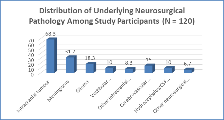
Figure 1 Distribution of Underlying Neurosurgical Pathology Among Study Participants (N = 120)
Table 3. Perioperative Changes in Optic Nerve Sheath Diameter (ONSD) Measurements (N = 120)
|
Time Point |
Right Eye ONSD (mm) Mean ± SD |
Left Eye ONSD (mm) Mean ± SD |
Mean Bilateral ONSD (mm) Mean ± SD |
p-value |
|
T0: Preoperative baseline |
5.12 ± 0.42 |
5.08 ± 0.39 |
5.10 ± 0.40 |
— |
|
T1: After induction of anaesthesia |
5.28 ± 0.44 |
5.24 ± 0.42 |
5.26 ± 0.43 |
<0.001 |
|
T2: After intubation |
5.46 ± 0.48 |
5.42 ± 0.45 |
5.44 ± 0.46 |
<0.001 |
|
T3: Intraoperative period |
5.38 ± 0.46 |
5.35 ± 0.44 |
5.37 ± 0.45 |
<0.001 |
|
T4: Postoperative period |
5.18 ± 0.41 |
5.15 ± 0.40 |
5.16 ± 0.40 |
0.041 |
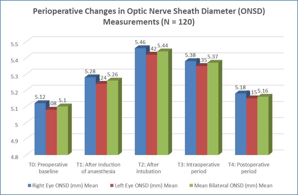
Figure 2 Perioperative Changes in Optic Nerve Sheath Diameter (ONSD) Measurements (N = 120)
Table 4. Comparison of ONSD Values According to Presence of Raised Intracranial Pressure-Related Clinical/Radiological Features (N = 120)
|
Parameter |
Present (n) |
ONSD (mm) Mean ± SD |
Absent (n) |
ONSD (mm) Mean ± SD |
p-value |
|
Headache |
54 |
5.48 ± 0.46 |
66 |
5.18 ± 0.39 |
<0.001 |
|
Vomiting |
32 |
5.56 ± 0.51 |
88 |
5.22 ± 0.40 |
<0.001 |
|
Visual symptoms |
26 |
5.62 ± 0.54 |
94 |
5.22 ± 0.40 |
<0.001 |
|
Hydrocephalus on imaging |
22 |
5.71 ± 0.52 |
98 |
5.23 ± 0.41 |
<0.001 |
|
Midline shift/mass effect |
36 |
5.68 ± 0.50 |
84 |
5.18 ± 0.38 |
<0.001 |
Table 5. Correlation of Maximum Perioperative ONSD with Clinical and Perioperative Variables (N = 120)
|
Variable |
Correlation Coefficient (r) |
p-value |
|
Age |
0.18 |
0.046 |
|
Tumour size |
0.52 |
<0.001 |
|
Duration of surgery |
0.29 |
0.001 |
|
Duration of anaesthesia |
0.31 |
<0.001 |
|
End-tidal CO₂ |
0.36 |
<0.001 |
|
Mean arterial pressure variation |
0.14 |
0.126 |
|
Postoperative neurological status |
-0.39 |
<0.001 |
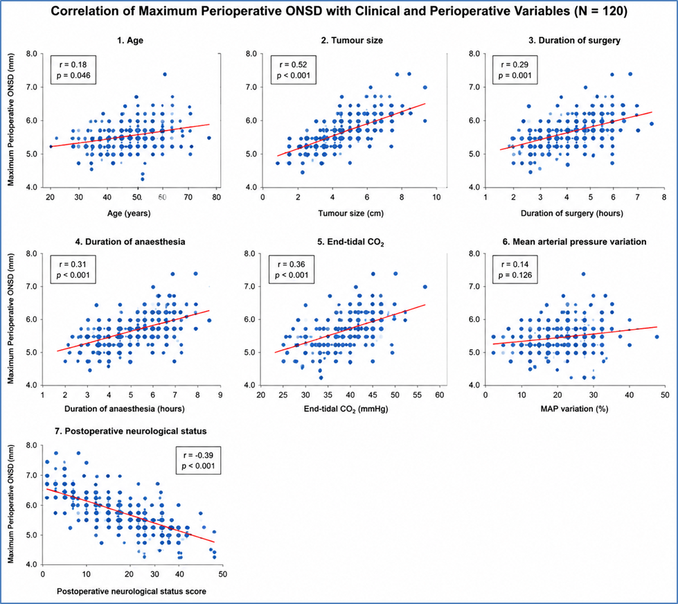
Figure 3 Correlation of Maximum Perioperative ONSD with Clinical and Perioperative Variables (N = 120)
Table 6. Diagnostic Performance of Perioperative ONSD for Prediction of Significant Intracranial Pressure Changes (N = 120)
|
Parameter |
Value |
|
Optimal ONSD cut-off value |
≥5.6 mm |
|
Sensitivity (%) |
82.5 |
|
Specificity (%) |
78.4 |
|
Positive predictive value (%) |
74.6 |
|
Negative predictive value (%) |
85.4 |
|
Overall accuracy (%) |
80.8 |
|
Area under ROC curve (AUC) |
0.86 |
|
95% Confidence interval |
0.79–0.92 |
|
p-value |
<0.001 |
DISCUSSION:
The present prospective observational study evaluated the role of optic nerve sheath diameter (ONSD) measurement as a non-invasive predictor of perioperative intracranial pressure (ICP) changes in 120 patients undergoing neurosurgical procedures. The study demonstrated significant perioperative variations in ONSD, with maximum enlargement observed after endotracheal intubation. An ONSD cut-off value of ≥5.6 mm showed good diagnostic performance for predicting significant ICP changes, with 82.5% sensitivity, 78.4% specificity, and an AUC of 0.86, supporting its clinical utility as a bedside neuromonitoring tool.
The mean age of study participants was 46.8 ± 12.6 years, with male predominance (60%) and a majority belonging to ASA II status (58.4%). Intracranial tumours constituted the most common pathology (68.3%), with meningioma being the predominant diagnosis (31.7%). Similar patient profiles have been reported in previous neurosurgical studies evaluating ONSD as an indirect marker of raised ICP. Benhur et al.[13] demonstrated dynamic perioperative changes in ONSD among patients undergoing intracranial tumour surgery and reported correlation with postoperative radiological findings.In the present study, mean bilateral ONSD increased significantly from 5.10 ± 0.40 mm at baseline to 5.26 ± 0.43 mm after induction and 5.44 ± 0.46 mm after intubation (p<0.001).
The subsequent reduction during surgery and postoperative period suggests that airway manipulation and anaesthetic-related physiological changes may transiently influence intracranial dynamics. The increase after intubation may be attributed to sympathetic stimulation, transient hypertension, increased cerebral blood flow, and alterations in intrathoracic pressure. Kim et al.[14] reported similar rapid changes in ONSD associated with variations in carbon dioxide levels, highlighting its sensitivity to cerebral haemodynamic alterations.Patients with clinical and radiological evidence of raised ICP demonstrated significantly higher ONSD values. Increased measurements were observed among patients with headache, vomiting, visual symptoms, hydrocephalus, and midline shift.
The highest ONSD values were noted in patients with hydrocephalus (5.71 ± 0.52 mm) and mass effect (5.68 ± 0.50 mm), supporting the concept that increased CSF pressure is transmitted through the optic nerve sheath. Robba et al.[15] confirmed a significant correlation between ultrasonographic ONSD measurements and invasive ICP values, supporting its role as a surrogate marker of intracranial hypertension. Berhanu et al.[16] similarly reported reliable diagnostic accuracy of ONSD ultrasonography, with optimal cut-off values ranging from 5.6 to 6.3 mm.A significant positive correlation was observed between maximum perioperative ONSD and tumour size (r=0.52, p<0.001), indicating the contribution of tumour burden and mass effect to increased ICP. ONSD also correlated significantly with duration of surgery, duration of anaesthesia, and end-tidal carbon dioxide levels, whereas mean arterial pressure variation showed no significant association.
These findings emphasise the importance of factors influencing cerebral blood flow and CSF dynamics rather than isolated systemic haemodynamic fluctuations.The diagnostic evaluation demonstrated that ONSD ≥5.6 mm predicted significant ICP changes with 74.6% positive predictive value, 85.4% negative predictive value, and 80.8% overall accuracy. These findings are consistent with previous evidence supporting ONSD as a rapid, repeatable, and non-invasive method for detecting intracranial pressure alterations. However, ONSD assessment remains operator-dependent and should complement rather than replace invasive monitoring and neuroimaging.
CONCLUSION:
The present study demonstrated that ultrasonographic measurement of optic nerve sheath diameter (ONSD) is a reliable, non-invasive method for detecting perioperative intracranial pressure changes in patients undergoing neurosurgical procedures. Significant variations in ONSD were observed during different perioperative phases, with maximum enlargement occurring after endotracheal intubation. Increased ONSD values showed significant association with tumour size, radiological markers of raised ICP, and perioperative physiological factors. An ONSD cut-off value of ≥5.6 mm demonstrated good diagnostic accuracy for predicting significant ICP changes, suggesting its potential role as a bedside neuromonitoring tool. ONSD assessment may serve as a valuable adjunct for early detection and perioperative management of intracranial hypertension.
Limitations
The study was conducted at a single centre with a relatively limited sample size, which may affect the generalizability of the findings. Direct comparison of ONSD measurements with invasive ICP monitoring was not feasible due to ethical and clinical considerations. Ultrasonographic ONSD assessment remains operator-dependent, and variations in technique may influence measurements. Further multicentric studies with larger populations and invasive ICP correlation are required to validate the findings.
REFERENCES:
1. Asthagiri AR, Parry DM, Butman JA, Kim HJ, Tsilou ET, Zhuang Z, et al. Neurofibromatosis type 2. Lancet. 2009;373(9679):1974-86.
2. Perry A, Giannini C, Raghavan R, Scheithauer BW, Banerjee R, Margraf L, et al. Aggressive phenotypic and genotypic features in pediatric and NF2-associated meningiomas: a clinicopathologic study of 53 cases. J Neuropathol Exp Neurol. 2001;60(10):994-1003.
3. Evans DG, Huson SM, Donnai D, Neary W, Blair V, Newton V, et al. A clinical study of type 2 neurofibromatosis. Q J Med. 1992;84(304):603-18.
4. Nunes FP, Merker VL, Jennings D, Caruso PA, di Tomaso E, Muzikansky A, et al. Bevacizumab treatment for meningiomas in NF2: a retrospective analysis of 15 patients. PLoS One. 2013;8(3):e59941.
5. Plotkin SR, Yohay KH, Nghiemphu PL, Dinh CT, Babovic-Vuksanovic D, Merker VL, et al. Brigatinib in NF2-related schwannomatosis with progressive tumors. N Engl J Med. 2024;390(24):2284-94.
6. Chang LS. Brigatinib causes tumor shrinkage in both NF2-deficient meningioma and schwannoma through inhibition of multiple tyrosine kinases but not ALK. PLoS One. 2021;16:e0252048.
7. Aboukais R. Causes of mortality in neurofibromatosis type 2. Br J Neurosurg. 2015;29(1):37-40.
8. Moreno ME, Del Carpio-O'Donovan R. Neuroimaging in the diagnosis and treatment of intracranial pressure disorders. Neurol Sci. 2023;44(3):845-58.
9. Kerscher SR, Zipfel J, Groeschel S, Bevot A, Haas-Lude K, Schuhmann MU, et al. Comparison of B-scan ultrasound and MRI-based optic nerve sheath diameter (ONSD) measurements in children. Pediatr Neurol. 2021;124:15-20.
10. Kerscher SR, Schöni D, Neunhoeffer F, Wolff M, Haas-Lude K, Bevot A, et al. The relation of optic nerve sheath diameter (ONSD) and intracranial pressure (ICP) in pediatric neurosurgery practice - Part II: influence of wakefulness, method of ICP measurement, intra-individual ONSD-ICP correlation and changes after therapy. Childs Nerv Syst. 2020;36(1):107-15.
11. Kerscher SR, Schöni D, Hurth H, Neunhoeffer F, Haas-Lude K, Wolff M, et al. The relation of optic nerve sheath diameter (ONSD) and intracranial pressure (ICP) in pediatric neurosurgery practice - Part I: correlations, age-dependency and cut-off values. Childs Nerv Syst. 2020;36(1):99-106.
12. Kerscher SR, Zipfel J, Haas-Lude K, Bevot A, Tellermann J, Schuhmann MU, et al. Transorbital point-of-care ultrasound versus fundoscopic papilledema to support treatment indication for potentially elevated intracranial pressure in children. Childs Nerv Syst. 2024;40(3):655-63.
13. Benhur A, Sharma J, Karna ST, Shrivastava A, Saigal S, Waindeskar VV, et al. Analysis of dynamic changes in optic nerve sheath diameter (ONSD) with ultrasound in post-craniotomy patients: trends and correlation with computed tomography ONSD and Glasgow Coma Scale in post-operative period. J Neurosci Rural Pract. 2022;13(4):676-683.
14. Kim JY, Min HG, Ha SI, Jeong HW, Seo H, Kim JU, et al. Dynamic optic nerve sheath diameter responses to short-term hyperventilation measured with sonography in patients under general anesthesia. Korean J Anesthesiol. 2014;67(4):240-245.
15. Robba C, Santori G, Czosnyka M, Corradi F, Bragazzi N, Padayachy L, et al. Optic nerve sheath diameter measured sonographically as non-invasive estimator of intracranial pressure: a systematic review and meta-analysis. Intensive Care Med. 2018;44(8):1284-1294.
16. Berhanu D, Cardoso Ferreira J, Abêgo Pinto L, Aguiar de Sousa D, Lucas Neto L, Tavares Ferreira J, et al. The role of optic nerve sheath ultrasonography in increased intracranial pressure: a systematic review and meta-analysis. J Neurol Sci. 2023;454:120853.