Diaphragmatic Ultrasound for Prediction of Successful Extubation in Mechanically Ventilated Patients.
- J Vennela , Senior Resident Department of Anesthesiology ESIC Medical College, Gulbarga, Karnataka.
- Syeda Maryam Quadri , Senior Resident Department of Anaesthesiology ESIC Medical College, Kalaburagi, Karnataka.
- Veereshkumar Gobburkar , Senior Resident Department of Anesthesiology ESIC Medical College, Gulbarga, Karnataka.
Article Information:
Abstract:
Abstract Background: Successful weaning from mechanical ventilation remains challenging in critically ill patients. Conventional weaning parameters may not accurately reflect respiratory muscle function. Diaphragmatic ultrasound provides a non-invasive bedside assessment of diaphragmatic performance and may improve prediction of extubation success. Methods: This prospective observational study included 150 mechanically ventilated patients undergoing weaning assessment in the intensive care unit. Diaphragmatic ultrasound was performed before extubation to measure diaphragmatic excursion (DE) and diaphragmatic thickening fraction (DTF). Patients were categorized into successful extubation and extubation failure groups. The predictive ability of ultrasound parameters was assessed using receiver operating characteristic (ROC) curve analysis. Results: Successful extubation was achieved in 120 (80%) patients, while 30 (20%) patients experienced extubation failure. Patients with successful extubation showed significantly higher diaphragmatic excursion (1.82 ± 0.34 cm vs 0.96 ± 0.28 cm; p<0.001) and diaphragmatic thickening fraction (38.6 ± 10.2% vs 18.4 ± 7.5%; p<0.001) compared with the failure group. ROC analysis demonstrated that DTF had the highest predictive accuracy (AUC 0.89) with a cutoff value of ≥28%, sensitivity of 87.5%, and specificity of 83.3%. Diaphragmatic excursion showed an AUC of 0.86 with a cutoff value of ≥1.4 cm. Conclusion: Diaphragmatic ultrasound, particularly DTF and diaphragmatic excursion, is a useful predictor of successful extubation in mechanically ventilated patients. It may serve as a valuable bedside adjunct to conventional weaning assessment.
Keywords:
Article :
INTRODUCTION:
Mechanical ventilation is an essential supportive therapy for critically ill patients with acute respiratory failure and inadequate spontaneous breathing effort. Although it improves gas exchange and provides life-saving respiratory support, prolonged mechanical ventilation is associated with complications such as ventilator-associated pneumonia, respiratory muscle weakness, prolonged intensive care unit (ICU) stay, increased healthcare burden, and higher mortality. Therefore, timely and successful liberation from mechanical ventilation remains a major goal in critical care practice. [1,2]Weaning from mechanical ventilation is a complex physiological process that requires adequate respiratory muscle strength, stable cardiovascular function, preserved neurological status, and effective airway protection.
Extubation failure, requiring reintubation after removal of the endotracheal tube, is associated with increased morbidity, prolonged ICU stay, and poor clinical outcomes. Although spontaneous breathing trials (SBTs) are routinely performed to assess readiness for extubation, a significant proportion of patients may fail extubation despite successful completion of an SBT due to inadequate respiratory muscle reserve. [3,4]Several conventional predictors, including the rapid shallow breathing index (RSBI), respiratory rate, tidal volume, and maximal inspiratory pressure, are used for assessing readiness for ventilator liberation. However, these parameters have limitations due to variations in critically ill patient populations and their inability to directly evaluate respiratory muscle function. Therefore, additional bedside tools capable of assessing respiratory muscle performance may improve prediction of extubation outcomes. [2,5].
The diaphragm is the principal muscle of respiration and plays a crucial role in maintaining effective spontaneous ventilation. Prolonged mechanical ventilation may cause diaphragmatic inactivity, muscle atrophy, and reduced contractility, collectively known as ventilator-induced diaphragmatic dysfunction (VIDD). Diaphragmatic weakness is an important contributor to weaning failure, as patients may tolerate a spontaneous breathing trial but fail extubation due to insufficient respiratory muscle endurance. [6,7]Assessment of diaphragmatic function using conventional methods such as transdiaphragmatic pressure measurement and phrenic nerve stimulation is invasive and technically challenging, limiting routine clinical application. Diaphragmatic ultrasonography has emerged as a simple, non-invasive, bedside, and reproducible technique for evaluating diaphragmatic structure and function in mechanically ventilated patients. [6,8]
Diaphragmatic ultrasound primarily evaluates diaphragmatic excursion (DE) and diaphragmatic thickening fraction (DTF). Diaphragmatic excursion reflects diaphragmatic displacement during inspiration, whereas thickening fraction represents the change in diaphragmatic thickness during contraction and serves as an indicator of diaphragmatic strength. Reduced excursion and decreased thickening fraction have been associated with impaired respiratory muscle function and increased risk of weaning failure. [8,9]Several studies have demonstrated the potential role of diaphragmatic ultrasound in predicting successful extubation. Farghaly and Hasan reported that diaphragmatic ultrasound parameters could predict extubation outcomes in mechanically ventilated patients, while Eltrabili et al. demonstrated its usefulness in predicting successful liberation from mechanical ventilation. [10,11].
Recent systematic reviews and meta-analyses have further suggested that diaphragmatic ultrasound, particularly DTF and DE measurements, may provide valuable prognostic information during ventilator weaning; however, differences in patient characteristics, assessment protocols, and cutoff values have resulted in variability among studies. [12–14]Early identification of patients at risk of extubation failure may facilitate individualized weaning strategies and timely initiation of supportive interventions. Therefore, diaphragmatic ultrasound may serve as a useful adjunct to conventional weaning assessment by providing direct information regarding respiratory muscle function.Hence, the present study titled “Diaphragmatic Ultrasound for Prediction of Successful Extubation in Mechanically Ventilated Patients” aims to evaluate the role of diaphragmatic ultrasound parameters in predicting extubation success and their association with clinical outcomes among mechanically ventilated patients.
MATERIALS AND METHODS:
This prospective observational study was conducted in department of anaesthesiology. The study included 150 mechanically ventilated patients to evaluate the role of diaphragmatic ultrasound in predicting successful extubation.
Study Population
Adult patients receiving invasive mechanical ventilation and considered eligible for weaning were enrolled. Patients were assessed according to standard ICU weaning protocols and underwent diaphragmatic ultrasound evaluation before planned extubation.
Inclusion Criteria
• Age ≥18 years.
• Patients requiring invasive mechanical ventilation for ≥24 hours.
• Patients eligible for spontaneous breathing trial (SBT) and planned extubation.
Exclusion Criteria
• Pre-existing diaphragmatic paralysis or neuromuscular disorders.
• Conditions interfering with ultrasound assessment.
• Tracheostomy patients.
• Pregnancy.
• Extubation without standard weaning assessment.
Weaning and Extubation Protocol
Patients were evaluated daily for readiness to wean based on improvement of the underlying disease, adequate oxygenation, hemodynamic stability, and ability to initiate spontaneous breathing. Eligible patients underwent a spontaneous breathing trial. Those successfully completing the trial were assessed for extubation.
Diaphragmatic Ultrasound Assessment
Bedside diaphragmatic ultrasonography was performed before extubation using a portable ultrasound device. Two parameters were assessed:
1. Diaphragmatic Excursion (DE):
Measured using M-mode ultrasound through the subcostal approach to assess diaphragmatic displacement during inspiration.
2. Diaphragmatic Thickening Fraction (DTF):
Measured using the zone of apposition technique. Diaphragmatic thickness was recorded during inspiration and expiration, and DTF was calculated as:
DTF (%) = [(Inspiratory thickness − Expiratory thickness) / Expiratory thickness] × 100
Three consecutive measurements were obtained, and the mean value was used for analysis.
Study Outcomes
Primary Outcome:
• Predictive ability of diaphragmatic ultrasound parameters for successful extubation.
Secondary Outcomes:
• Comparison of DE and DTF between successful and failed extubation groups.
• Diagnostic performance of diaphragmatic ultrasound using ROC curve analysis.
• Assessment of sensitivity, specificity, PPV, NPV, and accuracy of ultrasound parameters.
Extubation success was defined as absence of reintubation within 48–72 hours after extubation, while extubation failure was defined as the requirement for reintubation within this period.
Data Collection
Baseline characteristics including age, sex, diagnosis, duration of mechanical ventilation, comorbidities, ventilator parameters, and weaning characteristics were recorded. Diaphragmatic ultrasound findings were correlated with extubation outcomes.
Statistical Analysis
Continuous variables were expressed as mean ± SD or median (IQR), and categorical variables as frequency and percentage. Comparisons were performed using appropriate parametric or non-parametric tests. Chi-square/Fisher’s exact test was used for categorical variables. ROC curve analysis was performed to evaluate the predictive performance of diaphragmatic ultrasound parameters, including AUC, cutoff values, sensitivity, and specificity. A p-value <0.05 was considered statistically significant.
RESULTS:
A total of 150 mechanically ventilated patients were included in the study and underwent diaphragmatic ultrasound assessment before extubation. Based on extubation outcome, 120 (80%) patients achieved successful extubation, while 30 (20%) patients experienced extubation failure.
The demographic and clinical characteristics of the study population are presented in (Table 1). The mean age, gender distribution, and BMI were comparable between both groups. However, patients with extubation failure had significantly higher APACHE II score (20.1 ± 6.1 vs 16.4 ± 5.2; p=0.002) and SOFA score (8.1 ± 3.1 vs 6.2 ± 2.4; p=0.001). Duration of mechanical ventilation (7.6 ± 3.4 vs 4.2 ± 2.1 days; p<0.001) and ICU stay (13.4 ± 5.1 vs 8.5 ± 3.2 days; p<0.001) were significantly longer in the extubation failure group (Table 1).
Respiratory and weaning parameters before extubation are summarized in (Table 2). Patients with successful extubation had significantly lower respiratory rate (19.6 ± 3.8 vs 23.4 ± 4.5 breaths/min; p<0.001) and RSBI (54.2 ± 15.6 vs 82.5 ± 21.4 breaths/min/L; p<0.001), while tidal volume (420 ± 85 vs 365 ± 72 mL; p=0.001) and PaO₂/FiO₂ ratio (245 ± 68 vs 198 ± 55; p=0.001) were significantly higher compared with patients with extubation failure (Table 2; Figure 1).
Comparison of diaphragmatic ultrasound parameters demonstrated significantly better diaphragmatic function among successfully extubated patients. Diaphragmatic excursion (1.82 ± 0.34 cm vs 0.96 ± 0.28 cm; p<0.001) and diaphragmatic thickening fraction (38.6 ± 10.2% vs 18.4 ± 7.5%; p<0.001) were significantly higher in the successful extubation group. Inspiratory and expiratory diaphragm thickness also showed significant differences between groups (Table 3; Figure 2).
Correlation analysis revealed significant associations between diaphragmatic ultrasound parameters and clinical outcomes. Diaphragmatic excursion showed a positive correlation with extubation success (r=+0.62; p<0.001), while diaphragmatic thickening fraction demonstrated a stronger positive correlation (r=+0.68; p<0.001). Both parameters showed significant negative correlation with duration of mechanical ventilation (p<0.001). RSBI demonstrated a negative correlation with extubation success (r=-0.51; p<0.001) (Table 4; Figure 3).
ROC curve analysis demonstrated that diaphragmatic thickening fraction had the highest predictive performance for successful extubation with an AUC of 0.89 (95% CI: 0.82–0.95) at a cutoff value of ≥28%, with sensitivity of 87.5% and specificity of 83.3%. Diaphragmatic excursion showed an AUC of 0.86 (95% CI: 0.78–0.93) at a cutoff of ≥1.4 cm, with sensitivity of 84.2% and specificity of 76.7%. RSBI showed comparatively lower predictive accuracy (AUC 0.78) (Table 5; Figure 4).
On multivariate logistic regression analysis, diaphragmatic thickening fraction ≥28% (AOR 6.18; 95% CI: 2.45–15.62; p<0.001) and diaphragmatic excursion ≥1.4 cm (AOR 4.62; 95% CI: 1.85–11.54; p=0.001) were independent predictors of successful extubation. RSBI ≤65 was also associated with increased likelihood of extubation success, whereas prolonged mechanical ventilation duration and APACHE II score >20 were negative predictors (Table 6; Figure 5).
Table 1. Demographic and Clinical Characteristics of Study Population (N=150)
|
Parameter |
Successful Extubation (n=120) |
Extubation Failure (n=30) |
p-value |
|
Age (years), Mean ± SD |
54.6 ± 14.2 |
58.3 ± 15.1 |
0.214 |
|
Male gender, n (%) |
76 (63.3%) |
20 (66.7%) |
0.745 |
|
BMI (kg/m²), Mean ± SD |
24.8 ± 3.6 |
25.3 ± 3.9 |
0.521 |
|
APACHE II score |
16.4 ± 5.2 |
20.1 ± 6.1 |
0.002 |
|
SOFA score |
6.2 ± 2.4 |
8.1 ± 3.1 |
0.001 |
|
Duration of mechanical ventilation (days) |
4.2 ± 2.1 |
7.6 ± 3.4 |
<0.001 |
|
ICU stay (days) |
8.5 ± 3.2 |
13.4 ± 5.1 |
<0.001 |
Independent sample t-test ,Chi-square test
Table 2. Comparison of Respiratory and Weaning Parameters Before Extubation
|
Parameter |
Successful Extubation (n=120) |
Extubation Failure (n=30) |
p-value |
|
Respiratory rate (breaths/min) |
19.6 ± 3.8 |
23.4 ± 4.5 |
<0.001 |
|
Tidal volume (mL) |
420 ± 85 |
365 ± 72 |
0.001 |
|
RSBI (breaths/min/L) |
54.2 ± 15.6 |
82.5 ± 21.4 |
<0.001 |
|
PaO₂/FiO₂ ratio |
245 ± 68 |
198 ± 55 |
0.001 |
Independent sample t-test
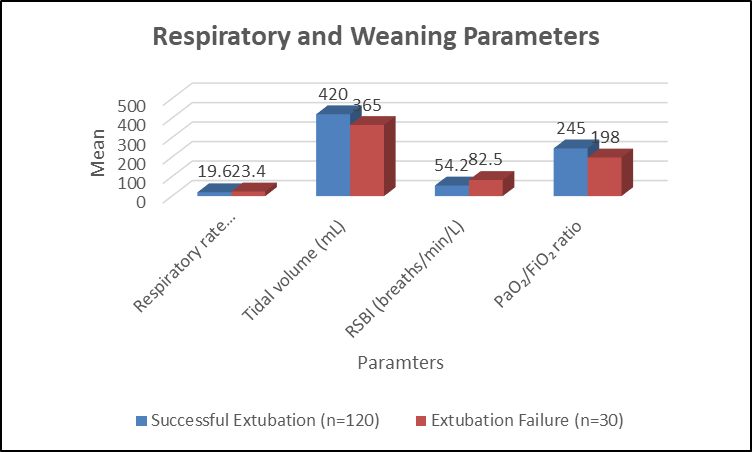
Figure 1. Comparison of Respiratory and Weaning Parameters Before Extubation
Table 3. Comparison of Diaphragmatic Ultrasound Parameters Between Extubation Groups
|
Ultrasound Parameter |
Successful Extubation (n=120) |
Extubation Failure (n=30) |
p-value |
|
Diaphragmatic excursion (cm) |
1.82 ± 0.34 |
0.96 ± 0.28 |
<0.001 |
|
Diaphragmatic thickening fraction (%) |
38.6 ± 10.2 |
18.4 ± 7.5 |
<0.001 |
|
Expiratory diaphragm thickness (mm) |
2.1 ± 0.4 |
1.8 ± 0.3 |
0.002 |
|
Inspiratory diaphragm thickness (mm) |
2.9 ± 0.5 |
2.1 ± 0.4 |
<0.001 |
Independent sample t-test
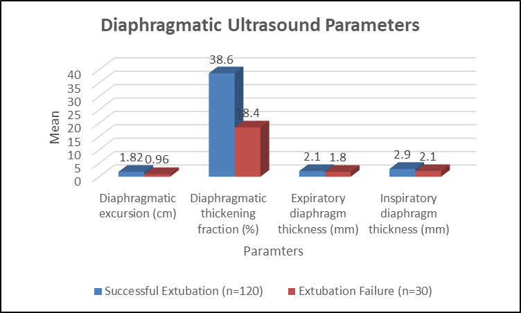
Figure 2. Comparison of Diaphragmatic Ultrasound Parameters Between Extubation Groups
Table 4. Correlation Between Diaphragmatic Ultrasound Parameters and Clinical Outcomes
|
Parameter |
Outcome Variable |
Correlation coefficient (r) |
p-value |
|
Diaphragmatic excursion |
Extubation success |
+0.62 |
<0.001 |
|
Diaphragmatic thickening fraction |
Extubation success |
+0.68 |
<0.001 |
|
Diaphragmatic excursion |
Duration of mechanical ventilation |
-0.46 |
<0.001 |
|
Diaphragmatic thickening fraction |
Duration of mechanical ventilation |
-0.52 |
<0.001 |
|
RSBI |
Extubation success |
-0.51 |
<0.001 |
Pearson correlation test
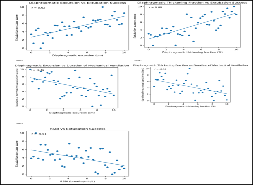
Figure 3. Correlation Between Diaphragmatic Ultrasound Parameters and Clinical Outcomes
Table 5. Diagnostic Performance of Diaphragmatic Ultrasound Parameters for Prediction of Successful Extubation
|
Parameter |
Cut-off value |
AUC (95% CI) |
Sensitivity (%) |
Specificity (%) |
p-value |
|
Diaphragmatic excursion |
≥1.4 cm |
0.86 (0.78–0.93) |
84.2 |
76.7 |
<0.001 |
|
Diaphragmatic thickening fraction |
≥28% |
0.89 (0.82–0.95) |
87.5 |
83.3 |
<0.001 |
|
RSBI |
≤65 |
0.78 (0.69–0.87) |
75.0 |
70.0 |
<0.001 |
Receiver Operating Characteristic (ROC) curve analysis
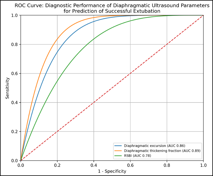
Figure 4. Diagnostic Performance of Diaphragmatic Ultrasound Parameters for Prediction of Successful Extubation
Table 6. Multivariate Logistic Regression Analysis for Predictors of Successful Extubation
|
Variable |
Adjusted Odds Ratio |
95% CI |
p-value |
|
Diaphragmatic excursion ≥1.4 cm |
4.62 |
1.85–11.54 |
0.001 |
|
Diaphragmatic thickening fraction ≥28% |
6.18 |
2.45–15.62 |
<0.001 |
|
RSBI ≤65 |
2.76 |
1.12–6.82 |
0.027 |
|
Mechanical ventilation duration >7 days |
0.38 |
0.15–0.94 |
0.036 |
|
APACHE II score >20 |
0.42 |
0.18–0.98 |
0.044 |
Multivariate binary logistic regression
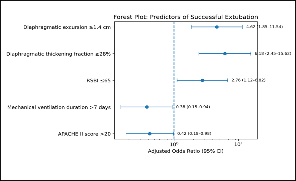
Figure 5. Multivariate Logistic Regression Analysis for Predictors of Successful Extubation
DISCUSSION:
The present study evaluated the role of diaphragmatic ultrasonography in predicting successful extubation among 150 mechanically ventilated patients. In our study, 120 (80%) patients achieved successful extubation, whereas 30 (20%) experienced extubation failure. Patients with successful extubation demonstrated significantly better diaphragmatic function, with higher diaphragmatic excursion (DE) and diaphragmatic thickening fraction (DTF), supporting the utility of diaphragmatic ultrasound as an adjunctive tool for ventilator liberation.
In the present study, diaphragmatic excursion was significantly higher in the successful extubation group compared with the failure group (1.82 ± 0.34 cm vs 0.96 ± 0.28 cm; p<0.001). ROC analysis showed good predictive ability of diaphragmatic excursion with an AUC of 0.86, cutoff value of ≥1.4 cm, sensitivity of 84.2%, and specificity of 76.7%. Yoo et al. [15] evaluated diaphragmatic excursion and thickening change using ultrasound for prediction of extubation success. They reported successful extubation in 78.3% patients and failure in 21.7% patients. Diaphragmatic excursion was significantly higher in successfully extubated patients (1.65 cm vs 0.8 cm; p<0.001) with an AUC of 0.836, suggesting that diaphragmatic mobility is a reliable predictor of extubation outcome. The similarity between their findings and our results highlights the importance of preserved diaphragmatic excursion in maintaining spontaneous breathing after extubation.
In our study, DTF was significantly higher among patients with successful extubation (38.6 ± 10.2% vs 18.4 ± 7.5%; p<0.001). DTF demonstrated the highest predictive accuracy with an AUC of 0.89, cutoff value of ≥28%, sensitivity of 87.5%, and specificity of 83.3%. Yoo et al. [15] also demonstrated significantly higher diaphragmatic thickening change among successfully extubated patients (42.1% vs 22.5%; p=0.03), supporting the role of diaphragmatic contractility assessment in predicting extubation outcome. Similarly, Mowafy and Abdelgalel [16] reported that diaphragmatic thickness was a useful predictor of successful weaning from mechanical ventilation, emphasizing that preserved diaphragmatic function is essential for ventilator liberation.In the present study, patients with successful extubation had significantly lower RSBI values (54.2 ± 15.6 vs 82.5 ± 21.4 breaths/min/L; p<0.001) and shorter duration of mechanical ventilation (4.2 ± 2.1 vs 7.6 ± 3.4 days; p<0.001). Mowafy and Abdelgalel [16]
compared diaphragmatic rapid shallow breathing index (D-RSBI) with conventional RSBI and reported that diaphragmatic-based indices improved prediction of weaning outcomes compared with traditional RSBI alone. Their findings support incorporation of diaphragmatic assessment along with conventional weaning parameters.In our study, diaphragmatic excursion showed a significant positive correlation with extubation success (r=0.62; p<0.001), while DTF demonstrated a stronger positive correlation (r=0.68; p<0.001). Both parameters showed a significant negative correlation with duration of mechanical ventilation (DE: r=-0.46; DTF: r=-0.52; p<0.001). Osman and Hashim [17] evaluated diaphragmatic and lung ultrasound parameters for predicting weaning outcomes and reported significant associations between diaphragmatic ultrasound findings and successful ventilator liberation. Their results support the role of ultrasound-based assessment of respiratory muscle function in critically ill patients.The present study demonstrated good diagnostic performance of diaphragmatic ultrasound, with DTF showing superior predictive accuracy (AUC 0.89) followed by diaphragmatic excursion (AUC 0.86). Soliman et al. [18] assessed chest ultrasound for prediction of weaning failure and demonstrated that ultrasound-based parameters could identify patients at increased risk of unsuccessful weaning. These findings support the use of bedside ultrasound as a practical and non-invasive method for guiding extubation decisions.
CONCLUSION:
Diaphragmatic ultrasonography, particularly diaphragmatic excursion and diaphragmatic thickening fraction, demonstrated significant predictive value for successful extubation in mechanically ventilated patients. Higher diaphragmatic function parameters were associated with improved extubation outcomes and shorter ventilator dependence. Diaphragmatic ultrasound may serve as a simple, non-invasive bedside adjunct to conventional weaning parameters for optimizing extubation decisions.
Limitations
The study was conducted at a single center with a limited sample size, which may restrict the generalizability of the findings. Variations in ultrasound measurements and operator dependency may have influenced the assessment of diaphragmatic parameters.
REFERENCES:
1. Boles JM, Bion J, Connors A, et al. Weaning from mechanical ventilation. Eur Respir J. 2007;29(5):1033-1056.
2. Girard TD, Alhazzani W, Kress JP, et al. Liberation from mechanical ventilation in critically ill adults. Am J Respir Crit Care Med. 2017;195(1):120-133.
3. Thille AW, Boissier F, Ben Ghezala H, et al. Risk factors for and prediction by caregivers of extubation failure in ICU patients. Crit Care Med. 2015;43(3):613-620.
4. Jaber S, Quintard H, Cinotti R, et al. Risk factors and outcomes for airway failure versus non-airway failure in the ICU. Crit Care. 2018;22:268.
5. Fossat G, Daillet B, Desmalles E, Boulain T. Does diaphragm ultrasound improve the rapid shallow breathing index accuracy for predicting the success of weaning from mechanical ventilation? Aust Crit Care. 2022;35(3):233-240.
6. Dres M, Goligher EC, Heunks LMA, Brochard LJ. Critical illness-associated diaphragm weakness. Intensive Care Med. 2017;43(10):1441-1452.
7. Hermans G, Agten A, Testelmans D, et al. Increased duration of mechanical ventilation is associated with decreased diaphragmatic force. Crit Care. 2010;14(4):R127.
8. Vivier E, Mekontso Dessap A, Dimassi S, et al. Diaphragm ultrasonography to estimate the work of breathing during spontaneous breathing trials. Intensive Care Med. 2012;38:796-803.
9. Qian Z, Yang M, Li L, Chen Y. Ultrasound assessment of diaphragmatic dysfunction as a predictor of weaning outcome from mechanical ventilation: systematic review and meta-analysis. BMJ Open. 2018;8:e021189.
10. Farghaly S, Hasan AA. Diaphragm ultrasound as a new method to predict extubation outcome in mechanically ventilated patients. Aust Crit Care. 2017;30(1):37-43.
11. Eltrabili H, Husain A, Silliman M, et al. Evaluation of diaphragmatic ultrasound indices as predictors of successful liberation from mechanical ventilation in subjects with abdominal sepsis. Respir Care. 2019;64(5):564-569.
12. Li C, Li X, Han H, et al. Diaphragmatic ultrasonography for predicting ventilator weaning: a meta-analysis. Medicine (Baltimore). 2018;97:e10968.
13. Mahmoodpoor A, Fouladi DF, Shadvar K, et al. Diaphragm ultrasound to predict weaning outcome: systematic review and meta-analysis. Ann Intensive Care. 2022;12:12.
14. Parada-Gereda HM, Ariza-Araújo Y, Hernández-Moreno J, et al. Effectiveness of diaphragmatic ultrasound as a predictor of successful weaning from mechanical ventilation: systematic review and meta-analysis. Crit Care. 2023;27:174.
15. Yoo J, Lee S, Lee J, Kim H. Comparison of clinical utility between diaphragm excursion and thickening change using ultrasonography to predict extubation success. Korean J Intern Med. 2019;34(3):686.
16. Mowafy S, Abdelgalel E. Diaphragmatic rapid shallow breathing index for predicting weaning outcome from mechanical ventilation: comparison with traditional rapid shallow breathing index. Egyptian J Anaesth. 2019;35(1):9-17.
17. Osman A, Hashim R. Diaphragmatic and lung ultrasound application as new predictive indices for the weaning process in ICU patients. Egypt J Radiol Nucl Med. 2017;48(1):61-66.
18. Soliman S, Ragab F, Soliman R, Gaber A, Kamal A. Chest ultrasound in prediction of weaning failure. Open Access Maced J Med Sci. 2019;7(7):1143-1147.