A Rare Case of Adrenal Myelolipoma Presenting As A Large Suprarenal Mass In A Male Patient.
- Varsha Verma , Senior Resident, Department of Pathology and Lab Medicine, All India Institute of Medical Sciences, Bhopal, Madhya Pradesh, India
- Deepti Joshi , Professor, Department of Pathology and Lab Medicine, All India Institute of Medical Sciences, Bhopal, Madhya Pradesh, India
- Kumar Madhavan , Associate Professor, Department of Department of Urology and Renal Transplantation, All India Institute of Medical Sciences, Bhopal, Madhya Pradesh, India
- Rutu Patel , Junior Resident, Department of Pathology and Lab Medicine, All India Institute of Medical Sciences, Bhopal, Madhya Pradesh, India
- Amanpreet Singh Guliani , Senior Resident, Department of Department of Urology and Renal Transplantation, All India Institute of Medical Sciences, Bhopal, Madhya Pradesh, India.
Article Information:
Abstract:
Background: Adrenal myelolipoma is a rare benign mesenchymal tumor composed of mature adipose tissue admixed with hematopoietic elements. Most lesions are asymptomatic and discovered incidentally during imaging studies. Large tumors may present with abdominal discomfort or compressive symptoms. We report a rare case of adrenal myelolipoma in a 47-year-old male who presented with chronic right hypochondriac pain and lower urinary tract symptoms. Radiological investigations revealed a large left suprarenal mass suggestive of adrenal myelolipoma. The patient underwent laparoscopic adrenalectomy, and histopathological examination confirmed the diagnosis. This case is unusual because of its association with a disorder of sexual development (DSD) and congenital adrenal hyperplasia due to CYP21A2 mutation. The report highlights the importance of clinicoradiological correlation and histopathological confirmation in adrenal masses.
Keywords:
Article :
INTRODUCTION:
Adrenal myelolipoma is a rare benign tumor of the adrenal gland composed of mature adipose tissue intermixed with hematopoietic elements.[1] It is usually detected incidentally during radiological investigations performed for unrelated abdominal conditions and therefore is commonly classified among adrenal incidentalomas.[2] The reported incidence has increased over the last decade because of the widespread use of ultrasonography, computed tomography (CT), and magnetic resonance imaging.[3]
Most adrenal myelolipomas are nonfunctional and asymptomatic; however, larger tumors may produce abdominal pain, flank discomfort, or symptoms due to compression of adjacent structures.[2] Rarely, giant lesions may undergo spontaneous hemorrhage resulting in acute abdomen.[4] The exact etiopathogenesis remains uncertain, although chronic stress, inflammation, degeneration, necrosis, and prolonged adrenocorticotropic hormone (ACTH) stimulation have been proposed as contributing factors.[1,5]
An important association has been described between adrenal myelolipoma and congenital adrenal hyperplasia (CAH), particularly in patients with poorly controlled steroid imbalance.[5] Chronic ACTH stimulation in CAH is believed to induce metaplastic changes within adrenal cortical cells, leading to tumor formation.[6] Patients with CAH may also present with associated disorders of sexual development because of abnormal steroidogenesis.[6]
Radiologically, adrenal myelolipoma typically appears as a well-circumscribed fat-containing suprarenal lesion on CT imaging.[3] Nevertheless, histopathological examination remains essential for definitive diagnosis and differentiation from other fat-containing retroperitoneal tumors including lipoma, angiomyolipoma, and liposarcoma.[1]
We present a rare case of adrenal myelolipoma in a genetically confirmed CAH patient with associated disorder of sexual development who presented with a large suprarenal mass.
CASE REPORT :
A 47-year-old male presented with complaints of dull aching pain in the right hypochondriac region for eight months. The pain was mild to moderate in intensity and was relieved with analgesics. The patient also complained of lower urinary tract symptoms, including weak urinary stream. There was no history of fever, hematuria, burning micturition, urinary retention, trauma, or weight loss.
The patient had a significant past history of genital reconstruction surgery for proximal penile hypospadias performed approximately 14 years earlier. There was also a childhood history suggestive of posterior urethral valve. He had previously received treatment for hepatitis C infection.
On examination, the patient had short stature with maintained secondary sexual characteristics. No gynecomastia was noted. Local genital examination revealed proximal penile hypospadias with bilateral testicular prosthesis. The abdomen was soft and non-tender with a healed lower abdominal scar.
Routine hematological and biochemical investigations were within acceptable limits. Hormonal evaluation and molecular genetic testing demonstrated homozygous CYP21A2 mutation, confirming congenital adrenal hyperplasia.
Contrast-enhanced CT (CECT) abdomen showing axial cut sections reveals a well-circumscribed mixed-density lesion measuring 7.2 × 7.0 × 8.5 cm arising from the left adrenal gland in the suprarenal region. The lesion contains substantial macroscopic fat density components and demonstrates no appreciable post-contrast enhancement. Splaying of the adrenal limbs is noted, with associated mass effect on the adjacent left kidney. No radiological evidence of local infiltration or vascular involvement is identified. These findings are consistent with an adrenal myelolipoma.
The patient underwent diagnostic laparoscopy with left laparoscopic adrenalectomy. Intraoperatively, a soft left adrenal mass approximately 9 cm in size was identified. No Müllerian structures were visualized. The postoperative period was uneventful.
Intra operative image during diagnostic laparoscopy delineating normal pelvic anatomy. Careful inspection of the pelvis failed to identify any Müllerian derivatives, including the uterus, bilateral fallopian tubes, and ovaries. These findings confirmed the absence of internal female genital structures.
Gross examination of the adrenalectomy specimen showed a well-capsulated grey-brown mass measuring 10 × 8 × 4.8 cm. Cut section revealed well-demarcated grey-yellow to reddish areas with residual golden-yellow adrenal cortex at the periphery.
Microscopically, the tumor was composed of mature adipose tissue admixed with trilineage hematopoietic elements including erythroid, myeloid, and megakaryocytic precursors in varying stages of maturation. Residual adrenal cortical tissue was identified at the periphery. No evidence of necrosis, atypia, mitosis, or malignancy was seen. These findings confirmed the diagnosis of adrenal myelolipoma.
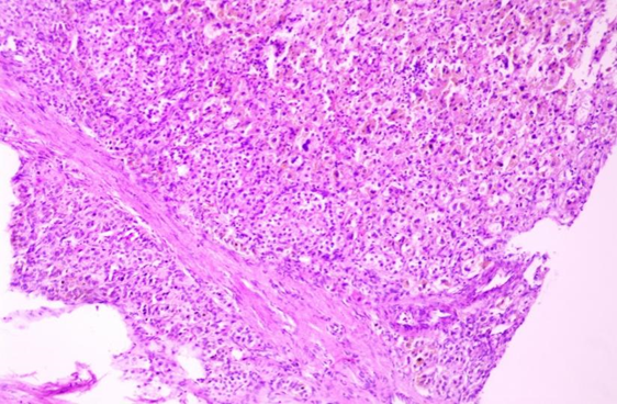
Figure 1: Low-power photomicrograph showing tumor composed of mixed hematopoietic elements and adipose tissue (H&E stain)
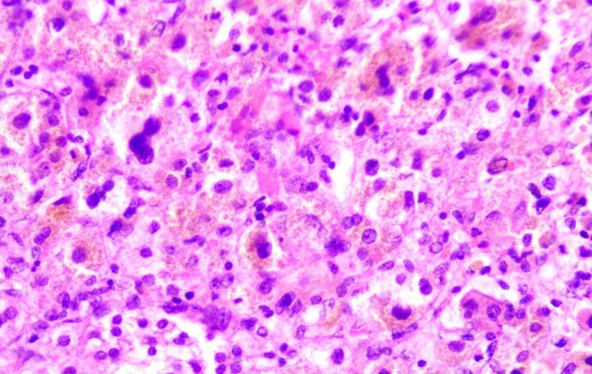
Figure 2: High-power view demonstrating hematopoietic precursors with mature adipocytes (H&E stain)
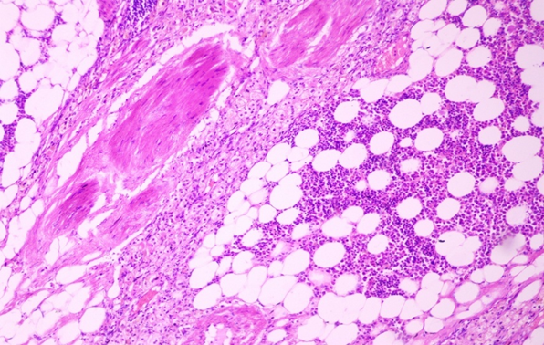
Figure 3: Histopathological section showing mature adipose tissue admixed with trilineage hematopoietic cells
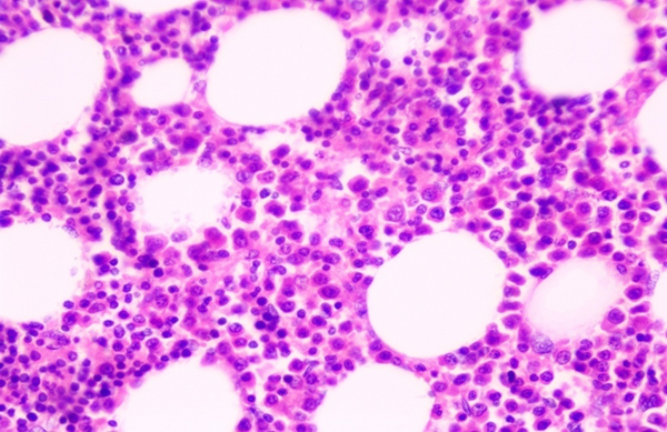
Figure 4: Microscopic image demonstrating erythroid and myeloid precursors within adipose tissue
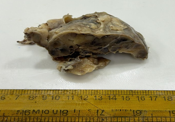
Figure 5: Gross specimen of adrenalectomy specimen showing external capsulated surface
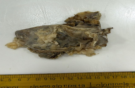
Figure 6: Gross photograph of adrenal mass with grey-yellow appearance
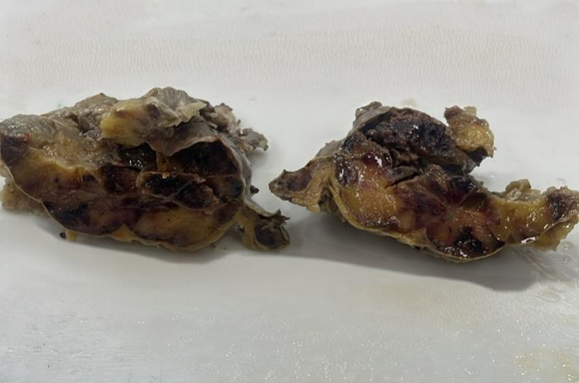
Figure 7: Cut section of adrenal mass showing yellow to reddish-brown areas consistent with myelolipoma
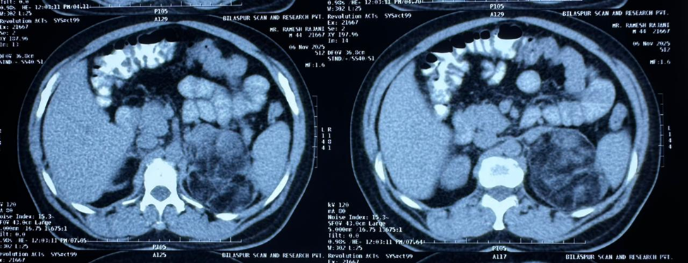
Figure 8: CECT abdomen showing left adrenal myelolipoma
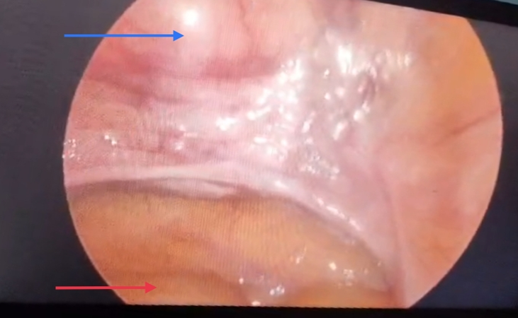
Figure 9: Diagnostic laparoscopy demonstrating absence of Müllerian structures
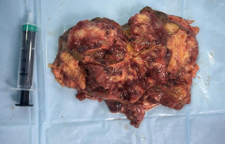
Figure 10: Cut section gross histopathology sample
DISCUSSION:
Adrenal myelolipoma is an uncommon benign adrenal neoplasm that is increasingly recognized because of improved radiological imaging techniques.[7] Although the majority of lesions are asymptomatic and discovered incidentally, symptomatic presentation is more frequently observed in tumors larger than 6 cm.[8] Patients may complain of abdominal pain, flank discomfort, or symptoms related to pressure on adjacent organs.[8]
The present case was unusual because of the coexistence of adrenal myelolipoma, congenital adrenal hyperplasia, and disorder of sexual development in an adult male patient. Several studies have demonstrated a strong association between chronic ACTH stimulation and development of adrenal myelolipoma in CAH patients.[9] Long-standing hormonal stimulation is believed to promote metaplastic transformation of reticuloendothelial cells within the adrenal cortex, resulting in myelolipomatous transformation of adrenal tissue.[10]
Computed tomography plays an important role in diagnosis because the presence of macroscopic fat within an adrenal lesion strongly favors adrenal myelolipoma.[11] In the present case, radiological imaging revealed a well-defined suprarenal mass with significant fat density and no post-contrast enhancement, findings consistent with previous reports.[11]
Histopathologically, adrenal myelolipoma is characterized by mature adipocytes admixed with trilineage hematopoietic elements including erythroid, myeloid, and megakaryocytic precursors.[7] The absence of atypia, necrosis, and increased mitotic activity confirms its benign nature and helps differentiate it from liposarcoma and other retroperitoneal fat-containing tumors.[12]
Management of adrenal myelolipoma depends on tumor size, symptomatology, and radiological characteristics.[8] Small asymptomatic lesions may be followed conservatively, whereas symptomatic or large tumors are usually treated surgically because of the risk of hemorrhage and compressive complications.[13] Laparoscopic adrenalectomy is currently considered a safe and effective treatment modality for most resectable adrenal myelolipomas.[13]
The present patient underwent successful laparoscopic adrenalectomy with uneventful postoperative recovery. This case highlights the importance of endocrine evaluation and histopathological confirmation in patients presenting with adrenal masses associated with congenital adrenal hyperplasia.
CONCLUSION:
Adrenal myelolipoma is a rare benign tumor that may present as a large suprarenal mass causing abdominal discomfort. Imaging studies play an important role in preoperative diagnosis; however, histopathological examination remains confirmatory. The coexistence of adrenal myelolipoma with congenital adrenal hyperplasia and DSD makes this case particularly uncommon. Early recognition and appropriate surgical management result in excellent prognosis.
REFERENCES:
1. Decmann Á, Perge P, Tóth M, Igaz P. Adrenal myelolipoma: a comprehensive review. Endocrine. 2018 Jan;59(1):7-15. doi: 10.1007/s12020-017-1473-4.
2. Shenoy VG, Thota A, Shankar R, Desai MG. Adrenal myelolipoma: Controversies in its management. Indian J Urol. 2015 Apr-Jun;31(2):94-101. doi: 10.4103/0970-1591.152807.
3. Albano D, Agnello F, Midiri F, Pecoraro G, Bruno A, Alongi P, et al. Imaging features of adrenal masses. Insights Imaging. 2019 Jan 25;10(1):1. doi: 10.1186/s13244-019-0688-8.
4. Ito I, Takehara K, Miyazaki Y, Tsuchiyama A, Mukae Y, Hayakawa K, et al. Spontaneous hemorrhage in adrenal myelolipoma treated with elective laparoscopic adrenalectomy following selective arterial embolization. IJU Case Rep. 2024 Dec 15;8(2):114-117. doi: 10.1002/iju5.12821.
5. Nermoen I, Falhammar H. Prevalence and Characteristics of Adrenal Tumors and Myelolipomas in Congenital Adrenal Hyperplasia: A Systematic Review and Meta-Analysis. Endocr Pract. 2020 Nov;26(11):1351-1365. doi: 10.4158/EP-2020-0058.
6. Speiser PW, Arlt W, Auchus RJ, Baskin LS, Conway GS, Merke DP, et al. Congenital Adrenal Hyperplasia Due to Steroid 21-Hydroxylase Deficiency: An Endocrine Society Clinical Practice Guideline. J Clin Endocrinol Metab. 2018 Nov 1;103(11):4043-4088. doi: 10.1210/jc.2018-01865.
7. Lam AK. Update on Adrenal Tumours in 2017 World Health Organization (WHO) of Endocrine Tumours. Endocr Pathol. 2017 Sep;28(3):213-227. doi: 10.1007/s12022-017-9484-5.
8. Calissendorff J, Juhlin CC, Sundin A, Bancos I, Falhammar H. Adrenal myelolipomas. Lancet Diabetes Endocrinol. 2021 Nov;9(11):767-775. doi: 10.1016/S2213-8587(21)00178-9.
9. Jacobson A, Koberlein E, Thomay A, Lombard CB, Adelanwa A, Lakhani DA, Smith KT. Congenital adrenal hyperplasia with associated giant adrenal myelolipoma, testicular adrenal rest tumors and primary pigmented nodular adrenocortical disease: A case report and brief review of the literature. Radiol Case Rep. 2021 Dec 28;17(3):710-716. doi: 10.1016/j.radcr.2021.12.031.
10. Shahshenas S, Yarmohammadi H, Khalaj A, Anisian A, Soltanipur M, Jalali Nadoushan M. Two Case Reports of Adrenal Myelolipoma: Insights into a Rare and Benign Mass. Case Rep Oncol. 2026 Mar 10;19(1):592-598. doi: 10.1159/000551367.
11. Chiapponi C, Santos DPD, Hartmann MJM, Schmidt M, Faust M, Wahba R, et al. Adrenal Surgery in the Era of Multidisciplinary Endocrine Tumor Boards. Horm Metab Res. 2022 May;54(5):294-299. doi: 10.1055/a-1808-7239.
12. Cho MH, Mandaliya R, Liang J, Patel M. A case report of symptomatic presacral myelolipoma. Medicine (Baltimore). 2018 Apr;97(15):e0337. doi: 10.1097/MD.0000000000010337.
13. Duarte Regalado CS, Guzmán Mejía JI, Gutiérrez Uvalle GE, Vargas Rodríguez AE, González Ledo J. A Case Report and Literature Review of Adrenal Myelolipoma. Cureus. 2023 Aug 9;15(8):e43240. doi: 10.7759/cureus.43240.