Role of High-Resolution Ultrasonography with Colour Doppler in the Evaluation of Acute Scrotal Pathologies: A Cross-Sectional Study.
- Rakesh Vijayvargiya , Professor, Department of Radiodiagnosis, MGM Medical College and M.Y. Hospital, Indore, Madhya Pradesh, India.
- Alka Agrawal , Professor and Head, Department of Radiodiagnosis, MGM Medical College and M.Y. Hospital, Indore, Madhya Pradesh, India.
- Manish Sarote , Junior Resident (PG RMO), Department of Radiodiagnosis, MGM Medical College and M.Y. Hospital, Indore, Madhya Pradesh, India.
- Anjana Kumari , Junior Resident (PG RMO), Department of Radiodiagnosis, MGM Medical College and M.Y. Hospital, Indore, Madhya Pradesh, India.
Article Information:
Abstract:
Background: Acute scrotal conditions often have similar clinical features but require different treatment. High-resolution ultrasonography (HRUS) with Colour Doppler can provide anatomical and vascular information for early diagnosis. Methods: This hospital-based cross-sectional study included 100 patients aged 1–70 years presenting with acute scrotal symptoms. Both testes were examined using a 7.5–15 MHz linear transducer. Testicular and extratesticular structures were assessed on B-mode imaging, followed by Colour Doppler evaluation of vascularity. Imaging findings were compared with surgical findings wherever available. Results: Hydrocele was the most common diagnosis (38%), followed by varicocele (20%), inguinal or inguinoscrotal hernia (14%), epididymal cyst (8%), testicular torsion (7%), and epididymo-orchitis or funiculitis (7%). Hydrocele was detected as either a primary or associated finding in 55% of patients. Absent intratesticular flow was observed in all seven cases of testicular torsion. The whirlpool sign was present in four torsion cases (57.1%), while an elevated testicular position was seen in two (28.6%). All seven inflammatory cases showed increased vascularity. Venous reflux during the Valsalva manoeuvre was demonstrated in all 20 varicocele cases. Bowel peristalsis was detected in six of 14 hernia cases. Conclusion: HRUS with Colour Doppler is a useful first-line method for evaluating acute scrotal pathologies. It helps distinguish testicular torsion from inflammatory conditions and supports timely surgical or conservative management.
Keywords:
Article :
INTRODUCTION:
Acute scrotal pain and swelling are common urological emergencies that require prompt diagnosis. The differential diagnosis includes testicular torsion, epididymo-orchitis, torsion of the testicular appendage, scrotal trauma, hydrocele, varicocele, inguinoscrotal hernia and testicular tumour. These conditions often have similar clinical features, making differentiation by clinical examination alone difficult. Misdiagnosis between testicular torsion and epididymo-orchitis has been reported in 30–50% of cases.1
Testicular torsion occurs due to twisting of the spermatic cord, resulting in reduced blood flow and progressive testicular ischaemia. Testicular salvage is closely related to the time between symptom onset and surgical detorsion. The salvage rate approaches 100% within 6 hours but falls to less than 10% after 24 hours.2 In contrast, epididymo-orchitis is generally treated with antibiotics and anti-inflammatory medication. Therefore, early differentiation between these conditions is essential to avoid testicular loss or unnecessary surgery.3 Other conditions, such as testicular rupture and haematocele following trauma, also require accurate imaging to guide appropriate management.4
High-resolution ultrasonography is the preferred initial imaging method for acute scrotal symptoms. It provides detailed assessment of the testes, epididymides, tunical coverings and spermatic cords. It is widely available, affordable, free from ionising radiation and allows comparison with the opposite side.5 Colour Doppler provides additional information regarding testicular and peritesticular blood flow. Absent or reduced intratesticular flow suggests torsion, whereas increased vascularity supports epididymo-orchitis. Its reported diagnostic accuracy for testicular torsion ranges from 86% to 100%.6,7
The present study assessed the role and diagnostic accuracy of high-resolution B-mode ultrasonography with Colour Doppler in identifying and differentiating acute scrotal pathologies and compared the imaging findings with surgical findings where available.
MATERIALS AND METHODS:
This hospital-based, cross-sectional observational study was conducted in the Department of Radiodiagnosis, M.G.M. Medical College and M.Y. Hospital, Indore, Madhya Pradesh, over one year after approval from the Institutional Scientific Review Board and Institutional Ethics Committee. A total of 100 consecutive patients aged 1–70 years who were referred for ultrasonographic evaluation of acute scrotal symptoms were included. Patients presenting with acute scrotal pain and/or swelling of less than 48–72 hours’ duration were eligible. Patients with previous scrotal surgery, chronic scrotal disease, scrotal trauma, or unwillingness to participate were excluded. Written informed consent was obtained from adult participants and from the parents or legal guardians of minors.
All examinations were performed with the patient in the supine position using a high-frequency linear-array transducer of 7.5–15 MHz. The highest suitable frequency was selected according to the required depth of penetration. Both testes were examined systematically in transverse and longitudinal planes. The unaffected side was used for comparison.
On B-mode ultrasonography, testicular size, volume, echogenicity, echotexture and integrity of the tunica albuginea were assessed. Testicular volume was calculated using the formula: length × breadth × height × 0.523. The epididymal head, body and tail were examined for enlargement and altered echotexture. The spermatic cord was traced from the inguinal canal to the epididymal tail and assessed for the whirlpool sign. The tunica vaginalis was evaluated for hydrocele, pyocele, haematocele or internal debris. Scrotal wall thickness was also recorded.
Colour Doppler examination was performed in both testes after B-mode assessment. Doppler settings were adjusted to detect low-velocity blood flow, with a pulse repetition frequency of 800–1000 Hz and a low wall filter of 50–100 Hz. Minimal transducer pressure was applied to avoid compression of small vessels. Intratesticular vascularity was classified as absent, markedly reduced, normal or increased by comparison with the opposite testis.
In suspected varicocele, the pampiniform plexus was assessed at rest and during the Valsalva manoeuvre in the standing position. In suspected appendicular torsion, the upper testicular pole was examined for an avascular nodule with surrounding hyperaemia. Spectral Doppler was used in selected equivocal cases to assess peak systolic velocity, end-diastolic velocity and resistive index. Imaging findings were compared with surgical findings wherever available (Figure 1). Data were entered into Microsoft Excel and analysed using Jamovi software. Categorical variables, including age group, side of involvement, clinical diagnosis, B-mode findings, and Colour Doppler findings, were summarised as frequencies and percentages. Diagnosis-specific imaging findings were calculated using the number of patients in the respective diagnostic group as the denominator. As the study findings were mainly descriptive, no inferential statistical test was applied.
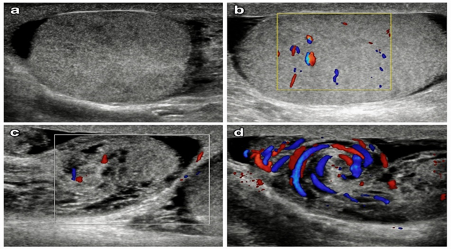
Figure 1. A 22-year-old man presenting with sudden-onset severe right scrotal pain that was not relieved by medication. (a) B-mode ultrasonography showing heterogeneous echotexture of the right testis. (b) Colour Doppler image showing altered intratesticular vascularity. (c) B-mode and Colour Doppler image showing a bulky, heterogeneous spermatic cord and epididymis. (d) Colour Doppler image demonstrating the whirlpool sign within the twisted spermatic cord, suggestive of testicular torsion.
RESULTS:
The largest proportion of participants belonged to the 21–30-year age group (24%), followed by the 31–40-year group (22%) and the 41–50-year group (20%). Left-sided involvement was slightly more common (47%) than right-sided involvement (42%), while 11% had bilateral disease. Most patients presented within 1–3 days of symptom onset (30%). Overall, 58% presented within the first three days, whereas only 6% reported symptoms lasting longer than 30 days (Table 1).
Table 1. Demographic and clinical characteristics of the study participants (N=100)
|
Demographic and clinical Characteristics |
Number |
Percentage (%) |
|
|
Age group (years) |
1–20 |
8 |
8.0 |
|
21–30 |
24 |
24.0 |
|
|
31–40 |
22 |
22.0 |
|
|
41–50 |
20 |
20.0 |
|
|
51–60 |
16 |
16.0 |
|
|
61–70 |
10 |
10.0 |
|
|
Side involved |
Right |
42 |
42.0 |
|
Left |
47 |
47.0 |
|
|
Bilateral |
11 |
11.0 |
|
|
Duration of symptoms |
0–12 hours |
10 |
10.0 |
|
12–24 hours |
18 |
18.0 |
|
|
1–3 days |
30 |
30.0 |
|
|
4–7 days |
24 |
24.0 |
|
|
8–30 days |
12 |
12.0 |
|
|
>30 days |
6 |
6.0 |
|
Hydrocele was the most common final diagnosis, observed in 38% of patients. This was followed by varicocele (20%), inguinal or inguinoscrotal hernia (14%), and epididymal cyst (8%). Testicular torsion and epididymo-orchitis or funiculitis were each diagnosed in 7% of cases. Torsion of the appendix testis, testicular infarction, testicular microlithiasis and undescended testis were uncommon, accounting for 1% each. Other or normal findings were recorded in the remaining 2% of patients (Table 2).
Table 2. Distribution of final diagnoses based on HRUS and Colour Doppler findings (N=100)
|
Final diagnosis |
Number |
Percentage (%) |
|
Hydrocele |
38 |
38.0 |
|
Varicocele |
20 |
20.0 |
|
Inguinal/inguinoscrotal hernia |
14 |
14.0 |
|
Epididymal cyst |
8 |
8.0 |
|
Testicular torsion |
7 |
7.0 |
|
Epididymo-orchitis/funiculitis |
7 |
7.0 |
|
Torsion of appendix testis |
1 |
1.0 |
|
Testicular infarction |
1 |
1.0 |
|
Testicular microlithiasis |
1 |
1.0 |
|
Undescended testis |
1 |
1.0 |
|
Other/normal findings |
2 |
2.0 |
|
Total |
100 |
100.0 |
B-mode ultrasonography showed normal testicular size and homogeneous echotexture in 72% of patients. Testicular enlargement was observed in 20%, while 16% had heterogeneous echotexture. Most patients (93%) had no focal or diffuse testicular parenchymal abnormality. Epididymal enlargement was identified in 22%, spermatic cord enlargement in 17%, and scrotal wall thickening in 26%. Hydrocele, either as a primary diagnosis or an associated finding, was detected in 55 patients. Among these, 37 (67.3%) had homogeneous anechoic fluid, while 18 (32.7%) had complex fluid containing internal echoes, septations or debris (Table 3).
Table 3. Principal B-mode ultrasonographic findings (N=100)
|
Sonographic characteristic |
Number |
Percentage (%) |
|
|
Testicular size |
Normal |
72 |
72.0 |
|
Enlarged |
20 |
20.0 |
|
|
Small |
8 |
8.0 |
|
|
Testicular echotexture |
Homogeneous |
72 |
72.0 |
|
Heterogeneous |
16 |
16.0 |
|
|
Hypoechoic |
7 |
7.0 |
|
|
Hyperechoic |
5 |
5.0 |
|
|
Testicular parenchymal abnormality |
No focal or diffuse abnormality |
93 |
93.0 |
|
Focal abnormality |
1 |
1.0 |
|
|
Diffuse abnormality |
6 |
6.0 |
|
|
Epididymal size |
Normal |
78 |
78.0 |
|
Enlarged |
22 |
22.0 |
|
|
Spermatic cord |
Normal |
83 |
83.0 |
|
Enlarged |
17 |
17.0 |
|
|
Scrotal wall thickness |
Normal (<4 mm) |
74 |
74.0 |
|
Thickened (>4 mm) |
26 |
26.0 |
|
|
Hydrocele detected as primary or associated finding |
Homogeneous/anechoic fluid |
37 |
67.3* |
|
Inhomogeneous/complex fluid |
18 |
32.7* |
|
Absent intratesticular blood flow was found in all seven patients with testicular torsion. The whirlpool sign was demonstrated in four cases (57.1%), while an elevated testicular position was seen in two (28.6%). Increased vascularity was present in all seven cases of epididymo-orchitis or funiculitis. All 20 varicocele cases showed venous reflux during the Valsalva manoeuvre; Grade III varicocele was the most frequent, occurring in 50% of these cases. Bowel peristalsis within the hernial sac was demonstrated in six of 14 hernia cases. Absent vascularity was found in the patient with testicular infarction, while reduced flow with surrounding hyperaemia was seen in the patient with torsion of the appendix testis (Table 4).
Table 4. Important diagnosis-specific HRUS and Colour Doppler findings
|
Diagnosis |
Cases (n) |
Important imaging finding |
Cases with finding n (%) |
|
Testicular torsion |
7 |
Absent intratesticular flow |
7 (100.0) |
|
Whirlpool sign |
4 (57.1) |
||
|
Elevated testicular position |
2 (28.6) |
||
|
Epididymo-orchitis/funiculitis |
7 |
Increased vascularity/hyperaemia |
7 (100.0) |
|
Varicocele |
20 |
Venous reflux during Valsalva manoeuvre |
20 (100.0) |
|
Grade I |
2 (10.0) |
||
|
Grade II |
8 (40.0) |
||
|
Grade III |
10 (50.0) |
||
|
Inguinal/inguinoscrotal hernia |
14 |
Bowel peristalsis within hernial sac |
6 (42.9) |
|
Testicular infarction |
1 |
Focal or diffuse absent vascularity |
1 (100.0) |
|
Torsion of appendix testis |
1 |
Reduced flow with surrounding hyperaemia |
1 (100.0) |
DISCUSSION:
High-resolution ultrasonography with Colour Doppler plays an important role in patients presenting with acute scrotal symptoms. Clinical findings often overlap, particularly in testicular torsion and epididymo-orchitis, although their management is different. In the present study, most patients were young or middle-aged adults, with the highest proportion in the 21–30-year age group. Left-sided disease was slightly more common than right-sided disease, partly because varicocele commonly affects the left side. Dogra et al. described ultrasonography as the main imaging method for assessing testicular and extratesticular lesions because it provides detailed anatomical and vascular information.8
Hydrocele was the most common diagnosis, accounting for 38% of patients. When primary and associated collections were considered together, scrotal fluid was present in 55%. Most hydroceles were homogeneous and anechoic, while some contained internal echoes, septations or debris. Kumar et al. reported hydrocele in 25 of 60 patients undergoing ultrasonographic evaluation of scrotal disease, which is comparable to the present findings.9 Differences in prevalence between studies may be related to age, patient selection and referral patterns. Varicocele was the second most common diagnosis. All 20 cases showed venous reflux during the Valsalva manoeuvre, including two subclinical cases, demonstrating the added value of Colour Doppler over clinical examination alone.
Testicular torsion was identified in seven patients. All showed absent intratesticular blood flow, while the whirlpool sign was demonstrated in four cases. Testicular enlargement, heterogeneous echotexture and an elevated position were useful additional findings. Vijayaraghavan reported that the twisted spermatic cord is a direct and reliable sonographic sign of torsion.10 In the large paediatric series by Yang et al., reduced or absent testicular flow was found in 91.3% of torsion cases.11 However, preserved flow does not completely exclude partial or early torsion. Imaging findings must therefore be interpreted with the symptoms and clinical examination, and surgery should not be delayed when clinical suspicion remains high.
All seven patients with epididymo-orchitis or funiculitis showed increased vascularity. Epididymal enlargement and scrotal wall thickening were also observed. Vijayaraghavan similarly found epididymal or testicular enlargement with increased Doppler flow in inflammatory disease.10 Agrawal et al. also showed that Colour Doppler helps distinguish epididymo-orchitis from testicular torsion and may prevent unnecessary surgical exploration.12
Overall, B-mode imaging defined the site and morphology of disease, while Colour Doppler demonstrated vascular changes. The study was limited by its single-centre design, small numbers of torsion and inflammatory cases, and lack of surgical confirmation in every patient.
CONCLUSION:
High-resolution ultrasonography with Colour Doppler is a reliable, non-invasive and radiation-free first-line imaging method for acute scrotal pathologies. It accurately characterises common conditions such as hydrocele, varicocele, hernia and epididymo-orchitis while helping distinguish inflammatory disease from vascular emergencies. Absent or reduced intratesticular flow enables early recognition of testicular torsion and supports urgent surgical referral. Its combined anatomical and vascular assessment guides appropriate treatment, reduces unnecessary exploration and improves patient outcomes, particularly when performed promptly after symptom onset in acute care.
REFERENCES:
1. Appelbaum L, Gaitini D, Dogra VS. Scrotal ultrasound in adults. Semin Ultrasound CT MR 2013;34(3):257- 73.
2. Rizvi SA, Ahmad I, Siddiqui MA, Zaheer S, Ahmad K. Role of color Doppler ultrasonography in evaluation of scrotal swellings: pattern of disease in 120 patients along review of literature. Urol J 2011;8(5):60-5.
3. Mirochnik B, Bhargava P, Dighe MK, Kanth N Ultrasound evaluation of scrotal pathology RadiolClin North Am. 2012;50(2):317-32.
4. Wright S, Hoffmann B. Emergency ultrasound of acute scrotal pain. Eur J Emerg Med. 2015;22(1):2-9.
5. Sommers D, Winter T .Ultrasonography evaluation of scrotal masses. RadiolClin North Am. 2014;52(6):1265-81.
6. Arjhansiri K, Vises N, Kitsukjit W. Sonographic evaluation of the intrascrotal disease. J Med Assoc Thai 2004;87 (Suppl 2):S161-7.
7. Siddiqui EH, Siddiqui S, Rasool G, Khan N. Scrotal Pathologies: Role of high resolution & Doppler ultrasound in evaluation. Professional Med J 2013;20(4):924-28.
8. Dogra VS, Gottlieb RH, Oka M, Rubens DJ. Sonography of the scrotum. Radiology. 2003;227(1):18-36.
9. Kumar K, Kumari M, Kumar V, Suman SK. Evaluation of Scrotal Pathologies by Ultrasound and Color Doppler. Cureus. 2023;15(3):e36776.
10. Vijayaraghavan SB. Sonographic differential diagnosis of acute scrotum: real-time whirlpool sign, a key sign of torsion. J Ultrasound Med. 2006;25(5):563-574.
11. Yang C Jr, Song B, Liu X, Wei GH, Lin T, He DW. Acute scrotum in children: an 18-year retrospective study. Pediatr Emerg Care. 2011;27(4):270-274.
12. Agrawal AM, Tripathi PS, Shankhwar A, Naveen C. Role of ultrasound with color Doppler in acute scrotum management. J Family Med Prim Care. 2014;3(4):409-412.