Nail Findings As Markers Of Systemic Disease.
- Ujjwal Jharia , Senior Resident, Department of Dermatology Venereology and Leprosy, Datia Medical College and Hospital, Datia (MP)
- Aayush Kapoor , Senior resident, Department of General Medicine, Datia Medical College and Hospital, Datia (MP)
Article Information:
Abstract:
Background: Nail abnormalities may provide important clues to systemic disease, yet they are often overlooked during routine examination. This study evaluated the spectrum of nail abnormalities in patients with systemic diseases and assessed their association with systemic disorders. Methods: This hospital-based observational cross-sectional study was conducted in the Department of Dermatology, Venereology and Leprosy, Datia Medical College and Hospital, Datia, Madhya Pradesh. A total of 100 adults with clinically or laboratory-confirmed systemic diseases were evaluated. Examination of the nail plate, matrix, bed, nail folds, and periungual skin was performed. Onychoscopy and nailfold capillaroscopy were undertaken when clinically indicated. Clinical and laboratory findings were analyzed using IBM SPSS Statistics. Associations were assessed using the chi-square or Fisher's exact test, with p<0.05 considered statistically significant. Results: Nail abnormalities were identified in 78% of participants. Leukonychia was the most frequent finding (24%), followed by clubbing (20%), onycholysis (15%), koilonychia (12%), Beau's lines (11%), and splinter hemorrhages (10%). Clubbing was significantly associated with respiratory disease (p=0.002), koilonychia with hematological disorders (p=0.001), half-and-half nails with renal disease (p=0.004), and Terry's nails with hepatobiliary disease (p=0.013). Nail abnormalities were more frequent with longer disease duration and greater disease severity. Onychoscopy provided additional characterization, while nailfold capillaroscopy demonstrated vascular abnormalities in connective tissue disorders. Conclusion: Nail abnormalities were common in patients with systemic diseases, and several characteristic findings demonstrated significant disease-specific associations. Systematic nail examination, supplemented by onychoscopy and capillaroscopy when appropriate, may serve as a simple, non-invasive adjunct for recognizing systemic disease and guiding further evaluation.
Keywords:
Article :
INTRODUCTION:
The skin provides important clues to systemic health, and the nails are particularly useful in identifying underlying systemic disorders. Nail examination is simple, non-invasive, and inexpensive, yet nail abnormalities are often overlooked in routine clinical practice. Careful assessment of nail morphology, color, texture, growth, and periungual structures may provide valuable evidence of cardiovascular, respiratory, gastrointestinal, hepatobiliary, renal, hematological, endocrine, rheumatological, and infectious diseases. [1]
The nail unit consists of the nail plate, matrix, bed, proximal and lateral nail folds, and hyponychium. [2] Because nail growth is slow, systemic disturbances affecting metabolism, nutrition, vascular supply, connective tissue, or cellular proliferation may produce changes that persist for weeks or months, making the nails a useful record of both acute and chronic disease processes. [3]
Nail abnormalities include nonspecific findings such as Beau’s lines, splinter hemorrhages, pitting, onycholysis, and koilonychia, as well as relatively characteristic manifestations such as clubbing, Terry’s nails, half-and-half nails, Muehrcke’s lines, and Mees’ lines. [4,5] Clubbing may occur in congenital heart disease, chronic pulmonary disorders, and gastrointestinal diseases, while splinter hemorrhages may suggest infective endocarditis or vasculitis. [6] Terry’s nails have been described in liver cirrhosis, congestive heart failure, and diabetes mellitus. [7,8] Koilonychia is classically associated with iron deficiency anemia, whereas half-and-half nails are frequently observed in chronic renal failure. [9] Muehrcke’s lines are associated with hypoalbuminemia, while Mees’ lines may occur in toxic exposures and systemic diseases. [10] Onycholysis and periungual abnormalities may provide clues to endocrine and autoimmune disorders. [11] Various infectious and respiratory disorders may also produce characteristic nail changes. [12-14]
Nail findings may additionally reflect disease severity, chronicity, nutritional status, and microvascular involvement. Dermoscopy and nailfold capillaroscopy have further increased the diagnostic value of nail examination, particularly in connective tissue and vascular disorders. [4] Therefore, systematic nail examination can complement routine clinical assessment and facilitate early recognition of systemic disease. The present study was undertaken to evaluate the spectrum of nail abnormalities in patients with systemic diseases and assess their association with underlying systemic disorders.
MATERIALS AND METHODS:
This hospital-based observational cross-sectional study was conducted in the Department of Dermatology, Venereology and Leprosy, Datia Medical College and Hospital, Datia, Madhya Pradesh, over a period of 12 months to evaluate the spectrum of nail abnormalities in patients with systemic diseases and assess their association with underlying systemic disorders. The study was conducted after obtaining approval from the Institutional Ethics Committee. Written informed consent was obtained from all participants. Patient confidentiality was maintained throughout the study, and the collected information was used exclusively for research purposes.
A total of 100 adult patients diagnosed with one or more systemic diseases and attending the outpatient or inpatient services during the study period were included. Institutional Ethics Committee approval was obtained before initiation of the study, and written informed consent was obtained from all participants.
Inclusion Criteria
Patients aged ≥18 years with a clinically or laboratory-confirmed systemic disease who were willing to undergo detailed nail examination and provided written informed consent were included in the study.
Exclusion Criteria
Patients with isolated primary nail disorders without an underlying systemic disease, significant occupational or traumatic nail changes that could interfere with assessment, those receiving treatment specifically for a nail disorder, and those unwilling to participate or provide informed consent were excluded.
Clinical Evaluation and Nail Examination
A structured proforma was used to record demographic details, presenting complaints, duration and type of systemic illness, relevant past and treatment history, comorbidities, and other clinical findings. A detailed examination of the nails was performed under adequate illumination. The nail plate, nail bed, nail matrix, proximal and lateral nail folds, and periungual skin were examined. The number and distribution of involved nails and the morphology of individual nail abnormalities were documented. Findings including clubbing, koilonychia, leukonychia, Beau's lines, splinter hemorrhages, onycholysis, pitting, Terry's nails, half-and-half nails, Muehrcke's lines, Mees' lines, periungual erythema, longitudinal melanonychia, and other nail changes were recorded. Standardized clinical photographs of the affected nails were obtained for documentation.
Onychoscopy and Nailfold Capillaroscopy
Onychoscopy was performed in patients with clinically evident nail abnormalities, particularly when characteristic dermoscopic features could aid further characterization or diagnosis. Nail plate and nail bed findings were assessed, and the observed patterns were documented with photographic records. Nailfold capillaroscopic examination was performed when clinically indicated, particularly in patients with suspected connective tissue or microvascular disorders. Findings such as capillary dilatation, microhemorrhages, capillary dropout, avascular areas, and abnormal capillary morphology were recorded where present. Polarized or non-polarized illumination was used as appropriate for assessment of the nail plate, nail bed, and nailfold structures.
Investigations
Relevant laboratory and disease-specific investigations were reviewed or performed when clinically indicated, particularly when the diagnosis or association between the nail finding and systemic disease was not clear on clinical examination alone. These included complete blood count, serum albumin, renal and liver function tests, blood glucose, thyroid profile, inflammatory markers, and other investigations guided by the underlying systemic condition.
Classification of Nail Findings
For analysis, the observed nail changes were categorized according to the principal systemic disease groups, including cardiovascular, respiratory, renal, hepatobiliary, hematological, endocrine, rheumatological/connective tissue, and infectious diseases. The frequency and distribution of individual nail abnormalities within each disease category were recorded.
Study Variables
The primary outcome variables were the type and frequency of nail abnormalities and their distribution across systemic disease categories. Secondary variables included age, sex, duration of systemic disease, disease severity, relevant comorbidities, and laboratory parameters. The association between specific nail findings and underlying systemic disorders was assessed.
Statistical Analysis
Data were entered into Microsoft Excel and analyzed using IBM SPSS Statistics. Continuous variables were expressed as mean ± standard deviation or median with interquartile range, as appropriate, while categorical variables were expressed as frequencies and percentages. Associations between categorical variables were assessed using the chi-square test or Fisher's exact test. Student's t-test or an appropriate non-parametric test was used for comparison of continuous variables. A p-value <0.05 was considered statistically significant.
RESULTS:
A total of 100 patients with systemic diseases were included in the study. The mean age of the study population was 47.2 ± 16.8 years, with an age range of 18–82 years. There were 56 males and 44 females, giving a male-to-female ratio of 1.27:1. The majority of patients belonged to the 41–60 years age group (42%), followed by 61–80 years (29%), 21–40 years (24%), and ≤20 years (5%). [Table 1, Figure 1]
Table 1. Demographic characteristics of the study population (N=100)
|
Variable |
Number (n) |
Percentage (%) |
|
Age group (years) |
|
|
|
≤20 |
5 |
5.0 |
|
21–40 |
24 |
24.0 |
|
41–60 |
42 |
42.0 |
|
61–80 |
29 |
29.0 |
|
Sex |
|
|
|
Male |
56 |
56.0 |
|
Female |
44 |
44.0 |
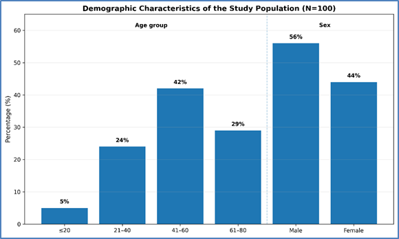
Figure 1: Demographic characteristics of the study population (N=100)
Spectrum of Systemic Diseases: Among the 100 patients, 24 (24%) had cardiovascular diseases, 18 (18%) had respiratory diseases, 17 (17%) had renal diseases, 12 (12%) had hepatobiliary diseases, 11 (11%) had hematological disorders, 10 (10%) had endocrine disorders, 5 (5%) had rheumatological/connective tissue diseases, and 3 (3%) had infectious diseases. [Table 2, Figure 2]
Table 2. Distribution of systemic diseases among study participants
|
Systemic disease category |
Number (n) |
Percentage (%) |
|
Cardiovascular diseases |
24 |
24.0 |
|
Respiratory diseases |
18 |
18.0 |
|
Renal diseases |
17 |
17.0 |
|
Hepatobiliary diseases |
12 |
12.0 |
|
Hematological disorders |
11 |
11.0 |
|
Endocrine diseases |
10 |
10.0 |
|
Rheumatological/connective tissue diseases |
5 |
5.0 |
|
Infectious diseases |
3 |
3.0 |
|
Total |
100 |
100.0 |
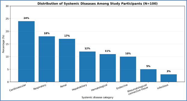
Figure 2: Distribution of systemic diseases among study participants
Spectrum of Nail Abnormalities: Nail abnormalities were identified in 78 (78%) patients, while 22 (22%) patients had no clinically significant nail changes. The most frequently observed abnormality was leukonychia, seen in 24 (24%) patients, followed by clubbing in 20 (20%), onycholysis in 15 (15%), koilonychia in 12 (12%), Beau's lines in 11 (11%), and splinter hemorrhages in 10 (10%). Other findings included pitting in 8 (8%), Terry's nails in 8 (8%), half-and-half nails in 7 (7%), Muehrcke's lines in 5 (5%), longitudinal melanonychia in 5 (5%), periungual erythema in 4 (4%), Mees' lines in 3 (3%), and yellow nail discoloration in 3 (3%). Some patients exhibited more than one nail abnormality. [Table 3, Figure 3]
Table 3. Spectrum of nail abnormalities observed among study participants
|
Nail abnormality |
Number (n) |
Percentage (%) |
|
Leukonychia |
24 |
24.0 |
|
Clubbing |
20 |
20.0 |
|
Onycholysis |
15 |
15.0 |
|
Koilonychia |
12 |
12.0 |
|
Beau’s lines |
11 |
11.0 |
|
Splinter hemorrhages |
10 |
10.0 |
|
Pitting |
8 |
8.0 |
|
Terry’s nails |
8 |
8.0 |
|
Half-and-half nails |
7 |
7.0 |
|
Muehrcke’s lines |
5 |
5.0 |
|
Longitudinal melanonychia |
5 |
5.0 |
|
Periungual erythema |
4 |
4.0 |
|
Mees’ lines |
3 |
3.0 |
|
Yellow nail discoloration |
3 |
3.0 |
|
No clinically significant nail abnormality |
22 |
22.0 |
Percentages for individual nail abnormalities were based on the total study population. Because some patients had multiple nail abnormalities, the percentages do not total 100%.
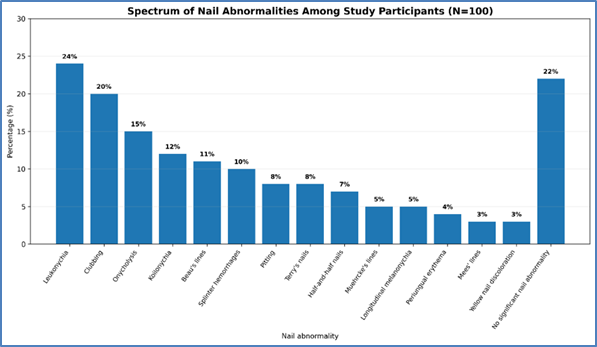
Figure 3: Spectrum of nail abnormalities observed among study participants
Nail Changes According to the Anatomical Component of the Nail Unit: The observed abnormalities were further classified according to involvement of the nail plate, nail matrix, nail bed, and nail fold. This classification allowed characterization of the predominant anatomical component affected in each systemic disease category. [Table 4, Figure 4]
Table 4. Nail changes according to systemic disease and anatomical component of the nail unit
|
Systemic disease category |
Nail plate findings, n (%) |
Nail matrix findings, n (%) |
Nail bed findings, n (%) |
Nail fold findings, n (%) |
|
Cardiovascular diseases (n=24) |
Leukonychia: 7 (29.2); Longitudinal melanonychia: 2 (8.3) |
Beau's lines: 4 (16.7) |
Clubbing: 7 (29.2); Splinter hemorrhages: 5 (20.8); Onycholysis: 3 (12.5) |
Periungual erythema: 2 (8.3) |
|
Respiratory diseases (n=18) |
Leukonychia: 5 (27.8); Longitudinal ridging: 2 (11.1) |
Beau's lines: 3 (16.7) |
Clubbing: 11 (61.1); Splinter hemorrhages: 2 (11.1); Onycholysis: 3 (16.7) |
Periungual erythema: 2 (11.1) |
|
Renal diseases (n=17) |
Half-and-half nails: 6 (35.3); Leukonychia: 4 (23.5) |
Beau's lines: 4 (23.5) |
Muehrcke's lines: 2 (11.8); Onycholysis: 4 (23.5) |
Periungual edema/erythema: 2 (11.8) |
|
Hepatobiliary diseases (n=12) |
Terry's nails: 5 (41.7); Leukonychia: 3 (25.0) |
Beau's lines: 2 (16.7) |
Muehrcke's lines: 2 (16.7); Onycholysis: 2 (16.7) |
Periungual erythema: 2 (16.7) |
|
Hematological disorders (n=11) |
Koilonychia: 8 (72.7); Leukonychia: 3 (27.3) |
Beau's lines: 3 (27.3) |
Nail-bed pallor: 4 (36.4); Splinter hemorrhages: 2 (18.2) |
Periungual pallor: 3 (27.3) |
|
Endocrine disorders (n=10) |
Onycholysis: 2 (20.0); Leukonychia: 2 (20.0) |
Pitting: 1 (10.0); Beau's lines: 2 (20.0) |
Onycholysis: 2 (20.0) |
Periungual erythema: 1 (10.0) |
|
Rheumatological/connective tissue diseases (n=5) |
Nail dystrophy: 1 (20.0); Longitudinal ridging: 2 (40.0) |
Pitting: 1 (20.0); Beau's lines: 1 (20.0) |
Onycholysis: 1 (20.0); Splinter hemorrhages: 1 (20.0) |
Dilated capillaries: 4 (80.0); Microhemorrhages: 2 (40.0); Capillary dropout: 2 (40.0); Avascular areas: 2 (40.0) |
|
Infectious diseases (n=3) |
Chromonychia: 1 (33.3); Leukonychia: 1 (33.3) |
Beau's lines: 1 (33.3) |
Onycholysis/subungual changes: 1 (33.3) |
Paronychia: 1 (33.3) |
Percentages were calculated within each systemic disease category. Because some patients had more than one nail abnormality, the frequencies within a category were not mutually exclusive.
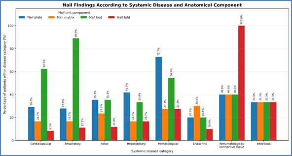
Figure 4. Nail changes according to systemic disease and anatomical component of the nail unit
Onychoscopic Findings: Onychoscopy was performed in patients with clinically evident nail abnormalities to further characterize nail plate, nail bed, and periungual changes. The onychoscopic findings were documented separately from clinical examination findings, while nailfold capillaroscopic abnormalities were recorded separately where capillaroscopy was indicated. [Table 5, Figure 5]
Table 5. Onychoscopic findings among study participants
|
Onychoscopic finding |
Number (n) |
Percentage (%) |
|
Pitting |
8 |
10.0 |
|
Onycholysis |
15 |
18.8 |
|
Subungual hyperkeratosis |
9 |
11.3 |
|
Chromonychia |
8 |
10.0 |
|
Microhemorrhages |
6 |
7.5 |
|
Dilated nailfold capillaries* |
4 |
5.0 |
|
Capillary dropout/avascular areas* |
2 |
2.5 |
Capillaroscopic findings were reported separately because they assess the nailfold microvasculature rather than the nail plate or nail bed itself.
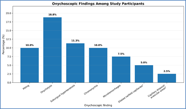
Figure 5: Onychoscopic findings among study participants
Nail Findings According to Systemic Disease: Clubbing was predominantly observed among patients with respiratory and cardiovascular diseases, occurring in 11 (61.1%) patients with respiratory disease and 7 (29.2%) patients with cardiovascular disease. Koilonychia was observed mainly in patients with hematological disorders, particularly those with anemia, where it was present in 8 (72.7%) patients.
Half-and-half nails were predominantly observed among patients with renal disease, being present in 6 (35.3%) of the 17 renal disease patients. Terry's nails were observed in 5 (41.7%) patients with hepatobiliary disease and 2 (8.3%) patients with cardiovascular disease. Muehrcke's lines were identified mainly among patients with renal and hepatobiliary disorders. Splinter hemorrhages were more frequent among patients with cardiovascular disease. [Table 6, Figure 6]
Table 6. Selected nail abnormalities according to systemic disease category
|
Nail finding |
Cardiovascular, n (%) |
Respiratory, n (%) |
Renal, n (%) |
Hepatobiliary, n (%) |
Hematological, n (%) |
|
Clubbing |
7 (29.2) |
11 (61.1) |
1 (5.9) |
1 (8.3) |
0 (0.0) |
|
Koilonychia |
1 (4.2) |
0 (0.0) |
1 (5.9) |
0 (0.0) |
8 (72.7) |
|
Terry's nails |
2 (8.3) |
0 (0.0) |
1 (5.9) |
5 (41.7) |
0 (0.0) |
|
Half-and-half nails |
0 (0.0) |
0 (0.0) |
6 (35.3) |
1 (8.3) |
0 (0.0) |
|
Muehrcke's lines |
1 (4.2) |
0 (0.0) |
2 (11.8) |
2 (16.7) |
0 (0.0) |
|
Splinter hemorrhages |
5 (20.8) |
2 (11.1) |
1 (5.9) |
1 (8.3) |
0 (0.0) |
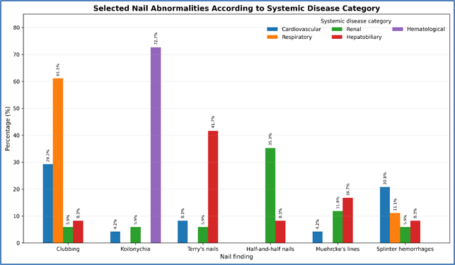
Figure 6: Selected nail abnormalities according to systemic disease category
Association Between Nail Abnormalities and Systemic Diseases: A statistically significant association was observed between clubbing and respiratory disease (p = 0.002) and between koilonychia and hematological disorders (p = 0.001). A significant association was also observed between half-and-half nails and renal disease (p = 0.004), and between Terry's nails and hepatobiliary disease (p = 0.013).
Leukonychia was observed across several systemic disease categories but did not demonstrate a statistically significant association with any single disease group (p = 0.184). Similarly, Beau's lines and onycholysis were relatively nonspecific and were observed across multiple systemic disorders. [Table 7]
Table 7. Association of selected nail abnormalities with systemic disease categories
|
Nail abnormality |
Systemic disease showing strongest association |
p-value |
Statistical significance |
|
Clubbing |
Respiratory disease |
0.002 |
Significant |
|
Koilonychia |
Hematological disorders |
0.001 |
Significant |
|
Half-and-half nails |
Renal disease |
0.004 |
Significant |
|
Terry's nails |
Hepatobiliary disease |
0.013 |
Significant |
|
Leukonychia |
No single predominant disease group |
0.184 |
Not significant |
|
Beau's lines |
No single predominant disease group |
>0.05 |
Not significant |
|
Onycholysis |
No single predominant disease group |
>0.05 |
Not significant |
Nail Abnormalities and Disease Duration: Nail abnormalities were more frequent among patients with a disease duration of >5 years, with abnormalities observed in 39 of 45 patients (86.7%), compared with 39 of 55 patients (70.9%) with a disease duration of ≤5 years. This difference was statistically significant (p = 0.048). Patients with more severe systemic disease also demonstrated a greater frequency of nail abnormalities compared with those having milder disease (89.7% vs. 65.1%; p = 0.006).
Table 8. Nail abnormalities according to duration and severity of systemic disease
|
Clinical characteristic |
Nail abnormality present, n (%) |
Nail abnormality absent, n (%) |
p-value |
|
Disease duration ≤5 years (n=55) |
39 (70.9) |
16 (29.1) |
0.048 |
|
Disease duration >5 years (n=45) |
39 (86.7) |
6 (13.3) |
|
|
Milder disease (n=63) |
41 (65.1) |
22 (34.9) |
0.006 |
|
More severe disease (n=37) |
33 (89.2) |
4 (10.8) |
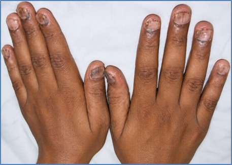
Figure 7: Fingernails with koilonychia and trachyonychia
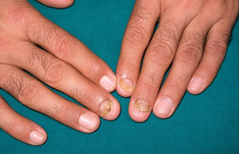
Figure 8: Onycholysis of fingernails in a patient with thyroid disorder
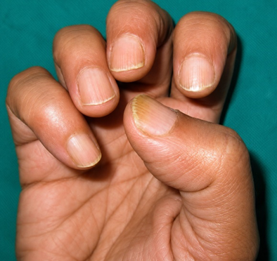
Figure 9: Longitudinal ridging with beading and fissuring associated with severe anemia
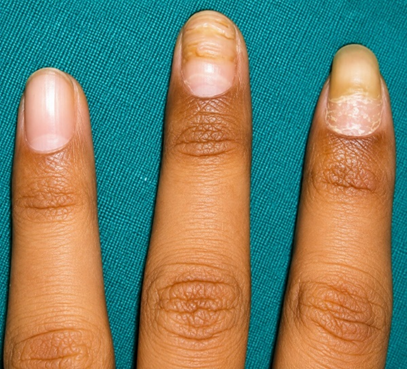
Figure 10: Multiple Beau’s lines in the middle finger and onychomadesis in the index finger
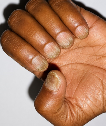
Figure 11: Trachyonychia in a patient with psoriatic arthritis without skin lesions
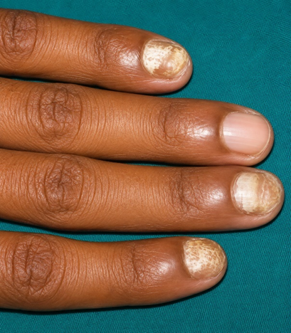
Figure 12: Superficial white onychomycosis
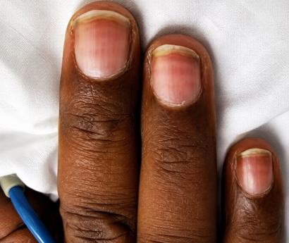
Figure 10: Muehrcke’s lines in a patient with end-stage renal disease
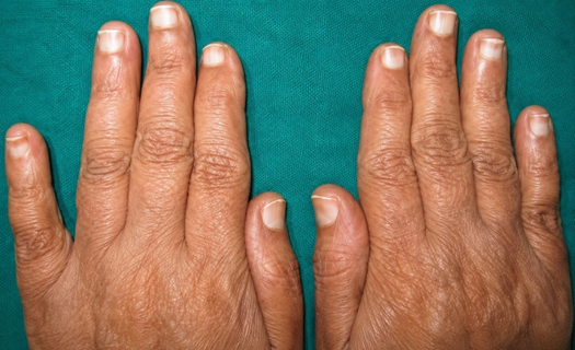
Figure 12: Terry’s nails in a patient with congestive heart failure
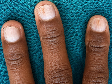
Figure 14: Splinter hemorrhage with bacterial endocarditis
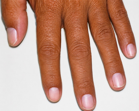
Figure 11: Half and half nails in a patient with chronic kidney disease
DISCUSSION:
The present study evaluated 100 patients with systemic diseases and identified clinically relevant nail abnormalities in 78% of participants. These findings reinforce the importance of the nail unit as a potential clinical indicator of systemic disease. Changes in nail colour, shape, texture, growth, and vascularity may occur because of alterations in nutrition, metabolism, circulation, inflammation, and connective tissue, making nail examination an important component of general physical examination. [15,16] The high frequency of nail abnormalities in the present study is particularly relevant because the study population consisted of patients with established systemic diseases rather than individuals presenting primarily with nail disorders.
The findings are comparable in principle with the earlier clinical study by Abraham et al., which evaluated 435 patients admitted to medical, surgical, and obstetric/gynecological wards and identified nail changes in 134 patients. Clubbing, longitudinal melanonychia, and platonychia were among the commonly reported findings. [17] Although the patient populations and disease spectrum differed, both studies demonstrate that systemic illnesses may produce clinically recognizable nail manifestations. In the present study, a broader spectrum of nail abnormalities was evaluated, with additional assessment using onychoscopy and nailfold capillaroscopy.
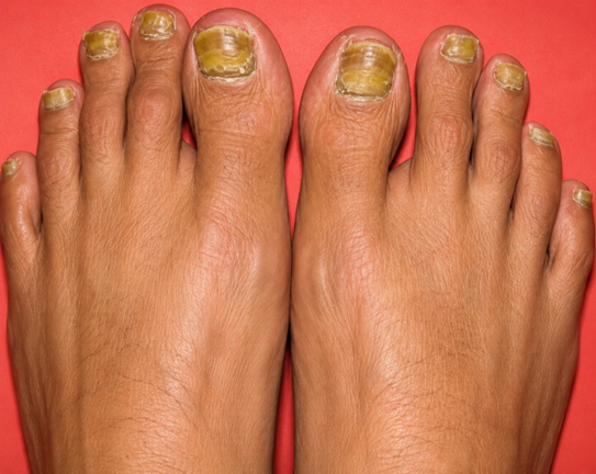
Leukonychia was the most frequent nail abnormality in the present study, occurring in 24% of patients, followed by clubbing (20%), onycholysis (15%), koilonychia (12%), Beau's lines (11%), and splinter hemorrhages (10%). Clubbing was observed in 20% of patients and demonstrated a significant association with respiratory disease, being present in 11 of 18 patients (61.1%) (p = 0.002). It was also observed in 7 of 24 patients (29.2%) with cardiovascular disease. This observation is consistent with established literature describing acquired clubbing predominantly in association with thoracic disease. Gollins and de Berker reported that acquired clubbing is most commonly associated with thoracic disorders, while broader reviews have established associations with pulmonary and cardiovascular diseases. [1,18] A recent systematic review published in 2025 also reported a strong relationship between clubbing and pulmonary or cardiovascular conditions. [19] Thus, the present findings highlight the diagnostic relevance of clubbing, particularly when it occurs in patients with respiratory symptoms. Although clubbing is not disease-specific, its identification should prompt appropriate evaluation for chronic pulmonary, cardiac, and other systemic disorders.
Splinter hemorrhages were present in 10% of the study population and were most frequent among patients with cardiovascular disease, occurring in 5 patients (20.8%). Although splinter hemorrhages are nonspecific and may result from trauma, they can also occur in systemic vascular and inflammatory disorders. Their recognition may therefore provide an additional clinical clue requiring correlation with cardiovascular and systemic findings. Reviews have described their association with endocarditis, vasculitis, and connective tissue disorders. [15,19]
Renal disease demonstrated a characteristic pattern of nail abnormalities. Half-and-half nails were observed in 6 of 17 patients (35.3%), showing a statistically significant association with renal disease (p = 0.004). Muehrcke's lines were also identified in 2 patients (11.8%). These findings are consistent with established descriptions of half-and-half nails in chronic renal failure and Muehrcke's lines in severe hypoalbuminemia, nephrotic syndrome, chronic renal disease, and altered protein balance. [1,15,16,18] Recognition of these findings may therefore support further assessment of renal function and nutritional or protein status.
Among patients with hepatobiliary disease, Terry's nails were observed in 5 of 12 patients (41.7%), with a statistically significant association (p = 0.013). Muehrcke's lines were also seen in 2 patients (16.7%). Terry's nails are classically associated with chronic liver disease, although they may also occur in systemic conditions such as heart failure and diabetes. [1,15,16,18] The relatively high frequency observed in the present study emphasizes the importance of careful inspection of the nail bed and distal nail plate in patients with chronic hepatobiliary disease. However, these findings should always be interpreted alongside clinical and biochemical evidence rather than considered diagnostic in isolation.
Koilonychia showed the strongest association with hematological disorders, occurring in 8 of 11 patients (72.7%) (p = 0.001). Nail-bed and periungual pallor were also observed in this group. This finding is consistent with the well-established association between koilonychia and iron deficiency and supports its usefulness as a clinical clue to nutritional deficiency and hematological disease. [15,18] In appropriate clinical settings, recognition of koilonychia may prompt further evaluation with complete blood count and iron studies, particularly in patients with pallor, fatigue, nutritional risk, or chronic blood loss.
Leukonychia was the most common abnormality overall but did not demonstrate a significant association with any single systemic disease category (p = 0.184). Similarly, Beau's lines and onycholysis occurred across several disease groups without significant disease-specific associations. This emphasizes that many nail abnormalities are nonspecific and may result from several local or systemic mechanisms. [1,15] Beau's lines, for example, represent temporary interruption of nail-matrix activity and may follow severe illness, nutritional stress, infection, or other systemic insults. [18,19] Consequently, such findings are best interpreted in relation to disease history, timing, distribution, associated nail changes, and laboratory findings.
Onychoscopy provided additional structural information beyond routine clinical examination. The commonest onychoscopic findings were onycholysis (18.8%), subungual hyperkeratosis (11.3%), pitting (10.0%), and chromonychia (10.0%), while microhemorrhages were observed in 7.5%. Warke et al. demonstrated the diagnostic value of detailed onychoscopic examination and reported characteristic patterns in disorders such as onychomycosis and psoriasis. [20] Their findings are methodologically relevant to the present study because they demonstrate that subtle abnormalities of the nail plate and subungual region can be better characterized using onychoscopy. Direct comparison of prevalence, however, is inappropriate because their cohort primarily consisted of dermatological nail diseases.
Nailfold capillaroscopy was particularly informative among patients with rheumatological or connective tissue disease. Dilated capillaries were observed in 4 of 5 patients (80%), while microhemorrhages, capillary dropout, and avascular areas were each present in 2 patients (40%). These findings are consistent with the observations of Warke et al., who documented microhemorrhages, dilated capillaries, capillary dropout, and avascular areas in systemic sclerosis and described characteristic capillary patterns in systemic lupus erythematosus and dermatomyositis. [20] The broader literature also recognizes nailfold vascular abnormalities as important clues to connective tissue diseases. [15,18] Thus, combining routine nail examination with capillaroscopy may improve recognition of microvascular abnormalities that are not readily apparent clinically.
An important finding was the relationship between nail abnormalities, disease duration, and disease severity. Nail changes were present in 86.7% of patients with disease duration greater than five years compared with 70.9% among those with disease duration of five years or less (p = 0.048). Similarly, nail abnormalities were more frequent in patients with severe systemic disease (89.2% vs. 65.1%; p = 0.006). These findings are biologically plausible because nail growth is slow and disturbances affecting the nail matrix or nail bed may persist for weeks or months following systemic insults. Recent reviews have also suggested that nail changes may reflect disease activity or severity and, in some circumstances, may precede other clinical manifestations. [15,19]
Overall, the present study supports the clinical value of systematic nail examination in patients with systemic disease. The strongest disease-specific associations were observed between clubbing and respiratory disease, koilonychia and hematological disorders, half-and-half nails and renal disease, and Terry's nails and hepatobiliary disease. These findings are broadly consistent with established literature. [1,15,18] At the same time, the nonspecific nature of leukonychia, Beau's lines, and onycholysis demonstrates that nail findings should not be interpreted in isolation. Their greatest clinical value lies in integrating morphology, distribution, disease duration, systemic manifestations, laboratory investigations, onychoscopy, and nailfold capillaroscopy. The findings therefore support the concept that the nail unit can serve as a useful, simple, non-invasive clinical marker of systemic disease and may provide additional information regarding chronicity, severity, and the need for targeted systemic evaluation.
CONCLUSION:
The present study demonstrated that nail abnormalities were common among patients with systemic diseases, with 78% showing clinically significant changes. Leukonychia was the most frequent abnormality, while characteristic findings showed stronger disease-specific associations. Clubbing was significantly associated with respiratory disease, koilonychia with hematological disorders, half-and-half nails with renal disease, and Terry's nails with hepatobiliary disease. Onychoscopy provided additional structural information, while nailfold capillaroscopy demonstrated vascular abnormalities in connective tissue disorders. These findings highlight the diagnostic value of systematic nail examination and support its routine incorporation into general clinical assessment for early recognition, appropriate evaluation, and long-term monitoring of systemic disease.
REFERENCES:
1. Gollins CE, de Berker D. Nails in systemic disease. Clin Med (Lond). 2021 May;21(3):166-169.
2. Haneke E. Surgical anatomy of the nail apparatus. Dermatol Clin 2006; 3:291–6
3. Baran R, de Berker DAR, Holzberg M, et al. Diseases of the nails and their management, 4th edn. Wiley-Blackwell, 2012.
4. Singh G. Nails in systemic disease. Indian J Dermatol Venereol Leprol 2011;77:646-651
5. Lawry M, Daniel CR 3 rd . Nails in systemic disease. In: Scher RK, Daniel CR, editors. Nails: Diagnosis, Therapy, Surgery. 3 rd ed. Philadelphia: Elsevier Science Limited; 2005. p. 147-69.
6. Fanning WL, Aronson M. Osler node, Janeway lesions and splinter haemorrhages. Arch Dermatol 1977; 133:648–9.
7. Stone OJ. Spoon nails and clubbing. Cutis 1975; 16:235–41
8. Holzberg M, Walker HK. Terry's nails: revised definitions and new correlations. Lancet 1984; 1:896–9.
9. Hogan GR, Jones B. The relationship of koilonychia and iron deficiency in infants. J Paediatr 1970;77:1054.
10. Meyerson MS, Scher RK. Nail signs of systemic disease. In: Callen JP, Jorizzo JL, Greer KE, Penneys NS, Piette WW, Zone JJ, editors. Dermatological signs of internal disease. 2 nd ed. Philadelphia: WB Saunders Co.; 1999. p. 368-75
11. Tunc S, Ertam I, Pirildar T, Turk T, Ozturk M, Doganavsargil E. Nail changes in connective tissue diseases: Do nail changes provide clues for the diagnosis? J Eur Acad Dermatol Venereol 2007;21:497-503.
12. Vignes S, Baran R. Yellow nail syndrome. Orphanet J Rare Dis 2017;12:1–10.
13. Sherber N, Wigley FM, Scher RK. Autoimmune disorders: nail signs and therapeutic approaches. Dermatol Ther 2007;20:17–30.
14. Maeda M, Kachi H, Takagi H. Haemorrhagic patterns of the cuticles distal to the proximal nail folds in the fingers of patients with systemic scleroderma. Eur J Dermatol 1997;7:191–6.
15. Rowe E, Zubek AE. Nail disorders as clues to systemic disease. Clin Dermatol. 2025;43(2):191-200.
16. Gregoriou S, Argyriou G, Larios G, Rigopoulos D. Nail disorders and systemic disease: What the nails tell us. J Fam Pract 2008;57:509 14.
17. Abraham S, Kamath N, Pai GS, Pinto J, Kuruvilla M. Nail changes in systemic diseases: a clinical study of 435 cases. Indian J Dermatol Venereol Leprol. 1998;64:144-145.
18. Singal A, Arora R. Nail as a window of systemic diseases. Indian Dermatol Online J. 2015;6(2):67-74.
19. Mehra S. Nail changes as indicators of systemic disease: a systematic review of correlations and diagnostic outcomes. Br J Dermatol. 2025;193(Suppl 1).237.
20. Warke N, Padhiar B. Nail changes in dermatological diseases: a cross-sectional study at a tertiary care hospital. J Onychol Nail Surg. 2024;1:13-23. doi:10.25259/JONS_2_2024.