Comparison Of Reflux Symptoms Index (Rsi) With Reflux Finding Score (Rfs) And Its Effectiveness In Management Of Laryngopharyngeal Reflux Disease (Lprd)
- Saurabh Yadav , PG Resident, third year Department of Otorhinolaryngology, Santosh Medical College and Hospital, Santosh deemed to be university, Ghaziabad, Uttar Pradesh.
- Tarun Malhotra , Head of the department and Professor, Department of Otorhinolaryngology, Santosh Medical College and Hospital, Santosh deemed to be university, Ghaziabad, Uttar Pradesh.
- Sushil Gaur , Professor, Department of Otorhinolaryngology, Santosh Medical College and Hospital, Santosh deemed to be university, Ghaziabad, Uttar Pradesh.
Article Information:
Abstract:
Background: Laryngopharyngeal reflux disease (LPRD) is an extra-esophageal manifestation of gastroesophageal reflux disease characterized by reflux of gastric contents into the larynx, causing chronic upper airway symptoms. Diagnosis remains challenging due to variable clinical presentation. This study aimed to compare the Reflux Symptom Index (RSI) with the Reflux Finding Score (RFS) and evaluate their effectiveness in management of LPRD. Methods: A hospital-based prospective study was conducted in the Department of ENT, Santosh Medical College and Hospital, Ghaziabad, among 240 patients with symptoms suggestive of LPRD. Patients were evaluated using RSI questionnaire and 70° rigid laryngoscopy. Laryngeal findings were assessed using RFS. Patients were followed up after treatment to assess symptom improvement and correlation between RSI and RFS. Results: The majority of patients belonged to the 41–60 years age group (35.8%), with female predominance (56.2%). Heartburn/regurgitation (82.5%), globus sensation (80.0%), and throat clearing (75.8%) were the most common symptoms. Erythema/hyperemia (79.2%) and diffuse laryngeal edema (57.5%) were the commonest RFS findings. RSI showed a significant positive correlation with RFS (Spearman ρ=0.349, p<0.001). Following treatment, mean RSI score significantly decreased from 20.59 ± 7.53 to 6.86 ± 6.46 (p<0.0001). Overall clinical improvement was observed in 79.2% of patients. Conclusion: RSI and RFS are valuable complementary tools for assessment and monitoring of LPRD. Their combined use improves diagnostic evaluation and treatment response assessment. Lifestyle modification and appropriate medical therapy provide significant symptom improvement, particularly among patients with good treatment compliance.
Keywords:
Article :
INTRODUCTION:
Laryngopharyngeal reflux disease (LPRD) is an increasingly recognized extra-esophageal manifestation of gastroesophageal reflux disease (GERD), characterized by the retrograde movement of gastric contents into the larynx, pharynx, and upper aerodigestive tract, resulting in chronic irritation and inflammation of the laryngeal mucosa.[1] Unlike typical GERD, LPRD frequently occurs without classical symptoms of heartburn and regurgitation, making diagnosis challenging. It commonly presents with hoarseness, chronic throat clearing, globus sensation, chronic cough, excessive throat mucus, and dysphagia, significantly affecting voice quality, daily functioning, and quality of life. [2,3]
The prevalence of LPRD varies widely due to differences in diagnostic criteria and population characteristics, with studies suggesting that reflux-related laryngeal symptoms account for a substantial proportion of patients presenting to otolaryngology clinics.[4] The disease has a multifactorial pathogenesis involving direct mucosal injury from gastric acid, pepsin, bile salts, and other refluxate components, along with impaired mucosal defense mechanisms and altered upper esophageal sphincter function.[5] Repeated exposure of the laryngeal mucosa to refluxate leads to edema, erythema, granuloma formation, posterior commissure hypertrophy, and vocal fold changes.[6]
Diagnosis of LPRD remains challenging because there is no single universally accepted gold standard investigation. Various diagnostic modalities, including 24-hour dual-probe pH monitoring, impedance-pH monitoring, endoscopic evaluation, and clinical scoring systems, have been used for diagnosis and assessment of treatment response. [7] However, invasive investigations are expensive, technically demanding, and not routinely available, resulting in increased interest in symptom-based and clinical assessment tools.[8]
The Reflux Symptom Index (RSI), introduced in 2002, is a validated patient-reported questionnaire designed to quantify the severity of symptoms associated with LPRD. It consists of nine symptoms, including hoarseness, throat clearing, excess throat mucus, swallowing difficulty, coughing after meals or lying down, breathing difficulties, troublesome cough, globus sensation, and heartburn/chest discomfort. Each symptom is scored from 0 to 5, with a total score of 45; a score greater than 13 is considered suggestive of LPRD. [9,10]
RSI provides a simple, non-invasive, and reproducible method for evaluating symptom severity and monitoring treatment outcomes. The Reflux Finding Score (RFS), developed is an objective endoscopic scoring system used to assess laryngeal findings associated with reflux.[11] It evaluates eight clinical parameters, including subglottic edema, arytenoid erythema, vocal fold edema, diffuse laryngeal edema, posterior commissure hypertrophy, granuloma formation, thick endolaryngeal mucus, and ventricular obliteration. An RFS score greater than 7 suggests the presence of LPRD. Unlike RSI, which reflects patient perception of symptoms, RFS provides clinician-based assessment of anatomical and inflammatory changes. [12,13]
Although RSI and RFS are widely used in clinical practice, the relationship between subjective symptoms and objective laryngeal findings remains controversial. Several studies have demonstrated a positive correlation between RSI and RFS, suggesting that symptom severity may correspond with laryngeal inflammatory changes, whereas others have reported poor correlation due to variations in symptom perception, disease duration, and individual sensitivity to reflux exposure.[14] Therefore, simultaneous assessment using both RSI and RFS may provide a more comprehensive evaluation of disease severity and therapeutic response.[15]
Management of LPRD primarily involves lifestyle modification, dietary measures, and pharmacological therapy, most commonly proton pump inhibitors (PPIs), sometimes combined with alginate preparations or other anti-reflux medications.[16] However, treatment response varies considerably, and objective evaluation of improvement remains difficult. Monitoring changes in RSI and RFS scores before and after therapy may help determine treatment effectiveness and guide long-term management strategies.[17]
Recent studies have emphasized the importance of a combined symptom and endoscopic approach rather than relying solely on subjective complaints or laryngoscopic findings. Comparison of RSI with RFS may help establish the effectiveness of these scoring systems in diagnosis, severity assessment, and follow-up of patients with LPRD.[18]
Understanding the correlation between patient-reported symptoms and objective laryngeal findings can improve diagnostic accuracy, reduce unnecessary investigations, and optimize therapeutic outcomes. Therefore, the present study, “Comparison of Reflux Symptoms Index (RSI) with Reflux Finding Score (RFS) and Its Effectiveness in Management of Laryngopharyngeal Reflux Disease (LPRD)”, is undertaken to evaluate the association between RSI and RFS, assess changes following treatment, and determine their role in monitoring disease response among patients with LPRD.
MATERIALS AND METHODS:
The present study was conducted as a hospital-based prospective observational study in the Department of Otorhinolaryngology (ENT) at Santosh Medical College and Hospital, Ghaziabad. The study period included recruitment of eligible patients attending the ENT outpatient department (OPD) and patients identified through health camps. The study was initiated after obtaining approval from the Institutional Ethics Committee. Written informed consent was obtained from all participants or their guardians before enrolment.
Study Population
Patients presenting with symptoms suggestive of laryngopharyngeal reflux disease (LPRD), including throat discomfort, voice changes, chronic throat clearing, globus sensation, cough, and swallowing complaints, were evaluated and included in the study after fulfilling the predefined eligibility criteria.
Inclusion Criteria
Patients fulfilling the following criteria were included:
1. Patients with symptoms suggestive of reflux disease.
2. Patients aged more than 12 years.
3. Patients with freely mobile vocal cords on rigid or fibreoptic laryngoscopic examination.
4. Patients presenting with throat and voice-related complaints for more than one month.
5. Patients or guardians willing to provide informed consent for participation.
Exclusion Criteria
Patients were excluded from the study if they had:
1. Active infections such as rhinitis or pharyngitis.
2. History of smoking or alcohol consumption.
3. Voice abuse.
4. History of allergy.
5. Previous laryngeal intervention or chronic systemic disease affecting voice and throat symptoms.
6. Age below 12 years.
7. Refusal to provide consent by the patient or guardian.
Study Methodology
After obtaining ethical clearance, all eligible patients satisfying the inclusion criteria were enrolled in the study. A detailed history was obtained, including demographic details, duration of symptoms, dietary habits, associated risk factors, and presenting complaints.
In the initial assessment, all participants were asked to complete the Reflux Symptom Index (RSI) questionnaire proposed by Belafsky et al. Patients with an RSI score of more than 10 were considered to have abnormal reflux-related symptoms and were further evaluated.
All selected patients underwent 70° rigid laryngoscopic examination. The laryngoscopic examination was performed by a single experienced ENT surgeon to maintain uniformity and reduce observer variability. The findings were assessed according to the Reflux Finding Score (RFS) classification described by Belafsky et al.
The correlation between subjective symptom severity assessed by RSI and objective laryngeal findings assessed by RFS was evaluated. Patients were followed up at 6 weeks and 3 months after initiation of treatment to assess clinical response and changes in RSI and RFS scores.
Assessment Tools
Reflux Symptom Index (RSI)
The RSI is a validated patient-reported symptom scoring system used for assessing the severity of LPRD symptoms. Patients were asked to grade the severity of nine reflux-related symptoms experienced during the previous month.
Each symptom was scored from 0 to 5, where:
• 0 = No problem
• 5 = Severe problem
The nine assessed symptoms included:
1. Hoarseness or voice problems.
2. Throat clearing.
3. Excess throat mucus or postnasal drip.
4. Difficulty swallowing food, liquids, or tablets.
5. Coughing after meals or while lying down.
6. Breathing difficulty or choking episodes.
7. Troublesome cough.
8. Sensation of a lump or foreign body in the throat.
9. Heartburn, chest pain, indigestion, or acid reflux sensation.
The total RSI score ranged from 0 to 45. An RSI score >10 was considered abnormal and suggestive of LPRD.
Reflux Finding Score (RFS)
The RFS is an objective endoscopic scoring system used to evaluate laryngeal findings associated with reflux disease. The following parameters were assessed during rigid laryngoscopy:
1. Subglottic edema
· Present: 2
· Absent: 0
2. Ventricular obliteration
· Partial: 2
· Complete: 4
3. Erythema/hyperemia
· Arytenoids only: 2
· Diffuse: 4
4. Vocal cord edema
· Mild: 1
· Moderate: 2
· Severe: 3
· Polypoid: 4
5. Diffuse laryngeal edema
· Mild: 1
· Moderate: 2
· Severe: 3
· Obstructing: 4
6. Posterior commissure hypertrophy
· Mild: 1
· Moderate: 2
· Severe: 3
· Obstructing: 4
7. Granuloma/granulation tissue
· Present: 2
· Absent: 0
8. Thick endolaryngeal mucus
· Present: 2
· Absent: 0
The total RFS score was calculated for each patient and correlated with RSI values.
Statistical Analysis
Data collected during the study were entered into Microsoft Excel and analyzed using the Statistical Package for Social Sciences (SPSS) software 21.
Categorical variables including age group, sex, residence, body mass index category, duration of symptoms, dietary habits, associated risk factors, treatment modality, treatment compliance, and clinical response were expressed as frequency and percentage.Continuous variables such as total RSI score and total RFS score were expressed as mean ± standard deviation.The association between categorical variables was analyzed using the Chi-square test. Fisher’s exact test was applied whenever the expected cell frequency was less than five.The correlation between RSI and RFS total scores was assessed using Pearson’s correlation coefficient for parametric data. A p-value <0.05 was considered statistically significant.
RESULTS:
A total of 240 patients with laryngopharyngeal reflux disease (LPRD) were included in the study and evaluated using the Reflux Symptom Index (RSI) and Reflux Finding Score (RFS).
The demographic profile, clinical characteristics, symptom and laryngoscopic findings, correlation between RSI and RFS, treatment pattern, compliance, and therapeutic response were analyzed.The largest proportion of patients belonged to the 41–60-year age group (35.8%), followed by 21–40 years (30.0%).
Females constituted 56.2% of the study population, while 43.8% were males. Most patients were from urban areas (60.4%). Nearly half (48.8%) had a normal BMI, whereas 39.2% were overweight. A considerable proportion had long-standing symptoms, with 42.1% reporting symptoms for 13–36 months and 36.7% for more than 36 months (Table 1).
Table 1. Demographic and baseline characteristics of the study population (n=240)
|
Characteristic |
Category |
n (%) |
|
Age group (years) |
≤20 |
26 (10.8) |
|
21–40 |
72 (30.0) |
|
|
41–60 |
86 (35.8) |
|
|
≥60 |
56 (23.3) |
|
|
Sex |
Female |
135 (56.2) |
|
Male |
105 (43.8) |
|
|
Residence |
Urban |
145 (60.4) |
|
Rural |
95 (39.6) |
|
|
BMI (kg/m²) |
Underweight (<18.5) |
11 (4.6) |
|
Normal (18.5–24.9) |
117 (48.8) |
|
|
Overweight (25–29.9) |
94 (39.2) |
|
|
Obese (≥30) |
18 (7.5) |
|
|
Duration of symptoms |
≤3 months |
13 (5.4) |
|
4–12 months |
38 (15.8) |
|
|
13–36 months |
101 (42.1) |
|
|
>36 months |
88 (36.7) |
Spicy/non-vegetarian food consumption was the most frequently identified dietary factor, reported by 73.3% of patients, followed by tea/coffee consumption (71.3%) and late-night meals (61.7%). Regarding etiopathology, reflux-dominant GERD was the predominant category, accounting for 49.2% of patients. Allergic overlap was observed in 17.5%, while laryngeal hypersensitivity and smoking/irritant-related causes accounted for 12.5% and 12.1%, respectively (Table 2, Figure 1).
Table 2. Dietary habits, risk factors and etiopathological profile (n=240)
|
Variable |
n (%) |
|
Dietary habits/risk factors |
|
|
Spicy/non-vegetarian food intake |
176 (73.3) |
|
Tea/coffee intake |
171 (71.3) |
|
Late-night meals |
148 (61.7) |
|
Alcohol intake |
66 (27.5) |
|
Smoking/pollution exposure |
48 (20.0) |
|
Etiopathological category |
|
|
Reflux-dominant GERD |
118 (49.2) |
|
Allergic overlap |
42 (17.5) |
|
Laryngeal hypersensitivity |
30 (12.5) |
|
Smoking/irritant-related |
29 (12.1) |
|
Mixed/uncertain |
21 (8.8) |
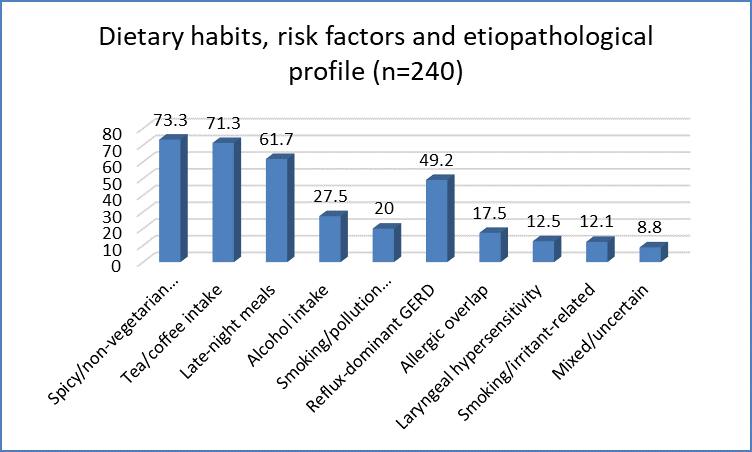
Figure 1 Dietary habits, risk factors and etiopathological profile (n=240)
Among the RSI symptoms, heartburn/regurgitation was the most frequent complaint (82.5%), followed by globus sensation (80.0%), chronic throat clearing (75.8%), and hoarseness/voice problems (75.0%). Excess throat mucus or postnasal drip was reported by 70.0%, while coughing after meals or lying down occurred in 65.0%. Breathing difficulty/choking was the least frequently reported symptom (12.5%). Heartburn/regurgitation also had the highest mean symptom score (2.40 ± 1.05), followed by globus sensation (2.35 ± 1.00) (Table 3).
Table 3. Distribution of Reflux Symptom Index symptoms (n=240)
|
RSI symptom |
Present, n (%) |
Mean score ± SD |
|
Hoarseness/voice problem |
180 (75.0) |
2.10 ± 1.00 |
|
Chronic throat clearing |
182 (75.8) |
2.25 ± 1.02 |
|
Excess throat mucus/PND |
168 (70.0) |
2.30 ± 1.08 |
|
Difficulty swallowing |
132 (55.0) |
1.90 ± 0.98 |
|
Cough after meals/lying down |
156 (65.0) |
2.05 ± 1.01 |
|
Breathing difficulty/choking |
30 (12.5) |
1.40 ± 0.75 |
|
Troublesome cough |
66 (27.5) |
1.85 ± 0.92 |
|
Globus sensation |
192 (80.0) |
2.35 ± 1.00 |
|
Heartburn/regurgitation |
198 (82.5) |
2.40 ± 1.05 |
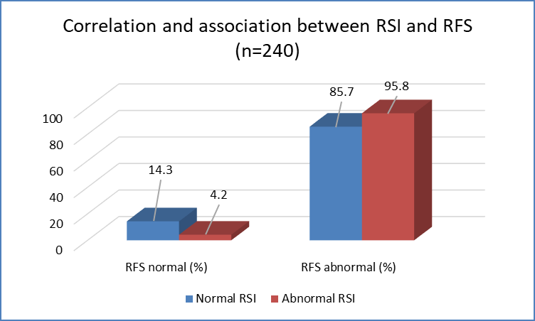
Figure 2 Distribution of Reflux Symptom Index symptoms (n=240)
On laryngoscopic examination, erythema/hyperemia was the most frequent RFS finding (79.2%), followed by diffuse laryngeal edema (57.5%), vocal cord edema (50.0%), and ventricular obliteration (43.3%). Thick endolaryngeal mucus was the least frequent finding (10.0%). According to overall RFS severity, 149 patients (62.1%) had mild disease, 56 (23.3%) had moderate disease, and 35 (14.6%) had severe disease (Table 4).
Table 4. Distribution of Reflux Finding Score components and RFS severity (n=240)
|
RFS parameter |
Present, n (%) |
Mean score ± SD |
|
Subglottic edema |
84 (35.0) |
0.50 ± 0.50 |
|
Ventricular obliteration |
104 (43.3) |
1.42 ± 0.89 |
|
Erythema/hyperemia |
190 (79.2) |
2.18 ± 0.92 |
|
Vocal cord edema |
120 (50.0) |
1.02 ± 0.61 |
|
Diffuse laryngeal edema |
138 (57.5) |
1.11 ± 0.66 |
|
Posterior commissure hypertrophy |
72 (30.0) |
1.43 ± 0.72 |
|
Granuloma/granulation |
37 (15.4) |
0.15 ± 0.36 |
|
Thick endolaryngeal mucus |
24 (10.0) |
0.54 ± 0.50 |
|
RFS severity grade |
n (%) |
|
|
Mild |
149 (62.1) |
|
|
Moderate |
56 (23.3) |
|
|
Severe |
35 (14.6) |
There was a positive and statistically significant correlation between total RSI and total RFS scores. Spearman's correlation coefficient was ρ=0.349 (p<0.001), while Pearson's correlation coefficient was r=0.370 (p<0.001), indicating that increasing symptom severity was associated with increasing severity of laryngoscopic findings. Among patients with an abnormal RSI, 95.8% also had an abnormal RFS compared with 85.7% of patients with a normal RSI. However, the categorical association between abnormal RSI and abnormal RFS did not reach statistical significance (p=0.078) (Table 5, Figure 3).
Table 5. Correlation and association between RSI and RFS (n=240)
|
Parameter |
Result |
p value |
|
Spearman correlation between total RSI and RFS (ρ) |
0.349 |
<0.001* |
|
Pearson correlation between total RSI and RFS (r) |
0.370 |
<0.001* |
|
RSI status vs RFS status |
RFS normal, n (%) |
RFS abnormal, n (%) |
|
Normal RSI (n=28) |
4 (14.3) |
24 (85.7) |
|
Abnormal RSI (n=212) |
9 (4.2) |
203 (95.8) |
|
Total |
13 (5.4) |
227 (94.6) |
|
Chi-square association |
— |
0.078 |
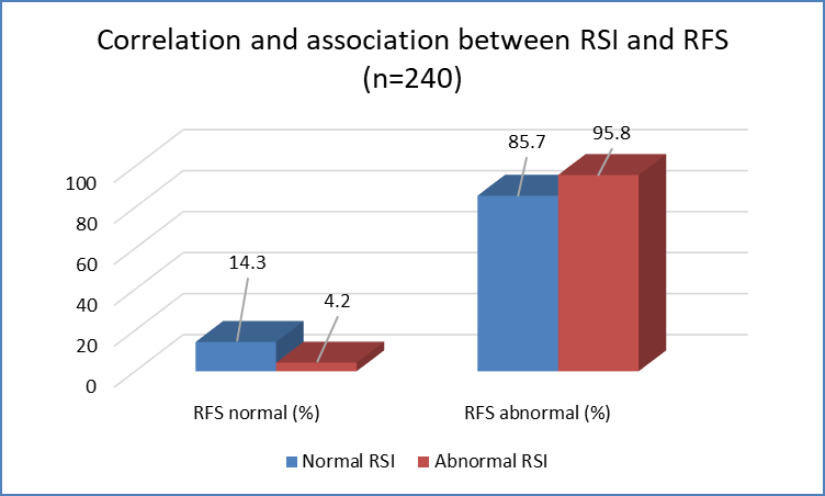
Figure 3 Correlation and association between RSI and RFS (n=240)
The most commonly prescribed initial treatment was PPI combined with alginate (49.6%), followed by PPI alone (23.3%). Good treatment compliance (≥80%) was observed in 51.2% of patients, moderate compliance in 34.2%, and poor compliance in 14.6%. Overall, 190 patients (79.2%) demonstrated clinical improvement, whereas 50 (20.8%) were classified as non-responders (Table 6).Clinical response showed a strong association with treatment compliance. All 123 patients with good compliance demonstrated improvement, compared with 76.8% of those with moderate compliance and only 11.4% of those with poor compliance. The association between compliance and treatment response was statistically significant (p<0.001) (Table 6).A substantial reduction in symptom burden was observed following treatment. The mean total RSI score decreased from 20.59 ± 7.53 at baseline to 6.86 ± 6.46 at follow-up, representing a mean reduction of 13.73 points. This improvement was statistically significant (p<0.0001) (Table 6).
Table 6. Treatment, compliance and clinical outcomes among study participants (n=240)
|
Parameter |
Category |
n (%) / Mean ± SD |
p value |
|
Initial treatment |
PPI + alginate |
119 (49.6) |
— |
|
PPI only |
56 (23.3) |
— |
|
|
PPI + alginate + H2RA |
33 (13.8) |
— |
|
|
Lifestyle modification only |
32 (13.3) |
— |
|
|
Compliance |
Good (≥80%) |
123 (51.2) |
— |
|
Moderate (50–79%) |
82 (34.2) |
— |
|
|
Poor (<50%) |
35 (14.6) |
— |
|
|
Clinical response |
Improved/responder |
190 (79.2) |
— |
|
Not improved/non-responder |
50 (20.8) |
— |
|
|
Response by compliance |
Good: 123/123 responders (100%) |
123 (100.0) |
<0.001* |
|
Moderate: 63/82 responders |
63 (76.8) |
||
|
Poor: 4/35 responders |
4 (11.4) |
||
|
RSI score |
Baseline |
20.59 ± 7.53 |
|
|
Follow-up |
6.86 ± 6.46 |
<0.0001* |
|
|
Mean reduction |
13.73 |
DISCUSSION:
Laryngopharyngeal reflux disease (LPRD) is a common extra-esophageal manifestation of gastroesophageal reflux disease caused by retrograde movement of gastric contents into the larynx and pharynx, resulting in chronic mucosal irritation and variable upper airway symptoms. Diagnosis remains challenging due to nonspecific symptoms and lack of a single definitive diagnostic test. The present prospective study evaluated 240 patients with suspected LPRD using the Reflux Symptom Index (RSI) and Reflux Finding Score (RFS), along with assessment of risk factors, treatment response, and patient compliance. In the present study, the majority of patients belonged to the 41–60 years age group (35.8%), followed by 21–40 years (30.0%). This finding indicates a higher occurrence of LPRD among middle-aged individuals. Similar observations were reported by Belafsky et al. [19] who described increased prevalence of LPR-related symptoms during the fourth to sixth decades of life. Reported a mean age of 51.2 years in a multicentric LPR study, with most patients belonging to the 40–60 years age group, supporting the present findings. A slight female predominance was observed in the current study, with females comprising 56.2% of cases. Similar female predominance has been reported by (approximately 58%) and Campagnolo et al. [20] (around 60%).
This may be related to greater healthcare-seeking behaviour, increased symptom perception, and possible hormonal influences affecting reflux susceptibility. Urban residence was observed in 60.4% of patients in the present study. Similar urban predominance was reported by Campagnolo et al. [20] suggesting that lifestyle-related factors such as irregular meals, caffeine intake, processed food consumption, and sedentary habits may contribute to reflux symptoms. Regarding BMI, 48.8% of patients had normal BMI, while 39.2% were overweight and 7.5% were obese. Qadeer et al. [21] reported higher RSI scores among patients with BMI >25 kg/m², indicating a relationship between increased body weight and reflux severity. Increased intra-abdominal pressure and altered gastroesophageal junction function may explain this association. A prolonged symptom duration was observed in the present study, with 42.1% of patients having symptoms for 13–36 months and 36.7% for more than 36 months. Similar findings were reported by where approximately 70% of patients had symptoms for more than one year before diagnosis.
Koufman et al. [22] emphasized that delayed diagnosis is common due to nonspecific clinical manifestations and overlap with other throat disorders. Dietary and lifestyle risk factors were frequently observed. Spicy/non-vegetarian food intake was reported by 73.3%, tea/coffee intake by 71.3%, and late-night meals by 61.7% of patients. Koufman et al. [22] reported similar dietary triggers in more than 70% of LPR patients, while highlighted caffeine intake and late-night eating as important contributors. Smoking and alcohol intake were present in 20.0% and 27.5% of patients respectively. The most common RSI symptoms in the present study were heartburn/regurgitation (82.5%), globus sensation (80.0%), chronic throat clearing (75.8%), and hoarseness (75.0%). Similar symptom patterns were reported by Belafsky et al. [19] and Campagnolo et al. [20] confirming the utility of RSI in evaluating symptom severity in LPRD. On laryngoscopic evaluation, erythema/hyperemia was the most frequent RFS finding (79.2%), followed by diffuse laryngeal edema (57.5%) and vocal cord edema (50.0%).
These findings are consistent with Belafsky et al. [19] who identified erythema and edema as common objective features of LPRD. Most patients had mild RFS severity (62.1%), followed by moderate (23.3%) and severe disease (14.6%), indicating that LPRD generally presents as a low-grade inflammatory condition. A significant positive correlation was observed between RSI and RFS scores in the present study (Spearman ρ=0.349, Pearson r=0.370; p<0.001). Similar findings were reported by Belafsky et al. [19] supporting the complementary role of symptom-based and endoscopic assessment in LPRD. Treatment resulted in significant clinical improvement, with 79.2% of patients showing response and mean RSI score decreasing from 20.59 ± 7.53 to 6.86 ± 6.46 (p<0.0001). Good compliance was strongly associated with better outcomes, with all patients having good compliance demonstrating improvement. Similar emphasis on combined medical therapy, dietary modification, and lifestyle changes was reported by Koufman et al. [22]
CONCLUSION:
Laryngopharyngeal reflux disease predominantly affects middle-aged individuals and is frequently associated with lifestyle-related risk factors such as dietary triggers, caffeine intake, and late-night meals. The Reflux Symptom Index (RSI) and Reflux Finding Score (RFS) demonstrated a significant positive correlation, indicating that subjective symptoms correlate with objective laryngeal findings. Combined assessment using RSI and RFS provides a useful approach for diagnosis, severity evaluation, and monitoring of treatment response in LPRD patients. Medical therapy along with lifestyle modification resulted in significant symptom improvement, with better outcomes observed among patients with good treatment compliance. RSI and RFS can serve as reliable, non-invasive clinical tools for routine assessment and follow-up of patients with suspected LPRD.
REFERENCES:
1. Brown J, Shermetaro C. Laryngopharyngeal reflux. In: StatPearls [Internet]. Treasure Island (FL): StatPearls Publishing; 2022.
2. Lechien JR, Akst LM, Hamdan AL, Schindler A, Karkos PD, Barillari MR, et al. Evaluation and management of laryngopharyngeal reflux disease: state of the art review. Otolaryngol Head Neck Surg. 2019;161(3):541-547.
3. Salihefendic N, Zildzic M, Cabric E. Laryngopharyngeal reflux disease (LPRD). Acta Clin Croat. 2017;56(3):541-548.
4. Lien HC, Wang HP, Chen CL. Diagnosis of laryngopharyngeal reflux: past, present, and future. Diagnostics (Basel). 2023;13(9):1643.
5. Al-Abbasi AM. Utility of Reflux Finding Score and Reflux Symptom Index in diagnosis of LPRD. Indian J Otolaryngol Head Neck Surg. 2021;73(3):417-424.
6. Joshi AA. Laryngopharyngeal reflux: clinical profile and management. Indian J Head Neck Surg. 2015;67(2):125-132.
7. Lechien JR, Saussez S. Management strategies for laryngopharyngeal reflux: review of current evidence. Respir Med Case Rep. 2021;33:101438.
8. Lien HC, Wang HP, Chen CL. Diagnostic advances in laryngopharyngeal reflux disease. Diagnostics (Basel). 2023;13(9):1643.
9. Belafsky PC, Postma GN, Koufman JA. The validity and reliability of the Reflux Finding Score (RFS). Laryngoscope. 2001;111(8):1313-1317.
10. Hutnik R, Suda A, Mehta K. Comparative evaluation of RSI and RFS improvements after treatment of LPRD. Am J Otolaryngol. 2020;41(5):102536.
11. Robbins J, Aviv JE. Normal esophageal physiology and laryngopharyngeal reflux. Otolaryngol Clin North Am. 2013;46(6):1093-1105.
12. StatPearls Authors. Laryngopharyngeal reflux. In: StatPearls [Internet]. Treasure Island (FL): StatPearls Publishing; 2024.
13. Cui N, Dai T, Liu Y, Wang YY, Lin JY, Zheng QF, et al. Laryngopharyngeal reflux disease: Updated examination of mechanisms, pathophysiology, treatment and association with gastroesophageal reflux disease. World J Gastroenterol. 2024;30(16):2209-2219.
14. Mao F, Zhang W. Anatomy of reflux: a growing health problem affecting structures of the upper aerodigestive tract. Anat Rec (Hoboken). 2006;289(11):1145-1152.
15. Fraser-Kirk K. Laryngopharyngeal reflux: a confounding cause of aerodigestive dysfunction. Aust Fam Physician. 2017;46(1):35-39.
16. Johnston N, Wells CW, Samuels TL, Blumin JH. Rationale for targeting pepsin in the treatment of reflux disease. Ann Otol Rhinol Laryngol. 2010;119(8):547-558.
17. Nilakhe S. A study of relationship between reflux symptom index and reflux finding score in patients with laryngopharyngeal reflux disease. J Voice. 2019;33(5):747-753.
18. Saha J. The importance of laryngoscopic findings as predictors of the treatment outcome in laryngopharyngeal reflux. Egypt J Otolaryngol. 2022;38(1):25.
19. Belafsky PC, Postma GN, Koufman JA. Validity and reliability of the reflux symptom index (RSI). J Voice. 2002;16(2):274-277.
20. Campagnolo AM, Priston J, Thoen RH, Medeiros T, Assunção AR. Laryngopharyngeal reflux: diagnosis, treatment, and latest research. Int Arch Otorhinolaryngol. 2014;18(2):184-191.
21. Qadeer MA, Swoger J, Milstein C, Hicks DM, Ponsky J, Richter JE, et al. Correlation between symptoms and laryngeal signs in laryngopharyngeal reflux. Laryngoscope. 2005;115(11):1945-1950.
22. Koufman JA, Sataloff RT, Toohill RJ. Laryngopharyngeal reflux: consensus report. J Voice. 1996;10(3):215-216.