Clinical Profile, Management Strategies, and Visual Outcomes in Patients with Ocular Trauma: A Prospective Study
- B. Karishma Munoli , Associate Professor, Department of Ophthalmology, RIMS, Raichur
- Ramanna , Assistant Professor, Department of Ophthalmology, RIMS, Raichur
- Vidyashri M , Private practice, Department of Ophthalmology, Shankarappagouda hospital, Yadgir
Article Information:
Abstract:
Background: Ocular trauma is an important and potentially preventable cause of visual impairment, particularly among young and economically active individuals. Early assessment and appropriate management are crucial for preserving vision and preventing complications. Objectives: To evaluate the clinical profile, patterns of injury, management strategies, visual outcomes, and factors associated with poor visual outcome among patients with ocular trauma. Materials and Methods: This prospective observational study included 100 patients presenting with ocular trauma. Demographic characteristics, mechanism and type of injury, ocular structures involved, time to presentation, clinical findings, and management strategies were recorded. Visual acuity was assessed at presentation and final follow-up. Factors associated with final visual outcome were analyzed, with p<0.05 considered statistically significant. Results: The 21–40-year age group constituted 43.0% of patients, and 74.0% were males. Workplace/occupational injuries were most frequent (32.0%), and blunt trauma was the commonest mechanism (39.0%). Closed-globe injuries constituted 62.0%, while 38.0% were open-globe injuries. Corneal injury was the most frequent clinical finding (52.0%). Conservative management was used in 55.0%, whereas 45.0% required surgery. Visual acuity ≥6/12 increased from 17.0% at presentation to 44.0% at final follow-up. Overall, 86.0% achieved a good visual outcome. Open-globe injury, delayed presentation >24 hours, lens involvement, vitreous haemorrhage, retinal detachment, RAPD, and endophthalmitis were significantly associated with poor visual outcome (p<0.05). Conclusion: Most patients achieved meaningful visual recovery following appropriate management. Early presentation and prompt treatment are essential, particularly in patients with high-risk clinical features.
Keywords:
Article :
INTRODUCTION:
Ocular trauma is an important and potentially preventable cause of visual impairment and blindness worldwide. It encompasses a wide spectrum of injuries, ranging from minor superficial lesions to severe disruption of ocular structures resulting in permanent visual disability[1]. Ocular injuries affect individuals across all age groups but are particularly important among children and working-age adults because of their long-term socioeconomic and functional consequences. Globally, millions of individuals experience ocular injuries annually, with a substantial proportion developing unilateral or bilateral visual impairment.[2,3]
The clinical profile of ocular trauma varies according to age, occupation, mechanism and circumstances of injury, nature of the offending agent, use of protective equipment, and time to presentation. Common causes include occupational and agricultural accidents, road traffic accidents, domestic injuries, sports-related trauma, and assault. Depending on the structures involved and integrity of the globe, ocular injuries may be classified as closed-globe or open-globe injuries[4]. Open-globe injuries represent particularly severe forms of ocular trauma and include rupture, penetrating injury, perforating injury, and injuries associated with intraocular foreign bodies. The Birmingham Eye Trauma Terminology (BETT) provides a standardized framework for classifying mechanical ocular injuries and facilitates uniform documentation and comparison of clinical outcomes.[5,6]
Visual prognosis following ocular trauma depends on the mechanism, anatomical location and severity of injury, presenting visual acuity, and associated ocular complications. Factors such as relative afferent pupillary defect, traumatic cataract, vitreous haemorrhage, retinal detachment, endophthalmitis, posterior-segment involvement, and retained intraocular foreign bodies are frequently associated with poorer outcomes.[7] The Ocular Trauma Score (OTS) incorporates presenting visual acuity and major adverse prognostic features to estimate the probability of final visual outcome and may assist in clinical decision-making and patient counselling.[7,8]
Management of ocular trauma requires prompt assessment and treatment tailored to the type and severity of injury. Minor injuries may be managed conservatively, whereas severe closed-globe and open-globe injuries may require urgent surgical intervention. Primary globe repair, traumatic cataract surgery, intraocular foreign-body removal, vitrectomy, retinal surgery, and secondary reconstructive procedures may be required.[9] Timely intervention and careful follow-up are essential because complications such as infection, secondary glaucoma, retinal detachment, proliferative vitreoretinopathy, and corneal scarring can adversely affect visual recovery.[10–13]
Despite advances in diagnostic imaging, microsurgical techniques, vitreoretinal surgery, and postoperative care, visual outcomes following ocular trauma remain highly variable. Prospective evaluation of the complete spectrum of ocular trauma can therefore provide valuable information regarding injury patterns, treatment practices, and determinants of visual recovery. Hence, the present prospective study was undertaken to evaluate the clinical and epidemiological profile of patients with ocular trauma, characterize the patterns and mechanisms of injury, assess the management strategies employed, determine subsequent visual outcomes, and identify factors associated with visual prognosis.
MATERIALS AND METHODS:
Study Design
The present study was conducted as a prospective observational study to evaluate the clinical profile, management strategies, and visual outcomes among patients presenting with ocular trauma.The study was conducted in the Department of Ophthalmology at a tertiary care hospital. Patients presenting to the ophthalmology outpatient department, emergency department, or referred from other departments with ocular trauma were screened for eligibility.
Study Population
The study population comprised patients presenting with ocular trauma during the study period. Both open-globe and closed-globe injuries, as well as other relevant forms of ocular trauma, were considered for inclusion according to the predefined eligibility criteria.A total of 100 patients with ocular trauma who fulfilled the eligibility criteria were included in the study.
Inclusion Criteria
Patients were included if they:
1. Presented with a history and clinical evidence of ocular trauma.
2. Had open-globe, closed-globe, or other clinically significant ocular injuries requiring ophthalmological evaluation.
3. Presented during the defined study period.
4. Were willing to participate in the study and undergo the required clinical evaluation and follow-up.
5. Provided written informed consent; for participants below the age of legal consent, consent was obtained from the parent or legal guardian.
Exclusion Criteria
Patients were excluded if they:
1. Had pre-existing severe ocular disease or visual impairment that could independently influence assessment of the visual outcome.
2. Had previous ocular surgery or ocular pathology that significantly interfered with evaluation of trauma-related visual outcome.
3. Had incomplete clinical information required for assessment of the ocular injury.
4. We’re unwilling to participate or provide informed consent.
5. Were unavailable for the required follow-up and final visual outcome assessment.
Data Collection
After enrolment, demographic and clinical information was recorded using a predesigned structured proforma. Data included age, sex, occupation, affected eye, place and circumstances of injury, mechanism of trauma, nature of the offending object, time interval between injury and presentation, and any treatment received before presentation.The ocular injury was categorized according to its mechanism and anatomical characteristics. Mechanical injuries were classified, wherever applicable, using the Birmingham Eye Trauma Terminology (BETT) system.
Clinical Evaluation
Each patient underwent a detailed ophthalmological evaluation. A comprehensive history regarding the injury was obtained, followed by examination of the affected and fellow eye.
Visual Acuity Assessment
Presenting visual acuity was assessed separately for each eye using an appropriate standard visual acuity chart whenever feasible. In patients with severe visual impairment, vision was documented as counting fingers, hand movements, perception of light, or no perception of light, as applicable.Best-corrected visual acuity was subsequently assessed during follow-up and at the final study visit.
Anterior Segment Examination
The anterior segment was examined using a torchlight and slit-lamp biomicroscopy whenever clinically feasible. The eyelids, conjunctiva, cornea, anterior chamber, iris, pupil, and lens were carefully evaluated. The presence of corneal or scleral wounds, hyphema, traumatic cataract, iris injury, anterior chamber abnormalities, or other trauma-related findings was recorded.
Pupillary Examination
Pupillary size and reaction were evaluated, and the presence or absence of a relative afferent pupillary defect (RAPD) was documented wherever assessment was possible.
Intraocular Pressure
Intraocular pressure was measured in patients in whom it was clinically appropriate and safe. Measurement was avoided in patients with suspected or confirmed open-globe injury until adequate globe repair had been performed.
Posterior Segment Examination
Posterior segment examination was performed using direct or indirect ophthalmoscopy whenever adequate visualization was possible. The vitreous, retina, macula, optic disc, and other posterior-segment structures were assessed for vitreous haemorrhage, retinal tears, retinal detachment, choroidal injury, optic nerve involvement, or other traumatic abnormalities.
Investigations
Investigations were performed according to the nature and severity of the ocular injury. B-scan ultrasonography was performed when posterior-segment visualization was obscured and globe integrity permitted its safe use.
Computed tomography of the orbit was performed when an intraocular or intraorbital foreign body, orbital fracture, or significant posterior orbital injury was suspected. Additional investigations were undertaken whenever clinically indicated.
Classification of Ocular Trauma
Ocular injuries were broadly categorized as open-globe and closed-globe injuries based on the integrity of the eyewall.
Closed-globe injuries included contusions and lamellar lacerations. Open-globe injuries included globe rupture and lacerations, with lacerations further categorized as penetrating injuries, perforating injuries, or injuries associated with an intraocular foreign body, in accordance with the BETT classification.
The anatomical structures and zones involved were documented wherever applicable.
Ocular Trauma Score
The Ocular Trauma Score (OTS) was calculated in patients for whom all necessary clinical parameters were available and in whom its application was appropriate. The score was determined using presenting visual acuity and relevant adverse prognostic factors, including globe rupture, endophthalmitis, perforating injury, retinal detachment, and relative afferent pupillary defect.Patients were categorized according to their OTS category, and the relationship between OTS and final visual outcome was evaluated.
Management
Management was individualized according to the type, severity, and anatomical extent of ocular trauma.
Conservative Management
Patients with injuries not requiring surgical intervention were managed conservatively with appropriate topical or systemic medications, wound care, protective measures, and observation according to the clinical condition.
Surgical Management
Surgical intervention was performed whenever clinically indicated. Procedures included primary repair of corneal or scleral wounds, removal of intraocular foreign bodies, anterior chamber reconstruction, traumatic cataract surgery, vitrectomy, retinal detachment surgery, and other reconstructive procedures as required.
Open-globe injuries were managed with appropriate stabilization followed by primary surgical repair at the earliest clinically feasible opportunity. Additional secondary procedures were performed according to subsequent clinical findings.
Follow-Up and Outcome Assessment
Patients were followed after initial treatment according to the clinical requirements of the injury. At each follow-up visit, visual acuity, anterior-segment findings, posterior-segment status, treatment response, and development of complications were assessed.
The primary outcome was the final visual acuity following management. Changes in visual acuity from presentation to final follow-up were recorded.
Secondary outcomes included the anatomical outcome, requirement for surgical intervention, number and type of procedures performed, and development of complications such as traumatic cataract, corneal opacity, secondary glaucoma, vitreous haemorrhage, retinal detachment, proliferative vitreoretinopathy, and endophthalmitis.
Factors potentially associated with final visual outcome, including presenting visual acuity, mechanism and type of injury, anatomical structures involved, time to presentation, posterior-segment involvement, associated complications, and management strategy, were evaluated.
Outcome Measures
The study assessed the following principal outcome measures:
• Demographic and clinical profile of ocular trauma patients.
• Mechanism, circumstances, and type of ocular injury.
• Anatomical structures involved and severity of injury.
• Presenting visual acuity.
• Conservative and surgical management strategies employed.
• Complications occurring during treatment and follow-up.
• Final visual acuity and change in vision following treatment.
• Association of clinical and injury-related factors with final visual outcome.
• Relationship between OTS category and final visual outcome, wherever applicable.
Statistical Analysis
The collected data were entered into a spreadsheet and analyzed using SPSS .21 statistical software. Continuous variables were summarized as mean ± standard deviation (SD) or median with interquartile range (IQR) depending on the distribution of data. Categorical variables were expressed as frequency and percentage.The normality of continuous variables was assessed using an appropriate normality test. Comparisons of continuous variables between two groups were performed using the independent-samples Student's t-test for normally distributed data and the Mann–Whitney U test for non-normally distributed data. Paired comparisons of presenting and final visual parameters were performed using the paired t-test or Wilcoxon signed-rank test, as appropriate.Categorical variables were compared using the Chi-square test or Fisher's exact test where expected cell frequencies were small. Associations between clinical characteristics, injury-related variables, management strategies, and final visual outcome were evaluated using appropriate univariate analyses. Where applicable, multivariable logistic regression analysis was performed to identify independent predictors of poor visual outcome, and results were reported as odds ratios with 95% confidence intervals.A p-value <0.05 was considered statistically significant.
RESULTS:
A total of 100 patients with ocular trauma were included in the study. The demographic and clinical characteristics of the study population are presented in Table 1. The majority of patients belonged to the age group of 21–40 years (43.0%), followed by 41–60 years (27.0%) and ≤20 years (21.0%), while only 9.0% were aged >60 years. Males constituted 74.0% of the study population, indicating a marked male predominance. The right eye was involved in 49.0% and the left eye in 48.0%, while bilateral involvement was observed in 3.0% of patients. Workplace or occupational injuries were the most common (32.0%), followed by injuries occurring at home (25.0%) and road/traffic-related injuries (19.0%). Regarding the time to presentation, 31.0% presented within 6 hours and 39.0% within >6–24 hours, indicating that 70.0% of patients presented within the first 24 hours (Table 1).The distribution of patients according to the mechanism and type of ocular injury is shown in Table 2. Blunt trauma was the most frequent mechanism of injury, accounting for 39.0% of cases, followed by sharp-object injuries (24.0%), foreign-body injuries (16.0%), and road traffic accidents (11.0%). Chemical and thermal injuries accounted for 6.0% and 2.0% of cases, respectively. Closed-globe injuries were more common than open-globe injuries (62.0% vs. 38.0%). Among the 62 closed-globe injuries, contusions constituted 75.8%, while lamellar lacerations accounted for 24.2%. Among the 38 open-globe injuries, penetrating injuries were the most common (50.0%), followed by globe rupture (26.3%), intraocular foreign-body injuries (15.8%), and perforating injuries (7.9%) (Table 2).
The ocular structures involved and major clinical findings at presentation are summarized in Table 3. Corneal injury was the most frequently observed finding, occurring in 52.0% of patients, followed by conjunctival injury in 44.0% and hyphema in 31.0%. Eyelid or adnexal injuries were present in 28.0%, while lens injury or traumatic cataract was documented in 27.0%. Posterior-segment manifestations included vitreous haemorrhage in 22.0%, retinal injury in 17.0%, and retinal detachment in 11.0%. Relative afferent pupillary defect (RAPD) was detected in 13.0%, intraocular foreign bodies in 6.0%, optic nerve injury in 5.0%, and endophthalmitis in 4.0% of patients (Table 3).
The management strategies employed are presented in Table 4 and illustrated in Figure 1. Conservative management was sufficient in 55.0% of patients, whereas 45.0% required surgical intervention. Primary corneal or scleral repair was performed in 25.0% of patients, making it the most frequently performed surgical procedure. Traumatic cataract surgery was required in 13.0%, vitrectomy in 12.0%, anterior chamber reconstruction in 7.0%, intraocular foreign-body removal in 6.0%, and retinal detachment surgery in 7.0%. Other reconstructive procedures were performed in 4.0% of patients, while 15.0% required a secondary surgical procedure during follow-up (Table 4; Figure 1).
A substantial improvement in visual acuity was observed from presentation to final follow-up, as demonstrated in Table 5 and Figure 2. At presentation, only 17.0% of patients had visual acuity ≥6/12; this proportion increased to 44.0% at final follow-up. Similarly, the proportion with visual acuity of 6/18–6/36 increased from 22.0% to 27.0%. In contrast, patients with severe visual impairment (<3/60 to perception of light) decreased markedly from 30.0% at presentation to 10.0% at final follow-up. The proportion with no perception of light also decreased from 7.0% to 4.0%. These findings demonstrated an overall improvement in visual status following appropriate management (Table 5; Figure 2).
Factors associated with final visual outcome are presented in Table 6 and depicted in Figure 3. Overall, 86.0% of patients achieved a good visual outcome, whereas 14.0% had a poor outcome. Poor visual outcome was considerably more frequent among patients with open-globe injuries than among those with closed-globe injuries (31.6% vs. 3.2%; p<0.001). Delayed presentation beyond 24 hours was associated with a poor outcome in 30.0% of affected patients (p<0.001). Lens involvement was also significantly associated with poor visual outcome (29.6%; p=0.004).
Posterior-segment and severe ocular findings showed particularly strong associations with unfavorable visual outcomes. Poor outcomes occurred in 36.4% of patients with vitreous haemorrhage (p<0.001), 54.5% of those with retinal detachment (p<0.001), and 53.8% of patients with RAPD (p<0.001). Endophthalmitis was associated with the highest proportion of poor outcomes, with 75.0% of affected patients experiencing an unfavorable visual result (p=0.001). Although 33.3% of patients with an intraocular foreign body had a poor outcome, this association was not statistically significant (p=0.15). Thus, open-globe injury, delayed presentation, lens involvement, vitreous haemorrhage, retinal detachment, RAPD, and endophthalmitis emerged as important factors associated with poor final visual outcome (Table 6; Figure 3).
Table 1. Demographic and Clinical Profile of Patients with Ocular Trauma (n=100)
|
Variable |
Category |
n (%) |
|
Age (years) |
≤20 |
21 (21.0) |
|
21–40 |
43 (43.0) |
|
|
41–60 |
27 (27.0) |
|
|
>60 |
9 (9.0) |
|
|
Sex |
Male |
74 (74.0) |
|
Female |
26 (26.0) |
|
|
Eye involved |
Right |
49 (49.0) |
|
Left |
48 (48.0) |
|
|
Bilateral |
3 (3.0) |
|
|
Place of injury |
Workplace/occupational |
32 (32.0) |
|
Home |
25 (25.0) |
|
|
Road/traffic |
19 (19.0) |
|
|
Agricultural field |
13 (13.0) |
|
|
Sports/recreational |
7 (7.0) |
|
|
Others |
4 (4.0) |
|
|
Time to presentation |
≤6 hours |
31 (31.0) |
|
>6–24 hours |
39 (39.0) |
|
|
>24–72 hours |
21 (21.0) |
|
|
>72 hours |
9 (9.0) |
Table 2. Distribution According to Mechanism and Type of Ocular Injury (n=100)
|
Injury characteristic |
Category |
n (%) |
|
Mechanism |
Blunt trauma |
39 (39.0) |
|
Sharp object |
24 (24.0) |
|
|
Foreign body |
16 (16.0) |
|
|
Road traffic accident |
11 (11.0) |
|
|
Chemical injury |
6 (6.0) |
|
|
Thermal injury |
2 (2.0) |
|
|
Others |
2 (2.0) |
|
|
Type of globe injury |
Closed-globe injury |
62 (62.0) |
|
Open-globe injury |
38 (38.0) |
|
|
Closed-globe injury (n=62) |
Contusion |
47 (75.8) |
|
Lamellar laceration |
15 (24.2) |
|
|
Open-globe injury (n=38) |
Rupture |
10 (26.3) |
|
Penetrating injury |
19 (50.0) |
|
|
Perforating injury |
3 (7.9) |
|
|
Intraocular foreign body |
6 (15.8) |
Table 3. Ocular Structures Involved and Major Clinical Findings (n=100)
|
Clinical finding |
n (%) |
|
Eyelid/adnexal injury |
28 (28.0) |
|
Conjunctival injury |
44 (44.0) |
|
Corneal injury |
52 (52.0) |
|
Scleral injury |
19 (19.0) |
|
Hyphema |
31 (31.0) |
|
Iris/pupillary injury |
21 (21.0) |
|
Lens injury/traumatic cataract |
27 (27.0) |
|
Vitreous haemorrhage |
22 (22.0) |
|
Retinal injury |
17 (17.0) |
|
Retinal detachment |
11 (11.0) |
|
Intraocular foreign body |
6 (6.0) |
|
Optic nerve injury |
5 (5.0) |
|
RAPD |
13 (13.0) |
|
Endophthalmitis |
4 (4.0) |
Table 4. Management Strategies Used in Patients with Ocular Trauma (n=100)
|
Management strategy |
n (%) |
|
Conservative management |
55 (55.0) |
|
Surgical management |
45 (45.0) |
|
Primary corneal/scleral repair |
25 (25.0) |
|
Traumatic cataract surgery |
13 (13.0) |
|
Intraocular foreign-body removal |
6 (6.0) |
|
Anterior chamber reconstruction |
7 (7.0) |
|
Vitrectomy |
12 (12.0) |
|
Retinal detachment surgery |
7 (7.0) |
|
Other reconstructive procedures |
4 (4.0) |
|
Required secondary surgery |
15 (15.0) |
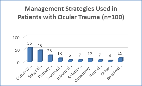
Figure 1 Management Strategies Used in Patients with Ocular Trauma (n=100)
Table 5. Comparison of Visual Acuity at Presentation and Final Follow-Up (n=100)
|
Visual acuity |
At presentation n (%) |
Final follow-up n (%) |
|
≥6/12 |
17 (17.0) |
44 (44.0) |
|
6/18–6/36 |
22 (22.0) |
27 (27.0) |
|
6/60–3/60 |
24 (24.0) |
15 (15.0) |
|
<3/60 to perception of light |
30 (30.0) |
10 (10.0) |
|
No perception of light |
7 (7.0) |
4 (4.0) |
|
Total |
100 (100.0) |
100 (100.0) |
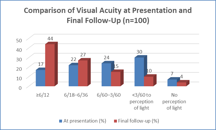
Figure 2 Comparison of Visual Acuity at Presentation and Final Follow-Up (n=100)
Table 6. Factors Associated with Final Visual Outcome (n=100)
|
Factor |
Good outcome n (%) |
Poor outcome n (%) |
p-value |
|
Overall |
86 (86.0) |
14 (14.0) |
— |
|
Open-globe injury (n=38) |
26 (68.4) |
12 (31.6) |
<0.001 |
|
Closed-globe injury (n=62) |
60 (96.8) |
2 (3.2) |
|
|
Delayed presentation >24 h (n=30) |
21 (70.0) |
9 (30.0) |
<0.001 |
|
Lens involvement (n=27) |
19 (70.4) |
8 (29.6) |
0.004 |
|
Vitreous haemorrhage (n=22) |
14 (63.6) |
8 (36.4) |
<0.001 |
|
Retinal detachment (n=11) |
5 (45.5) |
6 (54.5) |
<0.001 |
|
Intraocular foreign body (n=6) |
4 (66.7) |
2 (33.3) |
0.15 |
|
RAPD (n=13) |
6 (46.2) |
7 (53.8) |
<0.001 |
|
Endophthalmitis (n=4) |
1 (25.0) |
3 (75.0) |
0.001 |
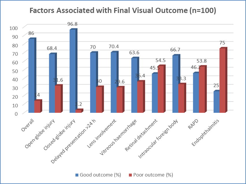
Figure 3 Factors Associated with Final Visual Outcome (n=100)
DISCUSSION:
The present prospective study evaluated the clinical profile, injury patterns, management, visual outcomes, and prognostic factors among 100 patients with ocular trauma. Young adults aged 21–40 years (43.0%) were most commonly affected, with a marked male predominance (74.0%). Similar demographic patterns have been reported in Indian studies, including Wagh et al.[14], who observed that 88.33% of patients were males, with the highest frequency among those aged 31–40 years. This predominance likely reflects greater occupational, outdoor, agricultural, and road-traffic exposure among young males.
Workplace/occupational injuries were the most common circumstances of trauma (32.0%), followed by injuries at home (25.0%), road/traffic injuries (19.0%), and agricultural injuries (13.0%). In contrast, Wagh et al.[14] reported road traffic accidents as a major cause, demonstrating geographical and occupational variation in ocular trauma. Blunt trauma (39.0%) was the most frequent mechanism in our study. Maurya et al.[15] similarly highlighted blunt trauma as an important cause of ocular morbidity.
Closed-globe injuries (62.0%) were more frequent than open-globe injuries (38.0%), consistent with Wagh et al.[14], who reported 80% closed-globe and 20% open-globe injuries. Corneal injury was the most frequent anatomical finding (52.0%), followed by conjunctival injury (44.0%), hyphema (31.0%), eyelid/adnexal injury (28.0%), and lens involvement (27.0%). Posterior-segment injuries included vitreous haemorrhage (22.0%) and retinal detachment (11.0%).
Most patients (70.0%) presented within 24 hours. Conservative management was sufficient in 55.0%, while 45.0% required surgery, including primary corneal/scleral repair (25.0%), traumatic cataract surgery (13.0%), vitrectomy (12.0%), retinal detachment surgery (7.0%), and intraocular foreign-body removal (6.0%).
Substantial visual improvement occurred following treatment. Visual acuity ≥6/12 increased from 17.0% at presentation to 44.0% at final follow-up, while severe visual impairment (<3/60 to perception of light) decreased from 30.0% to 10.0%. Overall, 86.0% achieved a good visual outcome, indicating that timely and appropriate management can provide meaningful visual rehabilitation.
Several factors were significantly associated with poor final vision. Poor outcomes occurred in 31.6% of open-globe injuries compared with 3.2% of closed-globe injuries (p<0.001). Delayed presentation >24 hours was associated with a 30.0% poor-outcome rate (p<0.001), while lens involvement was associated with poor outcome in 29.6% (p=0.004). Posterior-segment injuries carried particularly unfavorable prognoses: poor outcomes occurred in 36.4% with vitreous haemorrhage and 54.5% with retinal detachment (both p<0.001). RAPD was also strongly predictive, with 53.8% experiencing poor vision (p<0.001). Endophthalmitis, although uncommon (4.0%), was associated with poor outcome in 75.0% (p=0.001). These observations are consistent with established prognostic evidence for severe ocular trauma.
Overall, ocular trauma predominantly affected young males, with closed-globe injuries being more common and most patients achieving satisfactory visual recovery. However, open-globe injury, delayed presentation, lens involvement, vitreous haemorrhage, retinal detachment, RAPD, and endophthalmitis were important adverse prognostic factors. Early referral, prompt ophthalmological intervention, careful posterior-segment assessment, and preventive measures such as workplace protective eyewear are therefore essential for reducing avoidable visual morbidity.
CONCLUSION:
The present study demonstrated that ocular trauma predominantly affected young adult males, with occupational injuries, blunt trauma, and closed-globe injuries being the most common patterns. Most patients achieved significant visual improvement following timely and appropriate management. Open-globe injury, delayed presentation, lens involvement, vitreous haemorrhage, retinal detachment, RAPD, and endophthalmitis were significantly associated with poor visual outcomes. Early presentation, prompt diagnosis, appropriate intervention, and close follow-up are therefore essential for maximizing visual recovery. Preventive strategies, particularly workplace safety measures and protective eyewear, may further reduce trauma-related visual morbidity.
REFERENCES:
1. Blanch RJ, McMaster D, Patterson TJ. Management of open globe injury: a narrative review. Eye (Lond). 2024;38(16):3047-51.
2. Mir TA, Canner JK, Zafar S, Srikumaran D, Friedman DS, Woreta FA, et al. Characteristics of open globe injuries in the United States from 2006 to 2014. JAMA Ophthalmol. 2020;138(3):268-75.
3. Négrel AD, Thylefors B. The global impact of eye injuries. Ophthalmic Epidemiol. 1998;5(3):143-69.
4. Ozturk T, Cetin Dora G, Ayhan Z, Kaya M, Arikan G, Yaman A, et al. Etiology and visual prognosis in open globe injuries: results of a tertiary referral center in Turkey. Sci Rep. 2019;9(1):17977.
5. Li C, Fu Y, Liu S, Yu H, Yang X, Zhang M, et al. The global incidence and disability of eye injury: an analysis from the Global Burden of Disease Study 2019. EClinicalMedicine. 2023;62:102134.
6. Kuhn F, Morris R, Witherspoon CD, Heimann K, Jeffers JB, Treister G, et al. A standardized classification of ocular trauma. Ophthalmology. 1996;103(2):240-3.
7. Pieramici DJ, Sternberg P Jr, Aaberg TM Sr, Bridges WZ Jr, Capone A Jr, Cardillo JA, et al. A system for classifying mechanical injuries of the eye (globe). Am J Ophthalmol. 1997;123(6):820-31.
8. Kuhn F, Morris R, Witherspoon CD, Mester V. The Birmingham Eye Trauma Terminology system (BETT). J Fr Ophtalmol. 2004;27(2):206-10.
9. Kuhn F, Pieramici DJ, editors. Ocular trauma: principles and practice. New York: Thieme; 2002. p. 468.
10. Agrawal R, Wei HS, Teoh S. Prognostic factors for open globe injuries and correlation of ocular trauma score at a tertiary referral eye care centre in Singapore. Indian J Ophthalmol. 2013;61(9):502-6.
11. Fujikawa A, Mohamed YH, Kinoshita H, Matsumoto M, Uematsu M, Tsuiki E, et al. Visual outcomes and prognostic factors in open-globe injuries. BMC Ophthalmol. 2018;18(1):138.
12. Meng Y, Yan H. Prognostic factors for open globe injuries and correlation of ocular trauma score in Tianjin, China. J Ophthalmol. 2015;2015:345764.
13. Greenfield JA, Malek DA, Anant S, Antonietti M, Jammal A, Casella A, et al. A multi-center analysis of visual outcomes following open globe injury. Sci Rep. 2024;14(1):16638.
14. Wagh V, Tidake P. Clinical study and profile of ocular trauma: findings from a rural hospital in Central India. Cureus. 2022;14(7):e26915.
15. Maurya RP, Singh VP, Gautam S, Kumar A, Mishra CP, Jain P, et al. Study of epidemiology, clinical profile, visual outcome and prognostic factors of blunt ocular trauma in a teaching hospital. Indian J Clin Exp Ophthalmol. 2022;8(4):458-467.