Assessment of Dry Eye Disease After Cataract Surgery Using Tear Film Break-Up Time, Schirmer Test, and OSDI Score
- Vidyashri M , Private practice, Department of Ophthalmology, Shankarappagouda hospital, Yadgir
- Shruti Bidari , Department of Ophthalmology, KVG Medical College, Sullia
- Ramanna , Assistant Professor, Department of Ophthalmology, RIMS, Raichur
Article Information:
Abstract:
Background: Dry eye disease (DED) is a common ocular surface disturbance following cataract surgery and may adversely affect postoperative visual quality, ocular comfort, and patient satisfaction. This study assessed changes in DED after cataract surgery using Tear Film Break-Up Time (TBUT), Schirmer test, and Ocular Surface Disease Index (OSDI). Materials and Methods: This prospective observational study included 120 patients undergoing phacoemulsification with intraocular lens implantation. Dry-eye status was assessed preoperatively and at 1 week, 1 month, and 3 months postoperatively using TBUT, Schirmer test, and OSDI score. Changes across follow-up periods were analysed using appropriate repeated-measures statistical tests, and correlations between OSDI and objective parameters were assessed. A p-value <0.05 was considered statistically significant. Results: The mean age was 63.4 ± 8.7 years, and 53.3% were males. DED prevalence increased from 31.7% preoperatively to 83.3% at 1 week, before declining to 68.3% at 1 month and 42.5% at 3 months. Mean TBUT decreased from 11.42 ± 2.31 seconds preoperatively to 7.18 ± 2.06 seconds at 1 week, while Schirmer values decreased from 16.84 ± 4.21 to 11.26 ± 3.72 mm/5 min. Conversely, mean OSDI increased from 10.82 ± 6.34 to 27.46 ± 10.18 at 1 week. All parameters subsequently improved at 1 and 3 months (p<0.001). OSDI showed significant negative correlations with TBUT and Schirmer values. Conclusion: Cataract surgery was associated with significant transient worsening of DED, most pronounced at 1 week, followed by progressive recovery. Combined assessment using TBUT, Schirmer test, and OSDI facilitates comprehensive detection and monitoring of postoperative dry eye.
Keywords:
Article :
INTRODUCTION:
Cataract is one of the leading causes of visual impairment worldwide, particularly among older adults, and is characterized by progressive opacification of the crystalline lens resulting in deterioration of visual function. With increasing life expectancy and growth of the ageing population, the burden of cataract and the requirement for surgical treatment continue to increase.[1] Phacoemulsification with intraocular lens implantation has become the preferred surgical technique for cataract management because of its small incision, rapid visual rehabilitation, and favourable postoperative outcomes. Despite these advances, postoperative ocular surface disturbances, particularly dry eye disease (DED), remain an important concern and may adversely affect visual quality, ocular comfort, and patient satisfaction.[2]
DED is a multifactorial disorder characterized by loss of tear-film and ocular-surface homeostasis. Patients commonly experience dryness, burning, irritation, foreign-body sensation, grittiness, watering, ocular fatigue, and fluctuating or blurred vision.[3] Cataract surgery may induce new-onset DED or aggravate pre-existing ocular surface abnormalities, even when postoperative visual acuity is satisfactory. This is particularly relevant in elderly patients, who may already have reduced tear secretion, meibomian gland dysfunction, altered corneal sensitivity, and other age-related changes affecting the lacrimal functional unit.[4,5]
Several mechanisms contribute to the development of dry eye following cataract surgery. Clear corneal incisions may disrupt corneal sensory nerves and cause transient corneal hypoesthesia, thereby interfering with the neural reflex mechanisms responsible for blinking and lacrimal secretion[6]. Surgical manipulation, exposure to operating-microscope illumination, ocular surface irrigation, postoperative inflammation, and repeated administration of topical medications containing preservatives may further compromise ocular-surface integrity.[7,8] However, these factors can reduce tear secretion, destabilize the tear film, and increase postoperative dry-eye symptoms.
Assessment of postoperative DED requires consideration of both objective tear-film abnormalities and subjective symptoms, as clinical signs and patient-reported complaints may not always correlate. Tear Film Break-Up Time (TBUT) is a widely used clinical measure of tear-film stability; a shortened TBUT indicates increased tear-film instability.[9,10] The Schirmer test assesses aqueous tear production by measuring wetting of a standardized filter-paper strip and therefore provides information regarding the quantitative component of tear secretion. In contrast, the Ocular Surface Disease Index (OSDI) is a validated questionnaire that evaluates the severity of ocular symptoms, their effect on vision-related functioning, and the influence of environmental factors.[11-13]
The combined use of TBUT, Schirmer test, and OSDI score therefore enables comprehensive assessment of postoperative dry eye by evaluating tear-film stability, aqueous tear secretion, and patient-reported symptom burden.[14] Serial assessment before and after cataract surgery may help determine the magnitude and temporal pattern of ocular-surface changes and identify patients requiring early intervention.
Therefore, the present study aims to assess dry eye disease following cataract surgery using TBUT, Schirmer test, and OSDI score.
MATERIALS AND METHODS:
Study Design and Setting
This prospective observational study was conducted in the Department of Ophthalmology at a tertiary care hospital.
Study Population
The study included patients diagnosed with age-related cataract who underwent cataract surgery with phacoemulsification and posterior chamber intraocular lens implantation during the study period. Patients fulfilling the predefined eligibility criteria were enrolled consecutively.A total of 120 patients undergoing cataract surgery were included in the study. One eye from each participant was considered for analysis; therefore, 120 eyes of 120 patients were evaluated.
Inclusion Criteria
Patients aged 40 years or older with age-related cataract who were scheduled for uncomplicated phacoemulsification with intraocular lens implantation and who provided written informed consent were included in the study. Patients who were willing and able to complete the required postoperative follow-up assessments were considered eligible.
Exclusion Criteria
Patients with pre-existing severe dry eye disease, active ocular infection or inflammation, significant corneal pathology, glaucoma requiring multiple topical medications, previous ocular surgery or trauma, contact-lens use, eyelid abnormalities affecting tear-film distribution, or systemic conditions known to markedly influence tear production were excluded. Patients who developed significant intraoperative complications or were unable to complete the required follow-up were also excluded.
Preoperative Evaluation
A detailed history was obtained from each participant, including age, sex, presenting ocular complaints, duration of visual impairment, systemic comorbidities, medication history, and relevant ocular history. All patients underwent a comprehensive ophthalmological examination, including assessment of visual acuity, slit-lamp examination, intraocular pressure measurement, fundus examination wherever possible, and cataract grading.Baseline dry-eye status was assessed before cataract surgery using Tear Film Break-Up Time (TBUT), Schirmer test, and Ocular Surface Disease Index (OSDI) score. All assessments were performed under comparable environmental conditions to minimize variations in tear-film measurements.
Assessment of Tear Film Break-Up Time
Tear-film stability was assessed using the fluorescein Tear Film Break-Up Time (TBUT). A fluorescein strip moistened with sterile normal saline was gently applied to the inferior conjunctival fornix. The patient was instructed to blink several times to distribute the fluorescein uniformly over the ocular surface and was then asked to keep the eyes open.The tear film was examined using a slit lamp with cobalt-blue illumination. The interval between the last complete blink and the appearance of the first dry spot or discontinuity in the fluorescein-stained tear film was recorded in seconds. The test was repeated, and the average reading was used for analysis. A shorter TBUT indicated greater tear-film instability.
Schirmer Test
A Schirmer test was performed to assess aqueous tear secretion. A standardized Whatman filter-paper strip was folded at the marked end and placed in the lower conjunctival fornix at the junction of the middle and lateral thirds of the lower eyelid.The patient was instructed to keep the eyes gently closed during the test. After 5 minutes, the strip was removed, and the length of wetting was measured in millimetres. Lower Schirmer values indicated reduced aqueous tear production.
Ocular Surface Disease Index Assessment
Subjective dry-eye symptoms were assessed using the validated Ocular Surface Disease Index (OSDI) questionnaire. The questionnaire assessed ocular symptoms, limitations in vision-related activities, and symptoms associated with environmental conditions.
The OSDI score was calculated using the standard scoring method:
OSDI score = (Sum of scores for all questions answered × 25) / Total number of questions answeredThe final score ranged from 0 to 100, with higher scores indicating greater severity of dry-eye symptoms. The scores were interpreted as normal (0–12), mild (13–22), moderate (23–32), and severe (33–100) dry-eye symptoms.
Surgical Procedure
All patients underwent standard phacoemulsification with posterior chamber intraocular lens implantation under appropriate anaesthesia and aseptic precautions. A clear corneal incision was created, followed by continuous curvilinear capsulorhexis, hydrodissection, phacoemulsification of the nucleus, cortical aspiration, and implantation of a foldable intraocular lens in the capsular bag.At the completion of surgery, the corneal wounds were hydrated and checked for leakage. Postoperative topical medications were prescribed according to the institutional protocol. Efforts were made to maintain uniformity in the surgical technique and postoperative medication regimen among the study participants.
Postoperative Follow-Up
Patients were followed after cataract surgery at predetermined intervals. Dry-eye assessment was performed preoperatively and postoperatively at 1 week, 1 month, and 3 months.
At each follow-up visit, patients underwent clinical examination and assessment of:
• Tear Film Break-Up Time (TBUT)
• Schirmer test
• Ocular Surface Disease Index (OSDI) score
Changes in these parameters from preoperative baseline values were recorded and compared across the different postoperative follow-up periods. Any postoperative ocular complaints or complications were also documented.
Outcome Measures
The primary outcome was the change in dry-eye parameters following cataract surgery as determined by TBUT, Schirmer test, and OSDI score.
The secondary outcomes included the frequency and severity of postoperative dry eye, the temporal pattern of changes in dry-eye parameters during follow-up, and the relationship between subjective OSDI scores and objective TBUT and Schirmer test findings.
Data Collection
All demographic, clinical, surgical, and dry-eye assessment findings were recorded in a predesigned case-record form. Data were subsequently entered into a computerized database and checked for completeness and accuracy before statistical analysis.
Statistical Analysis
The collected data were entered into Microsoft Excel and analysed using SPSS.21statistical software. Continuous variables were expressed as mean ± standard deviation (SD) or median with interquartile range (IQR) according to the distribution of data, while categorical variables were presented as frequency and percentage.Normality of continuous variables was assessed using the Shapiro–Wilk test. Changes in TBUT, Schirmer test values, and OSDI scores across preoperative and postoperative follow-up periods were analysed using repeated-measures analysis of variance (ANOVA) for normally distributed data or the Friedman test for non-normally distributed data. Appropriate post-hoc pairwise comparisons were performed when an overall statistically significant difference was observed.Categorical variables were compared using the Chi-square test or Fisher's exact test, as appropriate. Correlations between OSDI scores and objective dry-eye parameters, including TBUT and Schirmer test values, were assessed using Pearson's or Spearman's correlation coefficient, depending on data distribution. A p-value <0.05 was considered statistically significant.
Ethical Considerations
The study was conducted in accordance with the principles of the Declaration of Helsinki. Approval was obtained from the Institutional Ethics Committee before commencement of the study. Written informed consent was obtained from all participants, and confidentiality of patient information was maintained throughout the study.
RESULTS:
A total of 120 patients undergoing cataract surgery were included in the study. The mean age of the study participants was 63.4 ± 8.7 years, with the majority belonging to the 60–69-year age group (43.3%). There were 64 (53.3%) males and 56 (46.7%) females. The right eye was operated on in 52.5% of patients. Diabetes mellitus and hypertension were present in 25.8% and 35.8% of participants, respectively. Nuclear cataract was the most frequent type (40.8%), followed by mixed (23.3%), cortical (20.8%), and posterior subcapsular cataract (15.0%). The demographic and clinical characteristics of the study participants are presented in Table 1.
Table 1. Demographic and Clinical Characteristics of Study Participants (n=120)
|
Variable |
Category |
n (%) / Mean ± SD |
|
Age (years) |
Mean ± SD |
63.4 ± 8.7 |
|
Age group |
40–49 years |
9 (7.5) |
|
50–59 years |
30 (25.0) |
|
|
60–69 years |
52 (43.3) |
|
|
≥70 years |
29 (24.2) |
|
|
Sex |
Male |
64 (53.3) |
|
Female |
56 (46.7) |
|
|
Eye operated |
Right |
63 (52.5) |
|
Left |
57 (47.5) |
|
|
Diabetes mellitus |
Present |
31 (25.8) |
|
Absent |
89 (74.2) |
|
|
Hypertension |
Present |
43 (35.8) |
|
Absent |
77 (64.2) |
|
|
Cataract type |
Nuclear |
49 (40.8) |
|
Cortical |
25 (20.8) |
|
|
Posterior subcapsular |
18 (15.0) |
|
|
Mixed |
28 (23.3) |
The prevalence of dry eye disease showed a marked increase following cataract surgery. Dry eye was present in 31.7% of patients preoperatively and increased to 83.3% at 1 week postoperatively. Thereafter, the prevalence progressively declined to 68.3% at 1 month and 42.5% at 3 months, indicating gradual recovery during postoperative follow-up. The temporal distribution of dry eye disease is presented in Table 2 and Figure 1.
Table 2. Prevalence of Dry Eye Disease at Different Follow-Up Periods (n=120)
|
Assessment time |
Dry eye present n (%) |
Dry eye absent n (%) |
Total |
|
Preoperative |
38 (31.7) |
82 (68.3) |
120 |
|
Postoperative 1 week |
100 (83.3) |
20 (16.7) |
120 |
|
Postoperative 1 month |
82 (68.3) |
38 (31.7) |
120 |
|
Postoperative 3 months |
51 (42.5) |
69 (57.5) |
120 |
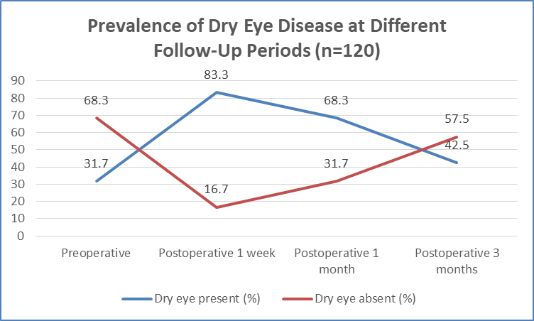
Figure 1 Prevalence of Dry Eye Disease at Different Follow-Up Periods (n=120)
Objective tear-film assessment demonstrated a similar postoperative pattern. The proportion of patients with abnormal TBUT increased from 24.2% preoperatively to 71.7% at 1 week, before decreasing to 54.2% at 1 month and 31.7% at 3 months. Similarly, abnormal Schirmer test findings increased from 18.3% preoperatively to 56.7% at 1 week, followed by a decline to 40.8% at 1 month and 24.2% at 3 months. Changes in both parameters across the follow-up period were statistically significant (p<0.001), as shown in Table 3.
Table 3. Distribution of Patients According to Abnormal TBUT and Schirmer Test at Different Follow-Up Periods (n=120)
|
Assessment time |
Abnormal TBUT n (%) |
Normal TBUT n (%) |
Abnormal Schirmer n (%) |
Normal Schirmer n (%) |
|
Preoperative |
29 (24.2) |
91 (75.8) |
22 (18.3) |
98 (81.7) |
|
Postoperative 1 week |
86 (71.7) |
34 (28.3) |
68 (56.7) |
52 (43.3) |
|
Postoperative 1 month |
65 (54.2) |
55 (45.8) |
49 (40.8) |
71 (59.2) |
|
Postoperative 3 months |
38 (31.7) |
82 (68.3) |
29 (24.2) |
91 (75.8) |
|
p-value |
<0.001 |
<0.001 |
The mean TBUT decreased significantly from 11.42 ± 2.31 seconds preoperatively to 7.18 ± 2.06 seconds at 1 week, followed by improvement to 8.63 ± 2.14 seconds at 1 month and 10.31 ± 2.22 seconds at 3 months (p<0.001). Similarly, the mean Schirmer test value decreased from 16.84 ± 4.21 mm/5 min to 11.26 ± 3.72 mm/5 min at 1 week, subsequently improving to 13.18 ± 3.86 mm/5 min and 15.32 ± 4.02 mm/5 min at 1 and 3 months, respectively (p<0.001). In contrast, the mean OSDI score increased from 10.82 ± 6.34 preoperatively to 27.46 ± 10.18 at 1 week, before declining to 20.14 ± 8.76 at 1 month and 13.68 ± 7.12 at 3 months (p<0.001). These changes are summarized in Table 4 and Figure 2.
Table 4. Comparison of Mean TBUT, Schirmer Test, and OSDI Scores Across the Study Period (n=120)
|
Dry-eye parameter |
Preoperative |
1 week |
1 month |
3 months |
p-value |
|
TBUT (seconds) |
11.42 ± 2.31 |
7.18 ± 2.06 |
8.63 ± 2.14 |
10.31 ± 2.22 |
<0.001 |
|
Schirmer test (mm/5 min) |
16.84 ± 4.21 |
11.26 ± 3.72 |
13.18 ± 3.86 |
15.32 ± 4.02 |
<0.001 |
|
OSDI score |
10.82 ± 6.34 |
27.46 ± 10.18 |
20.14 ± 8.76 |
13.68 ± 7.12 |
<0.001 |
Statistical test: Repeated-measures ANOVA.
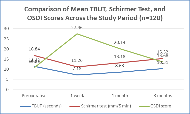
Figure 2 Comparison of Mean TBUT, Schirmer Test, and OSDI Scores Across the Study Period (n=120)
Assessment of dry-eye severity according to OSDI demonstrated that 68.3% of patients had normal scores preoperatively, which decreased to only 16.7% at 1 week. At the same time, moderate and severe dry eye increased from 8.3% and 2.5% preoperatively to 30.8% and 26.7% at 1 week, respectively. Subsequently, the proportion of patients with moderate and severe disease declined at 1 and 3 months, while the proportion with normal OSDI scores increased to 57.5% by 3 months. The distribution of dry-eye severity during follow-up is presented in Table 5.
Table 5. Distribution of Dry Eye Severity According to OSDI at Different Follow-Up Periods (n=120)
|
OSDI severity |
Preoperative n (%) |
1 week n (%) |
1 month n (%) |
3 months n (%) |
|
Normal (0–12) |
82 (68.3) |
20 (16.7) |
38 (31.7) |
69 (57.5) |
|
Mild (13–22) |
25 (20.8) |
31 (25.8) |
39 (32.5) |
30 (25.0) |
|
Moderate (23–32) |
10 (8.3) |
37 (30.8) |
27 (22.5) |
15 (12.5) |
|
Severe (33–100) |
3 (2.5) |
32 (26.7) |
16 (13.3) |
6 (5.0) |
|
Total |
120 (100) |
120 (100) |
120 (100) |
120 (100) |
A statistically significant negative correlation was observed between OSDI scores and both objective dry-eye parameters throughout the study period. The strongest correlation between OSDI and TBUT was observed at 1 week (ρ=−0.58, p<0.001), followed by 1 month (ρ=−0.53, p<0.001), 3 months (ρ=−0.42, p<0.001), and the preoperative assessment (ρ=−0.36, p<0.001). OSDI was also negatively correlated with Schirmer test values, with the strongest relationship at 1 week (ρ=−0.49, p<0.001). Thus, greater subjective dry-eye symptom severity was associated with lower tear-film stability and reduced tear secretion. The correlation findings are detailed in Table 6 and Figure 3.
Table 6. Correlation of OSDI Score With TBUT and Schirmer Test
|
Assessment time |
Variables correlated |
ρ |
p-value |
Interpretation |
|
Preoperative |
OSDI vs TBUT |
−0.36 |
<0.001 |
Moderate negative |
|
OSDI vs Schirmer test |
−0.31 |
0.001 |
Moderate negative |
|
|
1 week |
OSDI vs TBUT |
−0.58 |
<0.001 |
Moderate negative |
|
OSDI vs Schirmer test |
−0.49 |
<0.001 |
Moderate negative |
|
|
1 month |
OSDI vs TBUT |
−0.53 |
<0.001 |
Moderate negative |
|
OSDI vs Schirmer test |
−0.44 |
<0.001 |
Moderate negative |
|
|
3 months |
OSDI vs TBUT |
−0.42 |
<0.001 |
Moderate negative |
|
OSDI vs Schirmer test |
−0.37 |
<0.001 |
Moderate negative |
Statistical test: Spearman rank correlation.
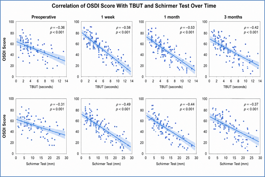
Figure 3 Correlation of OSDI Score With TBUT and Schirmer Test
DISCUSSION:
The present study evaluated changes in dry eye disease (DED) following cataract surgery using Tear Film Break-Up Time (TBUT), Schirmer test, and Ocular Surface Disease Index (OSDI). The principal finding was a significant worsening of both objective and subjective dry-eye parameters during the early postoperative period, particularly at 1 week, followed by progressive recovery at 1 and 3 months. This pattern supports the predominantly transient effect of cataract surgery on tear-film homeostasis.The mean age of participants was 63.4 ± 8.7 years, with 43.3% aged 60–69 years; 53.3% were males. This demographic profile was comparable with Ishrat et al.[15], who reported a mean age of 63.1 ± 8.3 years. Diabetes mellitus and hypertension were present in 25.8% and 35.8% of our patients, respectively. Miura et al.[9] identified age, sex, systemic diseases, pre-existing DED, meibomian gland dysfunction, preservative-containing eye drops, and surgical factors as potential contributors to postoperative DED.
DED was present in 31.7% preoperatively, increasing markedly to 83.3% at 1 week, before declining to 68.3% at 1 month and 42.5% at 3 months. A similar temporal pattern was reported by Ishrat et al.[15], with DED prevalence of 42% at 1 week, 15% at 1 month, and 9% at 3 months. Miura et al.[9] reported a pooled incidence of new-onset postoperative DED of 37.4% (95% CI: 22.6–52.3) among patients without pre-existing DED.Mean TBUT decreased significantly from 11.42 ± 2.31 seconds preoperatively to 7.18 ± 2.06 seconds at 1 week, subsequently improving to 8.63 ± 2.14 seconds at 1 month and 10.31 ± 2.22 seconds at 3 months (p<0.001).
Ishrat et al.[15] similarly observed a reduction from 15.8 seconds preoperatively to 8.7 seconds at 1 week, followed by recovery toward baseline by 3 months.Mean Schirmer values decreased from 16.84 ± 4.21 mm/5 min to 11.26 ± 3.72 mm/5 min at 1 week, followed by improvement to 13.18 ± 3.86 and 15.32 ± 4.02 mm/5 min at 1 and 3 months, respectively (p<0.001). Garg et al.[16] similarly demonstrated a marked postoperative reduction in Schirmer values at 1 week followed by substantial recovery.Subjective symptoms followed the same pattern. Mean OSDI increased from 10.82 ± 6.34 preoperatively to 27.46 ± 10.18 at 1 week, before decreasing to 20.14 ± 8.76 at 1 month and 13.68 ± 7.12 at 3 months (p<0.001). OSDI also demonstrated significant negative correlations with TBUT and Schirmer values, with the strongest correlations at 1 week, indicating that greater symptom severity was associated with poorer tear-film stability and reduced tear secretion.
CONCLUSION:
Cataract surgery was associated with a significant transient worsening of dry eye disease, with maximum deterioration observed during the first postoperative week. TBUT and Schirmer test values decreased significantly, while OSDI scores increased, indicating reduced tear-film stability, decreased tear secretion, and greater subjective symptoms. These parameters progressively improved at 1 and 3 months, although complete recovery to preoperative levels was not observed in all patients. Significant negative correlations between OSDI and both TBUT and Schirmer values supported the combined use of subjective and objective assessments. Early identification and appropriate management of postoperative dry eye may improve ocular comfort, visual rehabilitation, and patient satisfaction.
REFERENCES:
1. Hashemi H, Pakzad R, Yekta A, Aghamirsalim M, Pakbin M, Ramin S, et al. Global and regional prevalence of age-related cataract: a comprehensive systematic review and meta-analysis. Eye (Lond). 2020;34(8):1357-1370.
2. Song P, Wang H, Theodoratou E, Chan KY, Rudan I. The national and subnational prevalence of cataract and cataract blindness in China: a systematic review and meta-analysis. J Glob Health. 2018;8(1):010804.
3. Wang D, Tang T, Li P, Zhao J, Shen B, Zhang M, et al. The global burden of cataracts and its attributable risk factors in 204 countries and territories: a systematic analysis of the Global Burden of Disease Study. Front Public Health. 2024;12:1366677.
4. Magliano DJ, Boyko EJ, editors. IDF Diabetes Atlas. 10th ed. Brussels: International Diabetes Federation; 2021.
5. GBD 2021 Diabetes Collaborators. Global, regional, and national burden of diabetes from 1990 to 2021, with projections of prevalence to 2050: a systematic analysis for the Global Burden of Disease Study 2021. Lancet. 2023;402:203-234.
6. Kiziltoprak H, Tekin K, Inanc M, Goker YS. Cataract in diabetes mellitus. World J Diabetes. 2019;10(3):140-153.
7. Mrugacz M, Pony-Uram M, Bryl A, Zorena K. Current approach to the pathogenesis of diabetic cataracts. Int J Mol Sci. 2023;24(7):6317.
8. Naderi K, Gormley J, O'Brart D. Cataract surgery and dry eye disease: a review. Eur J Ophthalmol. 2020;30(5):840-855.
9. Miura M, Inomata T, Nakamura M, Sung J, Nagino K, Midorikawa-Inomata A, et al. Prevalence and characteristics of dry eye disease after cataract surgery: a systematic review and meta-analysis. Ophthalmol Ther. 2022;11(4):1309-1332.
10. Momin AA, Nikose AS, Thakre UD. Tear film dysfunction after clear corneal phacoemulsification in diabetics and non-diabetics. Indian J Ophthalmol. 2023;71(4):1517-1520.
11. Chung B, Choi M, Lee KY, Kim EK, Seo KY, Jun I, et al. Comparing dry eye disease after small incision lenticule extraction and laser subepithelial keratomileusis. Cornea. 2020;39(4):501-506.
12. Yahalomi T, Achiron A, Arnon R, Stanescu N, Pikkel J. Dry eye disease following LASIK, PRK, and LASEK: an observational cross-sectional study. J Clin Med. 2023;12(11):3761.
13. Ishrat S, Nema N, Chandravanshi SCL. Incidence and pattern of dry eye after cataract surgery. Saudi J Ophthalmol. 2019;33(1):34-40.
14. Luo L, Lin H, He M, Congdon N, Yang Y, Liu Y, et al. Clinical evaluation of three incision size-dependent phacoemulsification systems. Am J Ophthalmol. 2012;153(5):831-839.
15. Ishrat S, Nema N, Chandravanshi SCL. Incidence and pattern of dry eye after cataract surgery. Saudi J Ophthalmol. 2019;33(1):34-40.
16. Garg P, Gupta A, Tandon N, Raj P. Dry eye disease after cataract surgery: study of its determinants and risk factors. Turk J Ophthalmol. 2020;50(3):133-142.