Histomorphological Changes in Axillary Lymph Nodes Following Neoadjuvant Chemotherapy in Breast Carcinoma: Association with Pathological Nodal Response.
- Y. Sarasa Bharathi , Assistant Professor, Department of Pathology, Srinivasan Medical College Hospital, Trichy, Tamil Nadu, India.
- R. Lavanya , Assistant Professor, Department of Pathology, Srinivasan Medical College Hospital, Trichy, Tamil Nadu, India.
Article Information:
Abstract:
Background: Axillary lymph nodes exposed to neoadjuvant chemotherapy may show a spectrum of stromal, inflammatory and tumour-related alterations. Correct recognition of these changes is relevant when distinguishing residual metastasis from treatment-associated regression. Objective: To describe the histomorphological changes in axillary lymph nodes after neoadjuvant chemotherapy for invasive breast carcinoma and examine their association with post-treatment nodal status. Methods: This prospective observational study included 43 consecutive women with invasive breast carcinoma treated with neoadjuvant chemotherapy followed by breast and axillary surgery between February 2025 and February 2026 at a tertiary care hospital. Gross nodal characteristics, residual tumour and twelve predefined histomorphological features were recorded. Cases were grouped as ypN0 or ypN-positive (ypN+), and associations were assessed at a significance threshold of p<0.05. Results: The mean age was 51.4±11.2 years. Twenty-two patients (51.2%) were ypN0 and 21 (48.8%) had residual nodal tumour. Fibrosis was the most frequent nodal alteration (72.1%), followed by chronic inflammation (60.5%), hyalinization (55.8%) and hemosiderin-laden macrophages (51.2%). Fibrosis was more frequent in ypN0 than ypN+ cases (86.4% versus 57.1%; p=0.032), as was hyalinization (72.7% versus 38.1%; p=0.021). HER2-enriched and triple-negative tumours showed the highest descriptive ypN0 proportions, 66.7% and 64.3%, respectively. Among patients with Miller-Payne grade 4-5 breast response, 71.4% were ypN0. Conclusion: Fibrosis and hyalinization were significantly associated with post-treatment node-negative status. Careful appraisal of therapy-related nodal morphology may strengthen pathological interpretation of the treated axilla, although ypN staging remains dependent on identification of residual tumour and appropriate pretreatment nodal documentation.
Keywords:
Article :
INTRODUCTION:
Breast cancer remains the most frequently diagnosed malignancy among women worldwide and continues to contribute substantially to cancer mortality.[1] For patients with locally advanced or biologically aggressive disease, neoadjuvant systemic therapy is now integrated into multidisciplinary care. It can reduce tumour burden, facilitate breast conservation, downstage the axilla and provide an in vivo measure of treatment sensitivity that may influence subsequent systemic management.[2]
Pathological response after neoadjuvant therapy carries prognostic information, but its interpretation depends on how response is defined. Large pooled analyses have shown that eradication of invasive tumour from both the breast and regional lymph nodes has a stronger association with favourable long-term outcomes than breast response alone.[3] Response is also heterogeneous across tumour biology. High-grade, HER2-positive and triple-negative cancers generally achieve pathological complete response more often than luminal tumours, although the prognostic meaning of response differs among subtypes.[4,5] The treated surgical specimen presents a distinct diagnostic challenge.
Chemotherapy may leave no viable tumour yet produce fibrosis, hyalinization, foamy macrophages, hemosiderin deposition, necrosis, chronic inflammation or lymphoid depletion within previously involved lymph nodes. Conversely, small residual deposits can be obscured by a regressed or fibrotic nodal background. Earlier pathology guidance therefore emphasised systematic examination of post-neoadjuvant breast and axillary specimens.[6] More recent international recommendations have reinforced mapping, adequate sampling and explicit documentation of treatment effect and residual disease because variation in processing and reporting can alter response classification.[7,8]
Despite the clinical importance of post-treatment nodal status, axillary histomorphology has received less attention than primary breast tumour response, particularly in modest-sized prospective series from routine pathology practice. The present study was undertaken to characterise the gross and microscopic changes in axillary lymph nodes after neoadjuvant chemotherapy and to examine whether individual histomorphological features were associated with post-treatment node-negative status.
MATERIALS AND METHODS:
Study Design and Setting
A prospective observational study was conducted in the Department of Pathology of a tertiary care hospital from February 2025 to February 2026.
Study Population
All consecutive patients with invasive breast carcinoma who received neoadjuvant chemotherapy followed by axillary surgery during the study period were eligible. Patients with previous chemotherapy or radiotherapy, metastatic disease at presentation, or incomplete clinical or pathological records were excluded. The final study cohort comprised 43 patients.
Clinical and Tumour Variables
Age, menopausal status, presenting complaint, pretreatment clinical T and N categories, overall clinical stage, histological type and grade, immunohistochemistry-based molecular subtype, Ki-67 proliferation index, chemotherapy regimen and number of cycles were recorded. Receipt of anti-HER2 targeted therapy was recorded separately; because it was documented in only two patients, no treatment-specific response analysis was undertaken. Surgical variables included the type of breast operation, the axillary procedure and the number of lymph nodes retrieved.
Axillary Specimen Assessment
Axillary specimens were evaluated grossly for nodal size, consistency and cut-surface appearance. Microscopic assessment documented residual metastatic tumour and the presence or absence of fibrosis, hyalinization, necrosis, foam cell infiltration, hemosiderin-laden macrophages, chronic inflammation, granulomatous reaction, sinus histiocytosis, lymphoid depletion, germinal centre hyperplasia, vascular proliferation and calcification. Extracapsular extension and therapy-related cytological change were recorded among nodes containing residual tumour.
Outcome Definitions
The principal nodal outcome was post-treatment pathological nodal status, classified as ypN0 when no residual nodal metastasis was identified and ypN+ when residual tumour was present. Because pretreatment nodal metastasis was not pathologically documented in every patient, ypN0 was interpreted as post-treatment node-negative status and was not automatically equated with axillary pathological complete response. Residual tumour size was retained in the study-defined strata of <2 mm, 2-20 mm and >20 mm. Response in the breast was graded using the Miller-Payne system, in which grade 1 represents no meaningful reduction in cellularity and grade 5 denotes absence of viable invasive tumour.
Statistical Analysis
Continuous variables were summarised as mean±standard deviation, while categorical variables were expressed as frequency and percentage. Associations between individual histomorphological features and ypN status were examined using categorical tests, with p<0.05 considered statistically significant. Given the modest subgroup sizes, molecular subtype, histological grade, Ki-67 category and breast-response comparisons were treated as exploratory and reported descriptively.
Ethical Considerations
Written informed consent was obtained from all participants before enrolment. Participant confidentiality was maintained throughout the study, and the research was conducted in accordance with the principles of the Declaration of Helsinki.
RESULTS:
Clinical and Tumour Profile
All 43 participants were women. Their mean age was 51.4±11.2 years; 14 patients (32.6%) were aged 41-50 years, which was the largest age stratum. Twenty-five patients (58.1%) were postmenopausal. A breast lump was the presenting complaint in 37 (86.0%) cases. Before neoadjuvant chemotherapy, 19 patients (44.2%) had cT2 disease and 20 (46.5%) had cN1 disease. Clinically node-positive disease (cN1-cN3) was recorded in 38 of 43 patients (88.4%). Stage IIB was the most frequent pretreatment clinical stage (32.6%), followed by stage IIIA (27.9%) (Table 1).
Invasive ductal carcinoma of no special type accounted for 35 cases (81.4%). Grade II tumours were most common (48.8%), while 16 (37.2%) were grade III. Triple-negative carcinoma was the largest molecular group (32.6%), followed by luminal A (27.9%), HER2-enriched (20.9%) and luminal B (18.6%). A Ki-67 index of at least 20% was present in 28 patients (65.1%) (Table 1).
Table 1. Baseline clinical and tumour characteristics of the study cohort (n=43)
|
Variable |
Category |
n (%) |
|
Age, years |
≤40 |
9 (20.9) |
|
41-50 |
14 (32.6) |
|
|
51-60 |
12 (27.9) |
|
|
>60 |
8 (18.6) |
|
|
Mean±SD |
51.4±11.2 |
|
|
Menopausal status |
Premenopausal |
18 (41.9) |
|
Postmenopausal |
25 (58.1) |
|
|
Clinical presentation |
Breast lump |
37 (86.0) |
|
Nipple discharge |
3 (7.0) |
|
|
Skin changes |
2 (4.7) |
|
|
Other |
1 (2.3) |
|
|
Clinical T category |
cT1 |
4 (9.3) |
|
cT2 |
19 (44.2) |
|
|
cT3 |
13 (30.2) |
|
|
cT4 |
7 (16.3) |
|
|
Clinical N category |
cN0 |
5 (11.6) |
|
cN1 |
20 (46.5) |
|
|
cN2 |
13 (30.2) |
|
|
cN3 |
5 (11.6) |
|
|
Clinical stage |
IIA |
6 (14.0) |
|
IIB |
14 (32.6) |
|
|
IIIA |
12 (27.9) |
|
|
IIIB |
7 (16.3) |
|
|
IIIC |
4 (9.3) |
|
|
Histological type |
Invasive ductal carcinoma, NST |
35 (81.4) |
|
Invasive lobular carcinoma |
5 (11.6) |
|
|
Mixed ductal-lobular carcinoma |
2 (4.7) |
|
|
Other |
1 (2.3) |
|
|
Histological grade |
Grade I |
6 (14.0) |
|
Grade II |
21 (48.8) |
|
|
Grade III |
16 (37.2) |
|
|
Molecular subtype |
Luminal A |
12 (27.9) |
|
Luminal B |
8 (18.6) |
|
|
HER2-enriched |
9 (20.9) |
|
|
Triple-negative |
14 (32.6) |
|
|
Ki-67 proliferation index |
<20% |
15 (34.9) |
|
≥20% |
28 (65.1) |
|
|
Mean±SD |
28.6±14.3 |
|
|
NST: no special type; SD: standard deviation; HER2: human epidermal growth factor receptor 2 |
||
Treatment, surgery and gross nodal findings
The most frequently used neoadjuvant regimen was anthracycline based (37.2%), followed by sequential anthracycline and taxane therapy (32.6%). Twenty-five patients (58.1%) received five to six cycles, and the mean number of cycles was 5.2±1.4. Modified radical mastectomy was performed in 24 patients (55.8%), while 19 (44.2%) underwent breast-conserving surgery. Axillary lymph node dissection was undertaken in 32 patients (74.4%) and sentinel lymph node biopsy in 11 (25.6%). The mean nodal yield was 13.8±5.2; most specimens contained 10-15 nodes (46.5%) (Table 2).
On gross examination, lymph nodes measuring 5-10 mm constituted the largest category (44.2%). Firm consistency was observed in 21 cases (48.8%), and 10 (23.3%) showed a hard or fibrotic consistency. The cut surface was homogeneous in 41.9%, heterogeneous in 37.2% and necrotic or cystic in 20.9% (Table 2).
Table 2. Neoadjuvant treatment, surgical procedures and gross axillary nodal findings (n=43)
|
Variable |
Category |
n (%) |
|
Chemotherapy regimen |
Anthracycline based (AC/EC) |
16 (37.2) |
|
Taxane based |
8 (18.6) |
|
|
Sequential anthracycline + taxane |
14 (32.6) |
|
|
Anthracycline + taxane + platinum |
3 (7.0) |
|
|
Anti-HER2 targeted therapy with chemotherapy |
2 (4.7) |
|
|
Number of cycles |
3-4 |
12 (27.9) |
|
5-6 |
25 (58.1) |
|
|
>6 |
6 (14.0) |
|
|
Mean±SD |
5.2±1.4 |
|
|
Breast surgery |
Breast-conserving surgery |
19 (44.2) |
|
Modified radical mastectomy |
24 (55.8) |
|
|
Axillary surgery |
Sentinel lymph node biopsy |
11 (25.6) |
|
Axillary lymph node dissection |
32 (74.4) |
|
|
Total lymph nodes retrieved |
<10 |
9 (20.9) |
|
10-15 |
20 (46.5) |
|
|
>15 |
14 (32.6) |
|
|
Mean±SD |
13.8±5.2 |
|
|
Lymph node size |
<5 mm |
8 (18.6) |
|
5-10 mm |
19 (44.2) |
|
|
11-20 mm |
11 (25.6) |
|
|
>20 mm |
5 (11.6) |
|
|
Nodal consistency |
Soft |
12 (27.9) |
|
Firm |
21 (48.8) |
|
|
Hard/fibrotic |
10 (23.3) |
|
|
Cut surface |
Homogeneous |
18 (41.9) |
|
Heterogeneous |
16 (37.2) |
|
|
Necrotic/cystic |
9 (20.9) |
|
|
AC: doxorubicin and cyclophosphamide; EC: epirubicin and cyclophosphamide; HER2: human epidermal growth factor receptor 2 |
||
Histomorphological changes in axillary lymph nodes
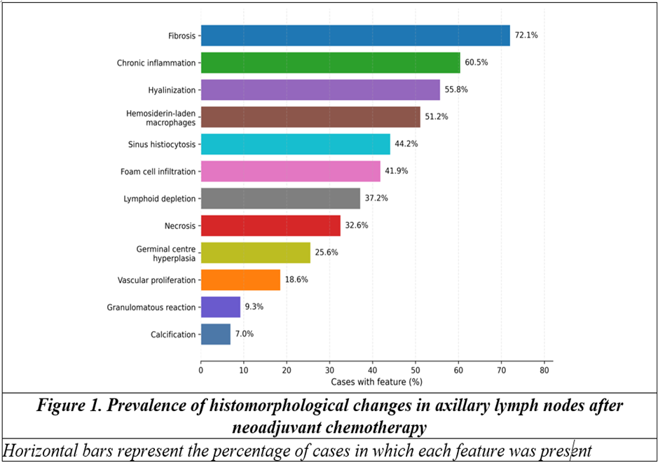
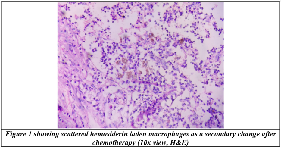
Fibrosis was the most prevalent microscopic alteration, present in 31 cases (72.1%). Chronic inflammation was identified in 26 (60.5%), hyalinization in 24 (55.8%), and hemosiderin-laden macrophages in 22 (51.2%). Sinus histiocytosis and foam cell infiltration were recorded in 44.2% and 41.9%, respectively. Granulomatous reaction and calcification were uncommon, occurring in 9.3% and 7.0% of cases (Table 3 and Figure 1).
Table 3. Frequency of histomorphological changes in axillary lymph nodes (n=43)
|
Histomorphological feature |
Present, n (%) |
Absent, n (%) |
|
Fibrosis |
31 (72.1) |
12 (27.9) |
|
Hyalinization |
24 (55.8) |
19 (44.2) |
|
Necrosis |
14 (32.6) |
29 (67.4) |
|
Foam cell infiltration |
18 (41.9) |
25 (58.1) |
|
Hemosiderin-laden macrophages |
22 (51.2) |
21 (48.8) |
|
Chronic inflammation |
26 (60.5) |
17 (39.5) |
|
Granulomatous reaction |
4 (9.3) |
39 (90.7) |
|
Sinus histiocytosis |
19 (44.2) |
24 (55.8) |
|
Lymphoid depletion |
16 (37.2) |
27 (62.8) |
|
Germinal centre hyperplasia |
11 (25.6) |
32 (74.4) |
|
Vascular proliferation |
8 (18.6) |
35 (81.4) |
|
Calcification |
3 (7.0) |
40 (93.0) |
Residual nodal tumour and pathological nodal response
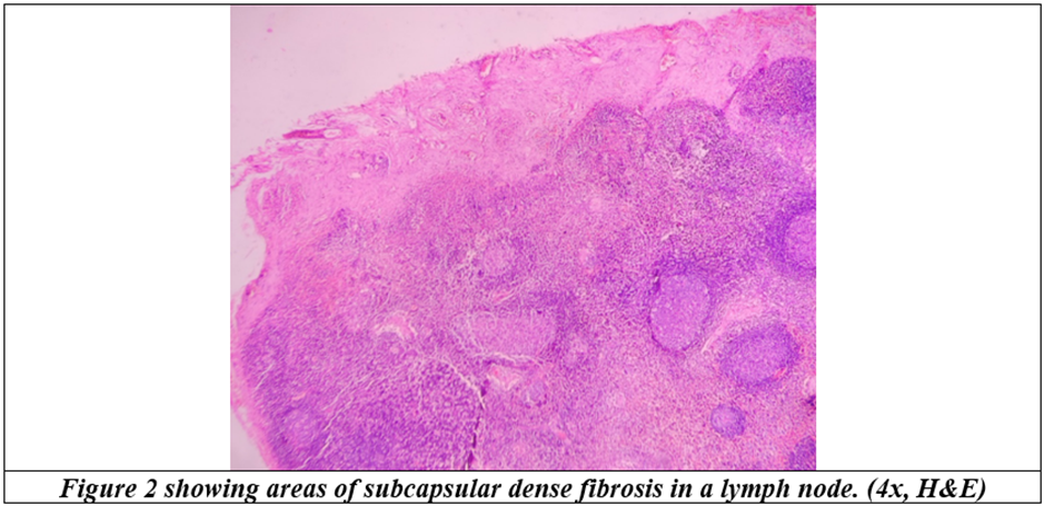
No residual nodal tumour was identified in 22 patients (51.2%), who were therefore classified as ypN0; the remaining 21 patients (48.8%) were ypN+ (Figure 2). Among the 21 ypN+ cases, residual deposits measured <2 mm in six, 2-20 mm in five and >20 mm in ten. Extracapsular extension was identified in seven of 21 ypN+ cases (33.3%). Therapy-related change in residual tumour cells was present in 18 of 21 (85.7%) and absent in three (14.3%) (Table 4).
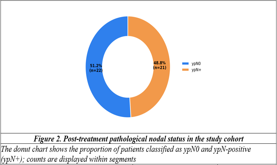
Table 4. Residual tumour characteristics and post-treatment nodal status
|
Parameter |
Category |
n/N (%) |
|
Post-treatment nodal status |
ypN0 |
22/43 (51.2) |
|
ypN+ |
21/43 (48.8) |
|
|
Residual tumour size among ypN+ |
<2 mm |
6/21 (28.6) |
|
2-20 mm |
5/21 (23.8) |
|
|
>20 mm |
10/21 (47.6) |
|
|
Extracapsular extension among ypN+ |
Present |
7/21 (33.3) |
|
Absent |
14/21 (66.7) |
|
|
Therapy-related change in residual tumour cells |
Present |
18/21 (85.7) |
|
Absent |
3/21 (14.3) |
|
|
Residual-tumour measurements are shown in the prespecified study strata. ypN+: residual nodal tumour present after neoadjuvant chemotherapy |
||
Association of nodal histomorphology with ypN status
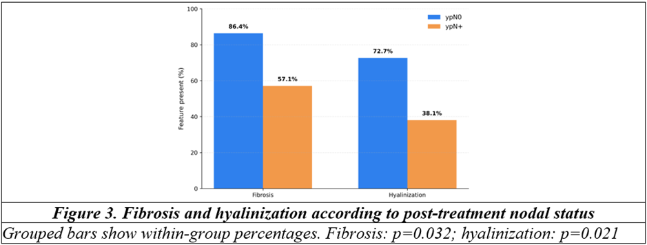
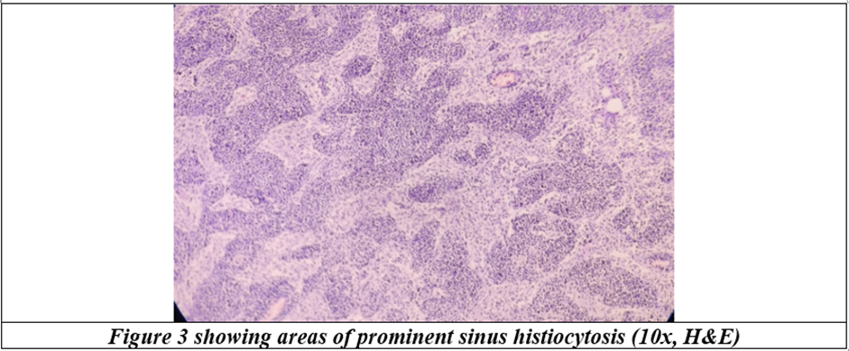
Fibrosis was present in 19 of 22 ypN0 cases (86.4%) compared with 12 of 21 ypN+ cases (57.1%), a statistically significant difference (p=0.032). Hyalinization showed a similar pattern, occurring in 72.7% of ypN0 and 38.1% of ypN+ cases (p=0.021). None of the remaining evaluated features reached the prespecified significance threshold, although foam cell infiltration, hemosiderin-laden macrophages and chronic inflammation were numerically more frequent in the ypN0 group (Table 5 and Figure 3).
Table 5. Association between selected histomorphological features and post-treatment nodal status
|
Feature present |
ypN0 (n=22) |
ypN+ (n=21) |
p-value |
|
Fibrosis |
19 (86.4) |
12 (57.1) |
0.032* |
|
Hyalinization |
16 (72.7) |
8 (38.1) |
0.021* |
|
Necrosis |
8 (36.4) |
6 (28.6) |
0.584 |
|
Foam cell infiltration |
12 (54.5) |
6 (28.6) |
0.085 |
|
Hemosiderin-laden macrophages |
14 (63.6) |
8 (38.1) |
0.094 |
|
Chronic inflammation |
16 (72.7) |
10 (47.6) |
0.091 |
|
Sinus histiocytosis |
12 (54.5) |
7 (33.3) |
0.162 |
|
Lymphoid depletion |
10 (45.5) |
6 (28.6) |
0.249 |
|
Germinal centre hyperplasia |
7 (31.8) |
4 (19.0) |
0.334 |
|
*Statistically significant at p<0.05. Percentages are calculated within each nodal-status group |
|||
Tumour biology and breast response in relation to ypN status
The ypN0 proportion was 66.7% in HER2-enriched tumours and 64.3% in triple-negative tumours, compared with 37.5% in luminal B and 33.3% in luminal A tumours (Table 6 and Figure 4). Grade III tumours had a higher descriptive ypN0 proportion than grade II and grade I tumours, 62.5%, 47.6% and 33.3%, respectively. Likewise, ypN0 occurred in 60.7% of tumours with Ki-67≥20% and 33.3% of those with Ki-67<20%. These patterns were considered exploratory.
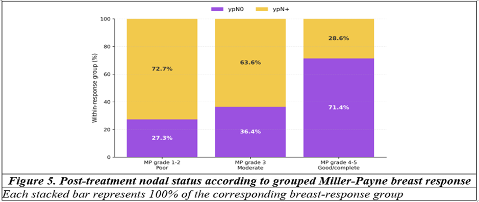
Miller-Payne grade 5 breast response was observed in 11 patients (25.6%), while grades 4 and 3 were recorded in 10 (23.3%) and 11 (25.6%), respectively. When grades 4-5 were grouped as good or complete breast response, 15 of 21 patients (71.4%) were ypN0. In contrast, only three of 11 patients (27.3%) with grade 1-2 response were ypN0 (Table 6 and Figure 5).
Table 6. Post-treatment nodal status according to molecular subtype, histological grade, Ki-67 and breast response
|
Variable |
Category |
ypN0, n/N (%) |
ypN+, n/N (%) |
|
Molecular subtype |
Luminal A |
4/12 (33.3) |
8/12 (66.7) |
|
Luminal B |
3/8 (37.5) |
5/8 (62.5) |
|
|
HER2-enriched |
6/9 (66.7) |
3/9 (33.3) |
|
|
Triple-negative |
9/14 (64.3) |
5/14 (35.7) |
|
|
Histological grade |
Grade I |
2/6 (33.3) |
4/6 (66.7) |
|
Grade II |
10/21 (47.6) |
11/21 (52.4) |
|
|
Grade III |
10/16 (62.5) |
6/16 (37.5) |
|
|
Ki-67 proliferation index |
<20% |
5/15 (33.3) |
10/15 (66.7) |
|
≥20% |
17/28 (60.7) |
11/28 (39.3) |
|
|
Breast response |
MP grade 1-2 |
3/11 (27.3) |
8/11 (72.7) |
|
MP grade 3 |
4/11 (36.4) |
7/11 (63.6) |
|
|
MP grade 4-5 |
15/21 (71.4) |
6/21 (28.6) |
|
|
MP: Miller-Payne; HER2: human epidermal growth factor receptor 2. These comparisons are descriptive |
|||
DISCUSSION:
The present prospective series documents a broad spectrum of treatment-associated changes in axillary lymph nodes after neoadjuvant chemotherapy. Slightly more than half of the patients were ypN0 at surgery, while residual metastatic tumour persisted in 48.8%. Fibrosis was the dominant morphological finding, followed by chronic inflammation, hyalinization and hemosiderin-laden macrophages. Most notably, fibrosis and hyalinization were significantly more frequent in ypN0 nodes. These findings suggest that stromal remodelling is not merely an incidental post-treatment appearance; it may represent a morphological footprint of nodal regression. Still, regression morphology must be interpreted alongside the direct search for viable tumour, not as a substitute for pathological nodal staging.
The Miller-Payne system was developed to grade reduction in primary breast tumour cellularity after chemotherapy and remains familiar in routine practice.[9] Sataloff and colleagues similarly emphasised separate evaluation of response in the breast and axillary nodes, acknowledging that the two compartments may not respond identically.[10] That distinction is visible in the current cohort. Good or complete breast response, represented by Miller-Payne grades 4-5, was accompanied by ypN0 in 71.4% of patients, whereas residual nodal disease remained in 28.6%. The result reinforces a practical point: an impressive breast response raises the probability of nodal clearance, but it cannot establish it.
Pathological response systems have evolved from descriptive grading toward quantitative assessment of residual disease. The Residual Cancer Burden framework integrates the dimensions and cellularity of the residual breast tumour with nodal burden, producing a continuous index that is strongly linked to outcome.[11] Although RCB could not be reconstructed in this series, the present findings support its underlying logic. The axilla contributes information that is distinct from the breast, and both residual tumour and evidence of regression should be documented systematically.
The observed frequency of fibrosis was higher than that reported in the Indian series by Chakrabarti et al., who described chemotherapy-associated morphological changes in breast and nodal specimens and found malignant cells in a subset of axillary nodes.[12] Direct numerical comparison is difficult because cohorts differ in stage distribution, treatment regimens, nodal sampling and definitions of individual histological changes. Agarwal et al. also demonstrated that post-neoadjuvant response varies when assessed using different grading systems, with complete response representing only one end of a broader continuum of partial and absent response.[13] The higher ypN0 proportion in the present cohort may partly reflect biological composition and treatment exposure, but another important issue is baseline nodal status: five patients were clinically cN0, and clinical nodal staging does not prove that every node-negative surgical specimen represents eradication of a previously documented metastasis.
The association of fibrosis and hyalinization with ypN0 is biologically plausible. Cytotoxic injury and tumour clearance can be followed by collagen deposition, stromal contraction and hyaline scarring. Histiocytic and inflammatory changes may accompany removal of cellular debris and altered blood products. Yet none of these features is specific in isolation. Fibrosis can occur in a node without known prior metastasis, and small residual deposits may remain within a markedly fibrotic node. Chung et al. showed that grading regression in metastatic axillary nodes can provide prognostic information beyond a simple positive-versus-negative classification.[14] Their work supports more explicit reporting of nodal treatment effect, particularly when pretreatment nodal involvement has been documented.
The molecular patterns in this cohort were consistent with the broader observation that HER2-enriched and triple-negative tumours are often more chemosensitive than luminal cancers. The ypN0 proportions were 66.7% and 64.3% in these two subgroups. Because the study was small and treatment exposure was heterogeneous, these values should be read as descriptive rather than definitive subtype estimates. The same caution applies to the higher ypN0 proportion among grade III tumours and tumours with Ki-67≥20%. High proliferative activity may increase susceptibility to cytotoxic chemotherapy, but patient-level multivariable modelling would require a larger cohort and internally consistent biomarker records.
Breast and axillary responses were directionally concordant. Morgan et al. reported that pathological response in the breast predicts axillary response among patients with biopsy-proven nodal disease, but the relationship is not absolute.[15] A nationwide Korean analysis likewise found a strong association between breast pathological complete response and axillary clearance, while showing that the residual nodal risk depends on initial clinical nodal burden.[16] In the present series, the predominance of ypN0 among Miller-Payne grade 4-5 tumours follows this pattern. Nevertheless, almost three in ten good or complete breast responders retained nodal tumour, which argues against assuming axillary response from breast morphology alone.
The study has practical relevance for pathology services in Indian tertiary hospitals, where post-neoadjuvant specimens are increasingly routine but preoperative nodal documentation, specimen mapping and access to comprehensive biomarker testing may vary. A structured nodal assessment that records residual tumour, the largest deposit, extracapsular extension and recognisable treatment effect can improve communication with the multidisciplinary team. It also makes the report more useful when adjuvant decisions depend on the presence of residual disease.
Several limitations merit attention. This was a single-centre study with 43 patients, restricting precision and subgroup analysis. Pretreatment nodal metastasis was not pathologically documented for every patient, so ypN0 should not be treated as synonymous with axillary pathological complete response across the entire cohort. Chemotherapy regimens were heterogeneous, and anti-HER2 targeted therapy was recorded in only two patients; this limited meaningful assessment of treatment-specific response in HER2-associated tumours. Interobserver reproducibility of histomorphological features was not assessed. The study also lacked survival follow-up, and the modest cohort did not support stable multivariable modelling.
CONCLUSION:
Axillary lymph nodes after neoadjuvant chemotherapy displayed diverse regressive and reactive changes. Fibrosis was the most common alteration, and both fibrosis and hyalinization were significantly associated with ypN0 status. HER2-enriched and triple-negative tumours, high Ki-67 tumours and good or complete Miller-Payne breast responders showed higher descriptive proportions of node-negative disease. These patterns support systematic documentation of therapy-related nodal morphology, while confirming that the final assessment must remain anchored to residual tumour detection, ypN staging and reliable pretreatment nodal information.
REFERENCES:
1. Bray F, Laversanne M, Sung H, Ferlay J, Siegel RL, Soerjomataram I, et al. Global cancer statistics 2022: GLOBOCAN estimates of incidence and mortality worldwide for 36 cancers in 185 countries. CA Cancer J Clin 2024;74(3):229-63. doi:10.3322/caac.21834.
2. Korde LA, Somerfield MR, Carey LA, Crews JR, Denduluri N, Hwang ES, et al. Neoadjuvant chemotherapy, endocrine therapy, and targeted therapy for breast cancer: ASCO guideline. J Clin Oncol 2021;39(13):1485-505. doi:10.1200/JCO.20.03399.
3. Cortazar P, Zhang L, Untch M, Mehta K, Costantino JP, Wolmark N, et al. Pathological complete response and long-term clinical benefit in breast cancer: the CTNeoBC pooled analysis. Lancet 2014;384(9938):164-72. doi:10.1016/S0140-6736(13)62422-8.
4. von Minckwitz G, Untch M, Blohmer JU, Costa SD, Eidtmann H, Fasching PA, et al. Definition and impact of pathologic complete response on prognosis after neoadjuvant chemotherapy in various intrinsic breast cancer subtypes. J Clin Oncol 2012;30(15):1796-804. doi:10.1200/JCO.2011.38.8595.
5. Houssami N, Macaskill P, von Minckwitz G, Marinovich ML, Mamounas E. Meta-analysis of the association of breast cancer subtype and pathologic complete response to neoadjuvant chemotherapy. Eur J Cancer 2012;48(18):3342-54. doi:10.1016/j.ejca.2012.05.023.
6. Pinder SE, Provenzano E, Earl H, Ellis IO. Laboratory handling and histology reporting of breast specimens from patients who have received neoadjuvant chemotherapy. Histopathology 2007;50(4):409-17. doi:10.1111/j.1365-2559.2006.02419.x.
7. Provenzano E, Bossuyt V, Viale G, Cameron D, Badve S, Denkert C, et al. Standardization of pathologic evaluation and reporting of postneoadjuvant specimens in clinical trials of breast cancer: recommendations from an international working group. Mod Pathol 2015;28(9):1185-201. doi:10.1038/modpathol.2015.74.
8. Bossuyt V, Provenzano E, Symmans WF, Boughey JC, Coles C, Curigliano G, et al. Recommendations for standardized pathological characterization of residual disease for neoadjuvant clinical trials of breast cancer by the BIG-NABCG collaboration. Ann Oncol 2015;26(7):1280-91. doi:10.1093/annonc/mdv161.
9. Ogston KN, Miller ID, Payne S, Hutcheon AW, Sarkar TK, Smith I, et al. A new histological grading system to assess response of breast cancers to primary chemotherapy: prognostic significance and survival. Breast 2003;12(5):320-7. doi:10.1016/S0960-9776(03)00106-1.
10. Sataloff DM, Mason BA, Prestipino AJ, Seinige UL, Lieber CP, Baloch Z. Pathologic response to induction chemotherapy in locally advanced carcinoma of the breast: a determinant of outcome. J Am Coll Surg 1995;180(3):297-306. PMID: 7874340.
11. Symmans WF, Peintinger F, Hatzis C, Rajan R, Kuerer H, Valero V, et al. Measurement of residual breast cancer burden to predict survival after neoadjuvant chemotherapy. J Clin Oncol 2007;25(28):4414-22. doi:10.1200/JCO.2007.10.6823.
12. Chakrabarti S, Mandal PK, Roy Chowdhury A, Das S. Consequence of neo-adjuvant chemotherapy on morphology of breast carcinoma: a systematic evaluation. Indian J Cancer 2016;53(1):29-33. doi:10.4103/0019-509X.180850.
13. Agarwal S, Pandey P, Ralli M, Chaturvedi V, Mittal K, Singh SP. Assessment of pathological response to neoadjuvant chemotherapy in locally advanced breast carcinoma using Sataloff grading system. Archive of Oncology 2019;25(2):13-8. doi:10.2298/AOO190128001A.
14. Chung YR, Woo JW, Ahn S, Kang E, Kim EK, Jang M, et al. Prognostic implications of regression of metastatic axillary lymph nodes after neoadjuvant chemotherapy in patients with breast cancer. Sci Rep 2021;11:12128. doi:10.1038/s41598-021-91643-z.
15. Morgan C, Stringfellow TD, Rolph R, Kovacs T, Kothari A, Pinder SE, et al. Neoadjuvant chemotherapy in patients with breast cancer: does response in the breast predict axillary node response? Eur J Surg Oncol 2020;46(4 Pt A):522-6. doi:10.1016/j.ejso.2019.11.498.
16. Ryu JM, Choi HJ, Park EH, Kim JY, Lee YJ, Park S, et al. Relationship between breast and axillary pathologic complete response according to clinical nodal stage: a nationwide study from Korean Breast Cancer Society. J Breast Cancer 2022;25(2):94-105. doi:10.4048/jbc.2022.25.e17.