Cholera Due to Vibrio cholerae O1 Ogawa Serotype: A Case Series from a Tertiary Care Hospital in Amravati, Maharashtra, India.
- Ranjana R. Khorgade , Associate Professor, Department of Microbiology, Government Medical College (GMC), Amravati, Maharashtra
- Syed Azizur Rahman , Professor, Department of General Medicine, Pacific Institute of Medical Sciences, Umarda, Udaipur, Rajasthan
- Nuzhat Firdos , Assistant Professor, Department of Microbiology, Dr. Panjabrao Deshmukh Memorial Medical College (Dr PDMMC), Amravati, Maharashtra
- Pawan S. Bahurupi , Tutor, Department of Community Medicine, Dr. Panjabrao Deshmukh Memorial Medical College (Dr PDMMC), Amravati, Maharashtra
Article Information:
Abstract:
Background: Cholera, an acute secretory diarrhoeal illness caused by toxigenic Vibrio cholerae serogroups O1 and O139, remains an important public health problem in India, particularly during the monsoon season, when compromised water and sanitation infrastructure facilitates transmission. Case Presentation: We describe three patients with acute watery diarrhoea presenting to a tertiary care teaching hospital in Amravati, Maharashtra, during June–July 2025: a 50-year-old woman with mild dehydration, a 9-year-old boy with severe dehydration requiring paediatric intensive care, and a 25-year-old woman with severe dehydration, hypotension, and tachycardia. Stool wet-mount microscopy showed characteristic darting motility in all three patients, and stool culture confirmed Vibrio cholerae O1, Ogawa serotype, in two of the three; the remaining patient's culture was negative, possibly reflecting prior antibiotic intake. Results: All patients were managed according to severity of dehydration — oral rehydration for mild dehydration (WHO Plan B) and intravenous Ringer's lactate under WHO Plan C, with antimicrobial therapy, for severe dehydration. Two patients improved and were discharged in stable condition; the child was discharged against medical advice after initial stabilisation. Conclusion: Cholera due to Vibrio cholerae O1, Ogawa serotype, should remain an important differential diagnosis for acute watery diarrhoea across paediatric and adult age groups in endemic regions, particularly during the monsoon season. Prompt dehydration assessment, stool microscopy and culture, and severity-based fluid and antimicrobial therapy are central to a favourable outcome.
Keywords:
Article :
INTRODUCTION:
Cholera is an acute, toxin-mediated secretory diarrhoeal illness caused by toxigenic strains of Vibrio cholerae, most commonly of serogroup O1. Globally, it causes an estimated 1.3–4.0 million cases and 21,000–143,000 deaths annually, chiefly where safe water and sanitation are lacking, and can progress to life-threatening dehydration within hours if untreated. [1,2]
India remains among the countries with the highest reported cholera burden, with outbreaks recurring seasonally during the monsoon. Maharashtra has repeatedly been among the states reporting the largest share of outbreaks, including previously documented Ogawa-serotype outbreaks in Yavatmal and Aurangabad districts. [4-7]
Vibrio cholerae O1 is further classified into Ogawa and Inaba serotypes; this distinction is relevant to surveillance and vaccine strain selection but does not influence clinical severity.[3] Diagnosis relies on stool wet-mount microscopy for darting motility, culture, and biochemical and serological confirmation, including the oxidase and string tests, IMViC reactions, and slide agglutination.[8] Timely confirmation and susceptibility testing are increasingly important given emerging antimicrobial resistance among circulating strains in India.[9]
Management is guided by dehydration severity: oral rehydration for mild-to-moderate disease and intravenous crystalloid resuscitation, typically Ringer's lactate, for severe dehydration, supplemented by a single-dose antimicrobial (commonly doxycycline or azithromycin) to shorten diarrhoea duration and vibrio excretion. [10-14] Acute kidney injury secondary to hypovolaemia is an increasingly recognised complication warranting close monitoring. [15,16]
We describe three cases of acute watery diarrhoea presenting to the Department of Microbiology of a tertiary care teaching hospital in Amravati, Maharashtra, during June–July 2025, to illustrate the clinical spectrum, laboratory diagnosis, and outcomes of cholera due to Vibrio cholerae O1, Ogawa serotype, in routine practice.
MATERIALS AND METHODS:
This case series was conducted in the Department of Microbiology of a tertiary care teaching hospital in Amravati, Maharashtra, during June–July 2025. Patients presenting with acute watery diarrhoea, vomiting, and clinical signs of dehydration were evaluated, and those meeting the departmental case definition for suspected cholera were included.
Fresh stool specimens were collected in sterile containers and transported promptly to the laboratory. Specimens were examined by wet-mount microscopy for darting motility and by Gram staining for preliminary morphology. Definitive identification used standard biochemical tests — oxidase and catalase tests, string test, and IMViC (indole, methyl red, Voges–Proskauer, citrate) reactions — alongside culture on thiosulfate–citrate–bile salts–sucrose (TCBS) agar. Confirmed isolates were serotyped by slide agglutination using polyvalent and monovalent antisera.[8] Dehydration severity was graded clinically according to WHO criteria (no / some / severe dehydration), and vital signs, general and systemic examination findings, and baseline complete blood count, serum electrolytes, and renal function were recorded on admission.
Case Presentation 1
A 50-year-old woman presented on 24 June 2025 with acute-onset watery diarrhoea and vomiting of 18 hours' duration, with 8–10 loose stool episodes and 2–3 vomiting episodes per day. She was afebrile (36.9 °C) and had no known comorbidities. She reported taking oral rehydration solution and an empirical oral antibiotic at home before admission; the specific agent was not documented.
On examination she was conscious and alert, with dry oral mucosa and mildly reduced skin turgor, consistent with “some dehydration” by WHO criteria. Vital signs were blood pressure 110/70 mmHg, pulse 96/min, respiratory rate 18/min, and SpO₂ 98%; cardiovascular, respiratory, and neurological examination was normal, and the abdomen was soft with hyperactive bowel sounds.
Wet-mount microscopy showed darting motility, and Gram staining revealed curved Gram-negative bacilli. Stool culture was negative, possibly reflecting the prior antibiotic intake, and no isolate was available for biochemical identification or serotyping. Complete blood count showed mild leukocytosis with a normal haemoglobin, and serum electrolytes showed mild hypokalaemia; renal function was normal.
Given the mild dehydration, she was managed with oral rehydration solution under WHO Plan B and supportive antiemetic therapy; intravenous fluids and antibiotics were not required. She improved clinically and was discharged in stable condition after a hospital stay of approximately 2 days, with complete recovery on follow-up; no complications were recorded. Exact discharge date was not documented.
Case Presentation 2
A 9-year-old boy was admitted to the paediatric intensive care unit with acute gastroenteritis of 12 hours' duration, with 12–15 stool episodes and 6–7 vomiting episodes per day. He was afebrile (37.1 °C) with no known comorbidities; there was a history of oral rehydration solution at home but no documented prior antibiotic use.
He was lethargic, with sunken eyes, dry oral mucosa, poor skin turgor, a weak pulse, and cold extremities — severe dehydration by WHO criteria. Vital signs were blood pressure 90/60 mmHg, pulse 128/min, respiratory rate 28/min, and SpO₂ 98%, with tachycardia on systemic examination; the abdomen was soft with hyperactive bowel sounds.
Wet-mount microscopy showed darting motility and Gram staining revealed curved Gram-negative bacilli. Stool culture confirmed Vibrio cholerae O1, Ogawa serotype, on standard biochemical testing (oxidase-, catalase-, and string test-positive; indole- and methyl red-positive; Voges–Proskauer-, citrate-, and urease-negative; motile) and serotyping. Complete blood count showed haemoconcentration with mild leukocytosis; electrolytes showed hyponatraemia and hypokalaemia, and renal function tests showed mild prerenal azotaemia.
He was resuscitated with intravenous Ringer's lactate under WHO Plan C and given azithromycin, together with zinc supplementation and an antiemetic; the specific dose, route, and duration of antibiotic therapy were not documented. No complications were recorded during the approximately 2-day recorded stay. Despite initial stabilisation, he was discharged against medical advice; the reason was not documented, and no further follow-up information was available.
RESULTS:
A 25-year-old woman from Mahuli Jahagir presented to the emergency department on 7 July 2025 with profuse watery diarrhoea and vomiting of 24 hours' duration — 15–20 stool episodes and 8 vomiting episodes per day. She was afebrile (36.8 °C) with no known comorbidities or documented prior medication use.
She was conscious but weak, with sunken eyes, cold extremities, and poor skin recoil — severe dehydration by WHO criteria — and was hypotensive (blood pressure 80/70 mmHg) and tachycardic (pulse 118/min; respiratory rate 24/min; SpO₂ 97%), with tachycardia and hypotension on systemic examination and a soft abdomen with hyperactive bowel sounds.
Wet-mount microscopy showed darting motility, and Gram staining revealed curved, comma-shaped Gram-negative bacilli. The isolate was oxidase-, catalase-, and string test-positive, with an IMViC pattern of indole- and methyl red-positive and Voges–Proskauer-, citrate-, and urease-negative, and was confirmed as Vibrio cholerae O1, Ogawa serotype, by culture and serotyping. Complete blood count showed haemoconcentration with mild leukocytosis; electrolytes showed hyponatraemia and hypokalaemia, and renal function tests showed mild prerenal azotaemia.
She was resuscitated with intravenous Ringer's lactate under WHO Plan C and given antimicrobial therapy (doxycycline or azithromycin per local protocol; the specific agent, dose, and duration administered to this patient were not documented), together with an antiemetic. She responded well, with no complications, and was discharged in stable condition after a hospital stay of approximately 3 days, with complete recovery on follow-up; exact discharge date was not documented.
Table 1. Summary of Clinical Presentation, Diagnosis, Treatment, and Outcome
|
Parameter |
Case 1 |
Case 2 |
Case 3 |
|
Age / Sex |
50 y / Female |
9 y / Male |
25 y / Female |
|
Presenting complaint |
Watery diarrhoea, vomiting (18 h) |
Diarrhoea, vomiting; PICU admission (12 h) |
Diarrhoea, vomiting, hypotension (24 h) |
|
Dehydration (WHO grade) |
Some dehydration |
Severe dehydration |
Severe dehydration |
|
Comorbidities |
None |
None |
None |
|
Diagnosis |
Culture-negative; wet mount/Gram stain suggestive |
V. cholerae O1, Ogawa |
V. cholerae O1, Ogawa |
|
Treatment |
ORS (WHO Plan B); no IV fluids/antibiotics |
IV Ringer's lactate (Plan C) + azithromycin |
IV Ringer's lactate (Plan C) + doxycycline/azithromycin |
|
Complications |
None |
None documented before LAMA |
None |
|
Outcome |
Improved; discharged stable; recovered |
Discharged against medical advice; follow-up unavailable |
Improved; discharged stable; recovered |
Abbreviations: IV, intravenous; LAMA, left/discharged against medical advice; ORS, oral rehydration solution; PICU, paediatric intensive care unit; WHO, World Health Organization.
Figures
The photographs below are representative laboratory images from this case series; the case records do not specify which patient each corresponds to.
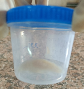
Figure 1. Rice-water appearance of a stool specimen.
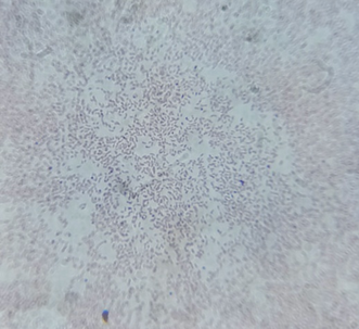
Figure 2. Gram-stained smear showing comma-shaped Gram-negative bacilli.
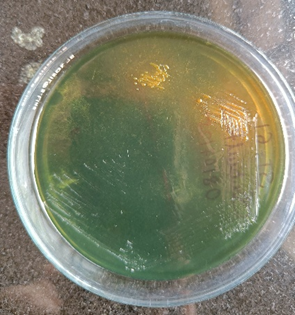
Figure 3. Sucrose-fermenting colonies of Vibrio cholerae on thiosulfate citrate bile salts sucrose (TCBS) agar.
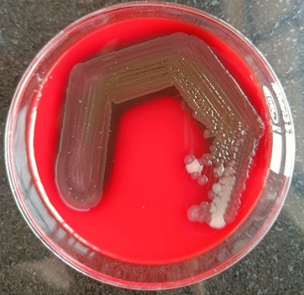
Figure 4. Haemolytic colonies of Vibrio cholerae on blood agar.
DISCUSSION:
This case series describes three patients with acute watery diarrhoea and varying dehydration severity presenting during the 2025 monsoon season in Amravati, Maharashtra, two of whom had culture-confirmed cholera due to Vibrio cholerae O1, Ogawa serotype. The principal findings were the seasonal clustering of cases, the predominance of the Ogawa serotype among confirmed isolates, a culture-negative case attributable to prior antibiotic intake, and a favourable clinical response to severity-based fluid and antimicrobial therapy in two severely dehydrated patients, one of whom was subsequently discharged against medical advice.
Muzembo et al. [4] found that Maharashtra accounted for 54 of 565 cholera outbreaks reported across India between 2011 and 2020, concentrated during June–September. This finding is consistent with our series, in which all cases presented during June–July 2025. Kanungo et al. [5] similarly reported that Maharashtra was among the states with the highest cholera burden between 1997 and 2006. Our findings are comparable, indicating that the state's endemicity has persisted over nearly two decades.
In a study by Kale et al. [6], an outbreak in Yavatmal district identified Vibrio cholerae O1, Ogawa serotype, biotype El Tor, as the causative organism, and Gopalkrishna et al.[7] reported the same serotype in Aurangabad. Both findings are consistent with our study, in which the two culture-confirmed isolates were also Ogawa serotype, supporting continued local predominance of this strain.
Gopalkrishna et al. [7] additionally found a stool culture positivity rate of only 13.1%, attributing this partly to antibiotic administration before sample collection. This finding is directly comparable to our study, in which the one culture-negative patient (Case 1) had a documented history of antibiotic intake before sampling, reinforcing this mechanism; however, our very small sample size precludes quantitative comparison of positivity rates.
Chatterjee et al. [9] found that Maharashtra was among the four states accounting for most antibiotic-resistance-associated cholera outbreaks in India. In contrast, our study did not perform antimicrobial susceptibility testing, so we could not assess whether resistance influenced our patients' course. This represents an important limitation given the resistance trends reported by Chatterjee et al. [9]
Regarding antimicrobial therapy, Khan et al. [13] found azithromycin achieved higher clinical success than erythromycin in children (76% versus 65%). Our paediatric patient, treated with azithromycin, showed initial stabilisation consistent with this efficacy, though discharge against medical advice precluded confirmation of durable response, unlike the completed follow-up in that trial. Saha et al. [11] similarly reported 73% clinical success with azithromycin in adults; our adult patient with severe dehydration responded well to doxycycline or azithromycin, broadly consistent with this evidence, although the exact agent used could not be confirmed from her records.
Vakrani et al. [15] found that 78% of cholera patients in a Bengaluru outbreak developed acute kidney injury, with 32.7% requiring haemodialysis. In contrast, our two severely dehydrated patients showed only mild prerenal azotaemia without progression to overt kidney injury, a difference plausibly explained by our smaller sample, earlier presentation, and prompt resuscitation. This is consistent with Qasem and Rabbani [16], who described reversible pre-renal injury following aggressive rehydration.
Overall, this series reinforces the continued circulation of Vibrio cholerae O1, Ogawa serotype, in Maharashtra, highlights antibiotic exposure as a determinant of culture yield, and supports the efficacy of severity-based rehydration and azithromycin-based therapy, while underscoring the need for routine antimicrobial susceptibility testing in future cases.
CONCLUSION:
Cholera due to Vibrio cholerae O1, Ogawa serotype, continues to cause clinically significant disease across paediatric and adult age groups in this region, ranging from mild, culture-negative illness to severe, life-threatening dehydration. Prompt clinical recognition, stool microscopy and culture, and severity-based fluid and antimicrobial therapy remain the cornerstones of a favourable outcome. Continued microbiological surveillance, including serotyping and antimicrobial susceptibility testing, is recommended to monitor local epidemiology and resistance patterns.
REFERENCES:
1. World Health Organization. Cholera [Internet]. Geneva: World Health Organization; 2024 [cited 2025 Aug]. Available from: https://www.who.int/news-room/fact-sheets/detail/cholera
2. Ali M, Nelson AR, Lopez AL, Sack DA. Updated global burden of cholera in endemic countries. PLoS Negl Trop Dis. 2015;9(6):e0003832. doi:10.1371/journal.pntd.0003832.
3. Global Task Force on Cholera Control. Cholera facts [Internet]. Geneva: Global Task Force on Cholera Control; 2024 [cited 2025 Aug]. Available from: https://www.gtfcc.org/about-cholera/cholera-facts/
4. Muzembo BA, Kitahara K, Debnath A, Ohno A, Okamoto K, Miyoshi SI. Cholera outbreaks in India, 2011-2020: a systematic review. Int J Environ Res Public Health. 2022;19(9):5738. doi:10.3390/ijerph19095738.
5. Kanungo S, Sah BK, Lopez AL, Sung JS, Paisley AM, Sur D, et al. Cholera in India: an analysis of reports, 1997-2006. Bull World Health Organ. 2010;88(3):185-91. doi:10.2471/BLT.09.073460.
6. Kale SS, Bulle PA, Deshmukh DG, Tankhiwale SS, Gujar VM. An outbreak of diarrhoeal disease of El Tor Vibrio cholerae O1 Ogawa in and around Yavatmal district, Maharashtra, India in 2018. Int J Community Med Public Health. 2020;7(4):1297-300. doi:10.18203/2394-6040.ijcmph20201089.
7. Gopalkrishna V, Joshi M, Viswanathan R, Malu G, Ganorkar N, Chavan N, et al. Cholera outbreak in Aurangabad, Maharashtra, western India. Indian J Med Res. 2019;150(6):640-3. doi:10.4103/ijmr.IJMR_87_19.
8. Global Task Force on Cholera Control. Public health surveillance for cholera: guidance document [Internet]. Geneva: Global Task Force on Cholera Control; 2024 [cited 2025 Aug]. Available from: https://www.gtfcc.org
9. Chatterjee P, Kanungo S, Bhattacharya SK, Dutta S. Mapping cholera outbreaks and antibiotic resistant Vibrio cholerae in India: an assessment of existing data and a scoping review of the literature. Vaccine. 2020;38 Suppl 1:A93-A104. doi:10.1016/j.vaccine.2019.12.003.
10. Centers for Disease Control and Prevention. Recommendations for the use of antibiotics for the treatment of cholera [Internet]. Atlanta (GA): Centers for Disease Control and Prevention; 2011 [cited 2025 Aug]. Available from: https://stacks.cdc.gov/view/cdc/21854
11. Saha D, Karim MM, Khan WA, Ahmed S, Salam MA, Bennish ML. Single-dose azithromycin for the treatment of cholera in adults. N Engl J Med. 2006;354(23):2452-62. doi:10.1056/NEJMoa054493.
12. Rahaman MM, Majid MA, Alam AKMJ, Islam MR. Effects of doxycycline in actively purging cholera patients: a double-blind clinical trial. Antimicrob Agents Chemother. 1976;10(4):610-2. doi:10.1128/AAC.10.4.610.
13. Khan WA, Saha D, Rahman A, Salam MA, Bogaerts J, Bennish ML. Comparison of single-dose azithromycin and 12-dose, 3-day erythromycin for childhood cholera: a randomised, double-blind trial. Lancet. 2002;360(9347):1722-7. doi:10.1016/S0140-6736(02)11680-1.
14. World Health Organization. The treatment of diarrhoea: a manual for physicians and other senior health workers. Geneva: World Health Organization; 2005.
15. Vakrani GP, Nambakam T. Retrospective study on acute kidney injury among cholera patients in an outbreak in Whitefield, Bengaluru. Int J Nephrol. 2021;2021:6682838. doi:10.1155/2021/6682838.
16. Qasem A, Rabbani SA. Acute kidney injury associated with cholera. Cureus. 2023;15(1):e34101. doi:10.7759/cureus.34101.
17. Legros D; Partners of the Global Task Force on Cholera Control. Global cholera epidemiology: opportunities to reduce the burden of cholera by 2030. J Infect Dis. 2018;218(Suppl 3):S137-S140. doi:10.1093/infdis/jiy486. Erratum in: J Infect Dis. 2019;219(3):509. doi:10.1093/infdis/jiy619.