Histomorphological Analysis Of Synovial Lesions.
- Shruthi , Assistant Professor, Department of Pathology, Navodaya Medical College and Research centre, Raichur, Karnataka.
- KK Suresh , Professor, Department of Pathology, JJM medical college, Davangere, Karnataka.
- Krupa Kadarappa , Assistant Professor , Department of Pathology , Kidwai Memorial Institute of Oncology, Bengaluru, Karnataka.
Article Information:
Abstract:
Background: Synovial tissue is a specialized mesenchymal tissue that is involved in a wide range of inflammatory, infectious, degenerative, metabolic, and tumor like processes affecting joints. Arthritis presenting with pain, swelling and stiffness is common across all age groups, and early cases with non specific clinical and laboratory findings can be difficult to classify. Histopathological examination of synovial biopsies can assist in establishing an early and specific diagnosis, particularly in monoarticular or clinically unclassified arthritis. Methods: This retrospective study was conducted in the Department of Pathology, JJM Medical College, Davangere, Karnataka, over a period of five years from June 2016 to May 2021. Seventy synovial biopsies obtained by open and arthroscopic methods were included. All non neoplastic and neoplastic synovial lesions were studied; extra articular synovial sarcoma and ganglion cysts were excluded. Relevant clinical data (age, sex, joint involved, symptoms, and provisional diagnosis) were retrieved from histopathology request forms. Specimens were fixed in 10% formalin, processed routinely, and stained with hematoxylin and eosin for histomorphological analysis. Results: The age of patients ranged from 25 days to 90 years, with a slight male predominance (male:female ratio 1.18:1). Knee joint was the most frequently involved site (54.3%), and joint swelling (92.8%) and pain (80%) were the commonest presenting symptoms. Based on morphology, lesions were grouped as degenerative, infectious, inflammatory, metabolic, benign tumor/tumor like conditions and others. Chronic non specific synovitis was the most common lesion, seen in 31 cases (44.3%), followed by tubercular arthritis in 13 cases (18.6%), rheumatoid arthritis in 5 cases (7.1%), and septic arthritis in 4 cases (5.7%). Less frequent lesions included synovial chondromatosis (4 cases, 5.7%), tenosynovial giant cell tumour (3 cases, 4.3%), Baker cyst (3 cases, 4.3%), lipoma arborescens (2 cases, 2.9%), bursitis (2 cases, 2.9%), and single cases of osteoarthritis, gouty arthritis, and traumatic arthritis. Characteristic histomorphological features, such as synovial hyperplasia, villous hypertrophy, granulomatous inflammation, lymphoplasmacytic infiltrate, and crystal induced giant cell reactions, aided in subclassification. Conclusion: Histomorphological evaluation of synovial biopsies is a valuable adjunct in the diagnosis of diverse synovial lesions and helps to distinguish infectious, inflammatory, degenerative, metabolic, and tumor like conditions. Correlation with clinical, radiological, biochemical, and serological findings remains essential to achieve an accurate and specific diagnosis, particularly in early or clinically unclassified arthritis.
Keywords:
Article :
INTRODUCTION:
Synovium is a specialized mesenchymal tissue that is essential for the appropriate function of the locomotor apparatus. It is the site for a series of pathologic processes that are characteristic, and in some cases specific, to this distinctive tissue. [1, 2]. Synovial membrane lines the inner surface of the capsule of a synovial joint and secretes synovial fluid which provides lubrication and eases movement. Synovial tissue can be obtained either by open surgery, by doing a blind needle biopsy, or at arthroscopy. [3] . Symptoms complex of pain, swelling and stiffness of joints labeled as arthritis is a common entity in clinical practice affecting all age groups. [4] A typical clinical presentation with negative routine investigations makes diagnosis of early arthritis difficult. It is in these cases that simple procedures such as synovial fluid examination and biopsy of the synovial tissue can provide an important clue to a possible diagnosis early in the course of the disease and to evaluate response to treatment if analysis is done at frequent intervals during the course of treatment. [4]
OBJECTIVES: To study histomorphological spectrum of synovial lesions from synovial biopsy.
MATERIALS AND METHODS:
The study was conducted in the department of pathology JJM medical college, Davangere, during the period of 5 years from June 2016 to May 2021. Relevant clinical information regarding age, sex, clinical features and provisional diagnosis were taken from the histopathology request forms.
• Inclusion criteria: All non-neoplastic and neoplastic synovial lesions diagnosed on histopathological examination.
• Exclusion criteria: Extra articular synovial sarcoma will be excluded. Ganglion cysts were excluded.
In this study, specimens were received as biopsies obtained by open method and by arthroscopic method. Gross examination of specimen was done and tissues were fixed in 10% formalin for 12-24 hours. Tissue bits processed routinely and stained with H&E for analysis and diagnosis.
RESULTS:
A total of 70 synovial biopsy specimens were received in present study. Based on morphological features these synovial lesions were classified into six groups which includes Degenerative, Infectious, Inflammatory, Metabolic, Benign tumor and tumor like condition and others.
TABLE1: INCIDENCE OFSYNOVIALLESIONS ANDGENDERDISTRIBUTION.
|
LESIONS |
CASES |
NO. OF CASES (%) |
MALE |
FEMALE |
M:F |
|
I. Degenerative |
|
|
|
|
|
|
Osteoarthritis (OA) |
1 |
(1.4%) |
0 |
1 |
0:1 |
|
II. Infectious |
|
|
|
|
|
|
Tb Arthritis |
13 |
(18.6%) |
3 |
10 |
1:3.3 |
|
Septic Arthritis |
4 |
(5.7%) |
2 |
2 |
1:1 |
|
III. Inflammatory |
|
|
|
|
|
|
Bursitis |
2 |
(2.9%) |
1 |
1 |
1:1 |
|
Cnss |
31 |
(44.3%) |
20 |
11 |
1.8:1 |
|
Rheumatoid Arthritis (Ra) |
5 |
(7.1%) |
2 |
3 |
1:1.5 |
|
IV. Metabolic |
|
|
|
|
|
|
Gout |
1 |
(1.4%) |
1 |
0 |
1:0 |
|
V. Benign Tumor And Tumor Like Conditions |
|
|
|
|
|
|
Lipoma |
2 |
(2.9%) |
0 |
2 |
0:2 |
|
Synovial Chondromatosis |
4 |
(5.7%) |
3 |
1 |
3:1 |
|
Tgct |
3 |
(4.3%) |
2 |
1 |
2:1 |
|
VI. Others |
|
|
|
|
|
|
Traumatic Arthritis |
1 |
(1.4%) |
1 |
0 |
1:0 |
|
Baker Cyst |
3 |
(4.3%) |
2 |
1 |
2:1 |
The cases age ranged from 25days baby to 75years. Highest incidence was noted in males 38 cases(54.2%) as compared to females 32 cases(45.7%)withaM:Fratio of 1.18:1.
TABLEII: AGEDISTRIBUTIONOFSYNOVIALLESIONS
|
AGE GROUP |
NUMBER OF CASES |
|
<1 |
5 |
|
1-10 |
3 |
|
11-20 |
6 |
|
21-30 |
5 |
|
31-40 |
8 |
|
41-50 |
13 |
|
51-60 |
19 |
|
61-70 |
7 |
|
71-80 |
3 |
|
81-90 |
1 |
|
TOTAL |
70 |
Commonest symptoms observed in present study were joint swelling in 65 cases(92.8%) followedbypainin56cases(80%).Kneewasthe commonestjointinvolved,contributingto about 38 cases(54.3%) followed by multiple joints in 10cases(14.3%).Among the 70 synovial biopsies, 31 (44.3%) cases were chronic non-specific synovitis (CNSS), the most common age group affected was between 51-60 years in 9 (29%) cases with male preponderance. Knee was the commonest joint involved. Microscopically all the cases showed focal or diffuse aggregates of lymphocytes in 24 cases (77.4%), macrophages in 10 cases (32.3%) and neutrophils in 2 cases.
Synovial cell hyperplasia and hypertrophy was seen in 21 cases (67.7%), synovial atrophy in 8 cases (25.8%) and villous hypertrophy was seen in 4 cases (12.9%). Fibrosis was seen in 3 cases (9.7%) and vascular congestion in 21 cases (67.7%).There were 13 cases (18.6%) of Tubercular arthritis with 3 males and 10 females. Most common age group affected between 41-60 years. Knee was the commonest joint involved. Microscopically all the cases showed granulomatous lesion consisting of epithelioid cells and diffuse infiltration of lymphocytes and sparse plasma cells.
Caseating necrosis were seen in 4 cases (30.8%) and noncaseating granuloma were seen in 9 cases (69.2%).Rheumatoid arthritis was seen in 5 cases (7.1%) with female predominance. Multiple joints were involved in all the cases. Microscopically all cases showed dense infiltration of lymphocytes and plasma cells, hyperplasia and hypertrophy of synoviocytes and villous hypertrophy. Lymphoid follicle formation were seen in four cases (80%), vascular proliferation was seen in one case, fibrin deposition in 3 cases (60%).Septic arthritis was seen in 4 cases (5.7%) with M:F ratio of 1:1. Knee was the commonest joint involved. Microscopically showed synovial tissue with subsynovial neutrophilic infiltrate and karyorrhectic debris.Synovial chondromatosis was seen in 4 cases (5.71%) with female predominance. Most common age group affected between 51-60 years and knee was the commonest joint involved. Microscopically all the cases showed cartilaginous fragments with one case showed enchondral ossification.
One case showed synovial hyperplasia and hypertrophy and Lymphocytic infiltrate seen in one case.Tenosynovial giant cell tumour was seen in 3 cases (4.28%), all of them were localized tenosynovial joint cell tumors. Male preponderance was seen and interphalangeal joint was the commonly involved in 2 cases. Microscopically multinucleated giant cells were seen in all the cases admixed with round to polygonal cells arranged in lobules in two cases and in sheets in one case in a fibrocollagenous stroma.Bakers cyst was seen in three patients (4.3%) with male preponderance. Knee joint was affected in all the cases. Microscopically the lesion showed fibrofatty tissue lined by low cuboidal to flattened epithelium and sparse lymphocytic infiltrate in stroma.Lipoma arborescence was seen in 2 cases (2.9%) with female preponderance.
Knee was the commonest joint affected. Microscopically one case showed synovial atrophy and hyperplasia in other. Subsynovial adipose tissue arranged in lobules with sparse lymphocytic infiltrate and congested blood vessels seen in both the cases.Bursitis was seen in two cases (2.9%) with female preponderance. Hip and knee joints were involved in one case each. Microscopically showed both the cases showed inflammatory infiltrate with lymphocytes in a fibrocollagenous stroma lined by flattened epithelium. One case showed neutrophilic infiltrate in the stroma.One case of osteoarthritis was encountered in a female patient aged 75 years. Microscopically showed synovial lining with subsynovial fat tissue and inflammatory cell infiltrate mainly lymphocytes and few plasma cells.
Areas of hemorrhage noted.One case of gouty arthritis was encountered in a male patient aged 3 years. Microscopically showed synovial lining with subsynovial large areas of amorphous eosinophilic needle like crystals surrounded by multinucleated giant cells, lymphocytes and plasma cells.One case of traumatic arthritis was encountered in a male patient aged 25 years. History of trauma was present. Microscopically showed hyperplasia of synoviocytes with dense mixed inflammatory infiltrate composed of predominantly of lymphocytes, few plasma cells, neutrophils and hemosiderin laden macrophages. Areas of hemorrhage seen.
MICROSCOPIC PICTURE
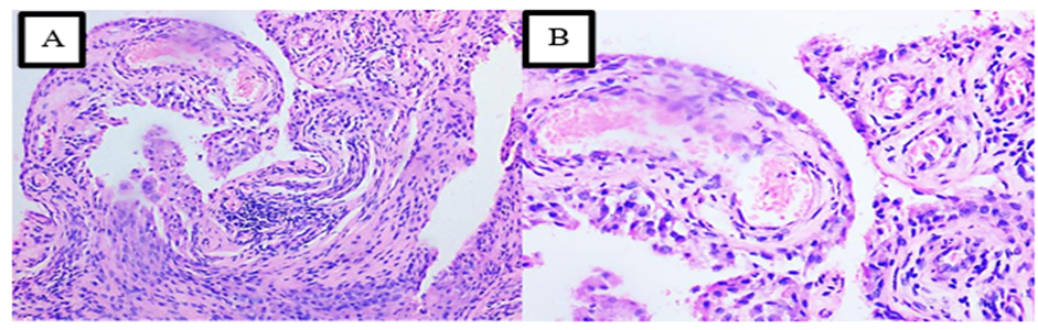
Figure 1: Chronic non-specific synovitis (H&E stain). (A) & (B) Low-power (100x) and high-power (400x) views showing synovial hyperplasia and villous hypertrophy with dense subsynovial lymphocytic infiltrate.
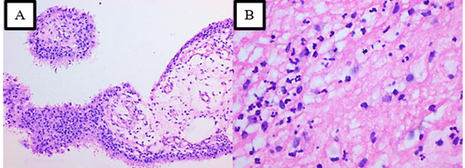
Figure 2: Septic arthritis (H&E stain). (A) Low-power (100x) view showing synovial hyperplasia and hypertrophy. (B) High-power (400x) view showing a predominantly neutrophilic infiltrate in the subsynovium.
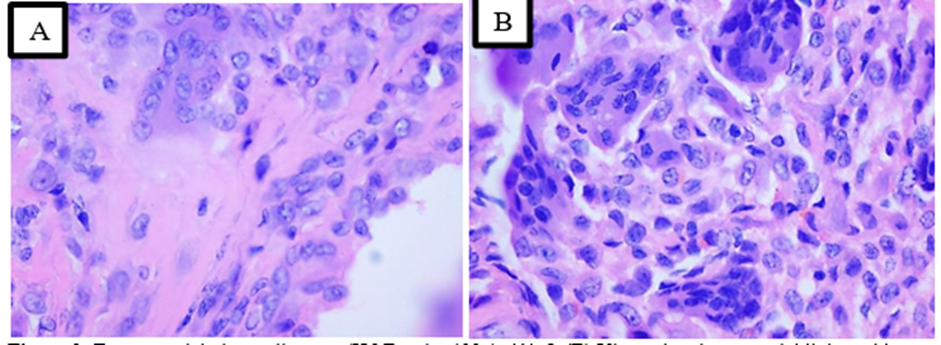
Figure 3: Tenosynovial giant cell tumor (H&E stain, 400x). (A) & (B) Views showing synovial lining with subsynovial multinucleated giant cells along with mononuclear round cells.
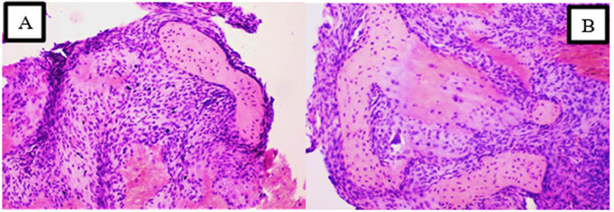
Figure 4: Synovial chondromatosis (H&E stain). (A) & (B) Low-power (200x) views showing lobules of cartilage in the synovium with focal synovial hyperplasia.
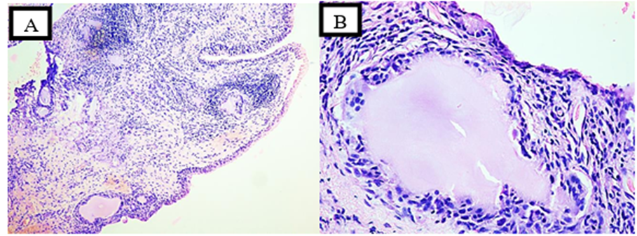
Figure 5: Gouty arthritis (H&E stain). (A) Low-power (100x) view showing synovium with multiple subsynovial areas of amorphous eosinophilic deposits. (B) High-power (400x) view showing the giant cell reaction surrounding the deposits.
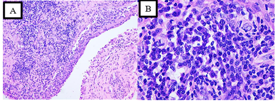
Figure 6: Rheumatoid arthritis (H&E stain). (A) Low-power (200x) view showing the papillary appearance of the synovium. (B) High-power (400x) view showing an intense lymphoplasmacytic infiltrate.
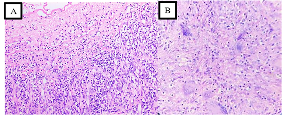
Figure 7: Tubercular arthritis (H&E stain, 100x). (A) View showing synovial lining with a dense subsynovial inflammatory infiltrate. (B) View showing aggregates of epithelioid cells with Langhans giant cells.
DISCUSSION:
The life expectancy of the general population of the country is increasing due to better living conditions and affordable health care. Hence, age related problems like arthritis are increasingly seen. [5] The unclassified arthritis, suspicion of neoplastic or granulomatous disease, deposition disease and persistence of infection in spite of negative synovial fluid culture, histopathological examination of synovial tissue will lead to correct diagnosis. [6]In the present study, 70 synovial biopsies were encountered over a period of 5 years, chronic non-specific synovitis was the commonest synovial lesion encountered comprising 31 patients (44.3%) followed by tubercular arthritis in 13 patients (18.6%), rheumatoid arthritis in 5 patients (7.1%) and septic arthritis in 4 patients (5.7%). Similar observation was seen in study by Jayanthi K.J et al. [5] Out of 204 samples 157 cases (73.36%) were non-specific synovitis, followed by tubercular arthritis in 15 cases (7%) and Vijay P.M et al. [7] were chronic non-specific synovitis was commonest (71%), followed by tubercular synovitis (18.07%).All 13 cases (18.6%) of tubercular arthritis histologically showed presence of granulomas. Caseating granulomas were seen in 4 cases (30.8%) and non-caseating granulomas in 9 cases (69.2%). Similar observation were seen in study done by Maithili et al. [8] and Jayanthi K.J et al. [5] were all the cases showed granuloma with Langhan’s type of giant cells.Aseptic arthritis due to an indolent infection is a challenge for timely diagnosis. [9]
It is mainly occurs in neonates and infants. [5] In the present study 4 cases (5.7%) of septic arthritis were encountered. Knee was the commonest joint involved with male to female ratio is 1:1. Routine laboratory investigation showed neutrophilic leukocytosis and microscopy showed predominant infiltration of neutrophils, synovial hypertrophy and hyperplasia and few lymphocytes. Similar observation were seen in a study done by Sakhuji AC et al. [10] in which 20% of septic arthritis was observed in the age group between 1-55 years with male predominance.Rheumatoid arthritis is polyarticular, symmetric and predisposed to involve the small joints of the hands and proximal fingers. [11] Diagnosis of rheumatoid arthritis was based on histopathological findings, serology for rheumatoid factor and clinical findings. In the present study 5 cases of rheumatoid arthritis were encountered constituting (7.1%). Two cases each were seen at age groups between 31-40 years and 51-60 years and one patient was at age group between 41- 50 years. In this study male preponderance noted which is not correlating with most literature studies. Possible explanation can be due to covid scenario, patients visiting to hospital were reduced. Multiple joints were commonly affected with knee was the commonest joint involved followed by wrist joint. A literature study done by Jayanthi K.J et al. [5] and Abhyankar et al. [12]
showed similar findings.In the present study, synovial hypertrophy and hyperplasia was seen in all 5 cases along with dense lymphoplasmacytic infiltrate, fibrosis and vascular proliferation and 4 cases showed lymphoid follicle formation with germinal center. Similar findings were observed in a study done by Rooney et al. [13]In this study, a total of three cases (4.28%) of tenosynovial giant cell tumors were encountered with male preponderance. Microscopically all three cases showed multinucleated giant cells admixed with round to polygonal cells arranged in lobules and in sheets in a fibrocollagenous stroma. Similar features were observed in a study done by Jayanthi K.J et al.
[5]Crystals induced arthritis includes that resulting from deposition of monosodium urate, calcium pyrophosphate dehydrate, calcium hydroxyapatite and calcium pyrophosphate in and around joints. [11] Knee is most common joint involved. In this study only one case of gouty arthritis was encountered in a male patient aged 3 years, presented with swelling, pain and stiffness of right knee joint. Microscopically showed synovial hypertrophy and hyperplasia with subsynovial large areas of amorphous eosinophilic needle like crystals surrounded by multinucleated giant cells, lymphocytes and plasma cells. Similar observations were seen in study done by Panage A et al. [2] and Gupta SJ et al. [14]In lipoma arborescence, fatty infiltration of subsynovial connective tissue is characteristically seen. [5] It possibly represents a secondary phenomenon following the degenerative process of the joint. [5, 15] In our study, there were 2 (2.85%) cases of lipoma arborescence with female predominance and microscopic features showed synovial atrophy in one case and hyperplasia in other. Subsynovial adipose tissue arranged in lobules with sparse lymphocytic infiltrate and congested blood vessels were seen in both the cases. In a literature study done by Panage et al.
[2] showed stromal replacement by adipose tissue upto synovial lining epithelium and moderate lymphoplasmocytic infiltrate.Synovial chondromatosis is a benign, typically self-limited process that may recur locally. [16] In our study 4 cases (5.71%) were encountered with female preponderance and microscopically all cases showed cartilaginous fragments with enchondral ossification in one case. Two cases showed synovial hyperplasia and hypertrophy and Lymphocytic infiltrate seen in two cases. Similar observations were seen in literature study done by Milgram JW et al. [16]Osteoarthritis is generally considered to be a non-inflammatory condition and is frequently referred to as degenerative joint disease. [2] The older publication suggest that synovial membrane is normal in osteoarthritis. [2, 15] In present study, One case of osteoarthritis was encountered in a female patient aged 75 years and histologically showed synovial hyperplasia and hypertrophy, subsynovial fat tissue and inflammatory cell infiltration mainly lymphocytes and sparse plasma cells. Similar features were observed in recent literature which showed the presence of a low grade synovitis in osteoarthritis and some cases have been described in which the histological appearance were similar to those seen in rheumatoid arthritis and chronic synovitides. [2, 17]Bursitis is a swelling or inflammation of a bursa, which is a synovium-lined, sac-like structure found throughout the body near bony prominences and between bones, muscles, tendons, and ligaments. [18]
In the present study, two cases of bursitis were encountered and microscopically both the cases showed lymphocytic infiltrate in a fibrocollagenous stroma lined by flattened epithelium. One case showed neutrophilic infiltrate which was reported as infective bursitis.In our study 3 cases (4.3%) of Baker cyst were encountered with male preponderance. Histologically all the three cases showed cyst wall consisting dense connective tissue lined by flattened epithelium with lymphocytic infiltrate. Such cystic swelling are frequently seen in cases of rheumatoid disease and are related to bursae, tendon sheath and joints, develops due to fluid exudation in to spaces lined by synovium as observed by David G. Palmer. [19]One case of traumatic arthritis was seen in present study in a male patient aged 25 years with history of trauma to the involved knee joint and histological features showed hyperplasia of synoviocytes with dense mixed inflammatory infiltrate.
CONCLUSION:
Histopathological study of synovial tissue is useful for diagnosing various inflammatory and non-inflammatory synovial conditions. [2] Synovial biopsies are indicated when only one joint is affected and helps in distinguishing various aetiologies such as infective, traumatic or crystal induced. [5] In many instances corroborating clinical, radiological, biochemical and serological examination becomes essential for making an accurate histopathological diagnosis. [7]
REFERENCES:
1. John O’Connell. Pathology of the synovium. AM J Clin Pathol. 2000;114:773-84.
2. Panage A, Cherian S, Naidu R, Abhani D. Histopathological analysis of synovial lesions. Annals Pathol Lab Med. 2018;5:276-82.
3. Abraham VT, Anand MG, Krishnagopal R. A study on the outcome of arthroscopic synovial biopsy and synovectomy in the management of synovitis of knee. Int J Res Orthop 2018 Sep;4:753-6.
4. Venkataraman M, Sathyadharan P. “Role of Needle Synovial Biopsy in Joint Diseases”. Journal of Evolution of Medical and Dental Sciences 2015;Vol.4, Issue 44, June 01; Page: 7626-7634, DOI: 10.14260/jemds/2015/117.
5. Jayanth K.J, Niranjan Gowda M.R*. Retrospective study of synovial biopsies- Tertiary centre experience. IP Archives of Cytology and Histopathology Research, April-June. 2019;4:128-32.
6. Gerlag MD, Tak PP. How to perform and analyse synovial biopsies. Best Practice & Research Clinical Rheumatology 2009;23:221-32.
7. Vijay PM, Doddikoppad MM. Clinicopathological study of Inflammatory Synovial Lesions. Int J Biol Med Res. 2011;2:882-8.
8. Maithili MK, Siddhi Gaurish Sinai Khandeparkar, Avinash R.Joshi, Sukhada R. Patil Pranoti V.Lengare. Clinicopathological study of synovial biopsies at tertiary care hospital and its diagnostic utility. Indian J Pathol Oncol 2017;4(4):595-9.
9. Legius B, Landuyt KV, Verschueren P, Westhovens R. Septic arthritis due to Staphylococcus warneri: a diagnostic challenge. Open Rheumatol J. 2012;6:310-11.
10. Sakhuja AC et al. Synovial fluid analysis and synovial biopsy in various types of arthritides. Ind J Orth 1981;15(2):157-61.
11. Hough, Aubrey J. Joints Chapter-7, in Anderson’s pathology, Vol.2, 10th Edition. Edt. Ivan, Damjanov & James Lindu, St.Louis; Mosby, 1996:2615.
12. Abhyankar SC, Vast RR, Shirdokar AB, Johari AN, Deodhar KP. A histopathological study of synovium in chronic joint diseases. Ind J Pathol Microbiol. 1987;30(1):1-5.
13. Rooney M, Condell D, Quinlan W, Daly L, Whelan A, Feighery C, Bresnihan B. Analysis of the histologic variation of synovitis in rheumatoid arthritis. Arthritis Rheum. 1988 Aug;31(8):956-63.
14. Gupta SJ. Crystal induced arthritis: An Overview. J Ind Rheumatol Assoc. 2002;10:5-15.
15. Rao S, Rajkumar A, Elizabeth MJ, Ganesan V, Kuruvilla S. Pathology of synovial lipomatosis and its clinical significance. J Lab Physicians 2011;3(2):84-8.
16. Milgram JW. Synovial osteochondromatosis: a histopathological study of thirty cases. J Bone Joint Surg Am 1977;59:792–801.
17. Inoque K, Shichikawa K, Nishioka J, Hirota S. Older age onset rheumatoid arthritis with or without osteoarthritis. Annals of the Rheumatic Diseases. 1987;46:908-11.
18. Williams CH, Jamal Z, Sternard BT. Bursitis. [Updated 2021 Aug 2]. In: StatPearls [Internet]. Treasure Island (FL): StatPearls Publishing; 2021 Jan-. Available from: https://www.ncbi.nlm.nih.gov/books/NBK513340/
19. Palmer G. David. Synovial cysts in Rheumatoid disease. Annals of internal medicine 1969:61-8.