A Study to Evaluate the Etiology in Couples Presenting to Fertility Clinic in a Tertiary Care Hospital.
- Saumya Tayal , Postgraduate Resident, Department of Obstetrics & Gynaecology, Shri Ram Murti Smarak Institute of Medical Sciences, Bareilly, Uttar Pradesh, India.
- Shashi Bala Arya , Professor & Head, Department of Obstetrics & Gynaecology, Shri Ram Murti Smarak Institute of Medical Sciences, Bareilly, Uttar Pradesh, India.
- Ruchica Goel , Assistant Professor, Department of Obstetrics & Gynaecology, Shri Ram Murti Smarak Institute of Medical Sciences, Bareilly, Uttar Pradesh, India.
- Preeti Singh , Assistant Professor, Department of Obstetrics & Gynaecology, Shri Ram Murti Smarak Institute of Medical Sciences, Bareilly, Uttar Pradesh, India.
Article Information:
Abstract:
Introduction: Infertility is a multifactorial reproductive health problem affecting both male and female partners and requires comprehensive etiological evaluation for appropriate management. Objective: To evaluate the etiological profile of infertility among couples attending a tertiary-care fertility clinic. Methodology: A hospital-based cross-sectional observational study was conducted among 313 infertile couples attending the Department of Obstetrics and Gynaecology, SRMS IMS, Bareilly, from April 2024 to September 2025. Detailed history, clinical examination, transvaginal sonography, hysterosalpingography, hysteroscopy, laparoscopy, and semen analysis were performed. Data were analyzed using descriptive statistics. Results: Primary infertility was observed in 66.77% couples, while secondary infertility accounted for 33.23%. Female-factor infertility was the most common etiology (40.65%), followed by combined male–female factors (29.03%) and only male-factor infertility was (19.35%). Ovarian and tubal pathologies were the predominant female cause for infertility. Tubal block (47%), endometriosis (44%), pelvic adhesions (52%), and uterine abnormalities (26%) were frequently identified on advanced evaluation. Abnormal semen parameters were identified in 48.39% semen samples on routine analysis, of these oligospermia and azoospermia being the commonest abnormalities. Conclusion: Infertility is predominantly multifactorial , with substantial contributions from both female and male factors. Comprehensive couple-based evaluation is essential for accurate etiological diagnosis and management. Multifactorial causes of infertility needs timely referral and special attention to specialized care and further management by ART techniques.
Keywords:
Article :
INTRODUCTION:
Procreation is one of the most classical hallmarks of living life and the inability to conceive is not simply labeled as infertile, but often interpreted as an infertility crisis due to its diverse cultural, psychological and social implications. The World Health Organization (WHO) describes infertility as a disease of the reproductive system or inability to have a clinical pregnancy after 12 months or more of frequent sexual intercourse without birth control.1 Infertility is classified into two types: Primary Infertility which occurs when a couple has not been able to achieve a successful pregnancy despite of cohabitating for 12 consecutive ovulatory cycles. Secondary Infertility refers to a delay in conception for a couple who have previously conceived, but the pregnancy may not have been successful. This examination may include cases of miscarriage, ectopic pregnancy and other pregnancy complications. As per the recent statistics around 2023, around 12-13 percent of the reproductive age couples in the United States is infertile, that is, one out of every six couples. The governments and other organizations that reported these estimates include the Centers of Disease Control and Prevention (CDC) and the American Society of Reproductive Medicine (ASRM).2
Unexplained infertility (UI) represents approximately 30 % of infertility cases in couples, according to recent epidemiological studies. The term is given when the normal diagnostics do not show any recognizable abnormalities in the reproductive systems of both partners. By definition, UI is a diagnosis of exclusion, which is made only after standard tests, including tests of ovulatory activity, tubal patency, uterine morphology, and semen analysis, have been found to be normal. However, even though it has been extensively used clinically, there is no standardized diagnostic protocol of UI, thus creating diagnostic variability.3
The International Committee of Monitoring Assisted Reproductive Technologies (ICMART) defines UI as infertility in couples with seemingly normal ovarian reserve, regular ovulatory activity, patent fallopian tubes, normal uterine and cervical morphology and normal pelvic morphology, and a satisfactory coital frequency. In the case of the male partner, the definition includes normal testicular functioning, normal anatomy of the genital tract, and normative semen parameters. More importantly, the determination of UI is inherently tied to the diagnostic modalities of a particular kind and their availability in the clinical environment. This investigation aims at assessing the clinical presentation, as well as determining the etiological factors underlying infertility in married couples who attend the outpatient department of a tertiary referral centre.
OBJECTIVE
To evaluate the etiology in couples presenting to fertility clinic in a tertiary care hospital.
METHODOLOGY:
A hospital-based cross-sectional observational study was conducted in the Department of Obstetrics and Gynaecology at Shri Ram Murti Smarak Institute of Medical Sciences (SRMS IMS), Bareilly, Uttar Pradesh, over a period of 18 months from 1 April 2024 to 30 September 2025. The study included couples attending the infertility clinic who fulfilled the inclusion criteria, including females aged 21–45 years and male partners aged 25–50 years diagnosed with either primary or secondary infertility. A minimum sample size of 303 participants was calculated using the standard prevalence-based formula with a 20% attrition rate. Data were collected using a semi-structured, pre-designed, and pre-tested case information proforma that included sociodemographic details, reproductive and obstetric history, medical and surgical history, lifestyle factors, clinical examination findings, and investigation records for both partners.
Comprehensive clinical evaluation and investigations were performed for both partners, including hormonal assays, imaging studies, semen analysis as per WHO 5th edition guidelines, and endoscopic procedures whenever indicated. Based on clinical and investigative findings, infertility was categorized into female factor, male factor, combined factor, or unexplained infertility. Ethical approval was obtained from the Institutional Ethics Committee of SRMS IMS, Bareilly, and written informed consent was taken from all participants. A pilot study was conducted on 10% of the sample to assess feasibility and reliability of the study tool. Data were entered into Microsoft Excel and analysed using SPSS version 23.0, with descriptive statistics such as mean, standard deviation, frequencies, and percentages used for data interpretation and presentation through tables, graphs, and charts.
RESULTS:
A total of 313 infertile couples fulfilling the inclusion criteria were enrolled in the present hospital-based cross-sectional observational study conducted at the Department of Obstetrics and Gynaecology, SRMS IMS, Bareilly. Detailed clinical evaluation and relevant investigations including ultrasonography, hysterosalpingography, hysteroscopy, laparoscopy, and semen analysis were performed to determine the etiological profile of infertility.
The majority of female partners belonged to the 26–30 years age group (37.38%), whereas most male partners were aged 31–35 years (33.23%). Duration of infertility of 3–5 years was most commonly observed in 40.89% couples. With respect to body mass index, most female and male partners had normal BMI, although overweight status was also frequently observed among both groups. Baseline demographic and clinical characteristics of infertile couples are summarized in (Table 1). Primary infertility constituted the predominant type of infertility, accounting for 66.77% cases, while secondary infertility was observed in 33.23% women (Figure 1). Clinical evaluation among female partners revealed adnexal pathology in 45.69% and uterine abnormalities in 42.49% women. Lower genital tract infection and pelvic inflammatory disease were identified in 38.98% cases. Transvaginal sonography demonstrated normal pelvic findings in 46.96% women, while polycystic ovaries represented the most common abnormality (20.77%), followed by ovarian cysts (10.54%) and fibroid uterus (8.63%). Endometrial thickness assessment showed normal or optimal endometrium in 72.84% women. Hysterosalpingography findings revealed normal tubal patency in 53.33% women, whereas unilateral and bilateral tubal block were detected in 21.90% and 24.76% cases, respectively (Table 2).
Among the 63 women who underwent hysteroscopic and laparoscopic evaluation, bilateral tubal ostial patency was observed in 63.49%, while unilateral and bilateral ostial block were noted in 26.98% and 9.52% women, respectively. Intrauterine adhesions, submucous fibroids, and endometrial polyps were identified in 26.98%, 23.81%, and 15.87% women, respectively. Laparoscopic findings demonstrated left-sided tubal abnormalities in 44.44% and right-sided abnormalities in 36.51% women. Tubal block, hydrosalpinx, endometriosis, and pelvic adhesions were also frequently observed, indicating a substantial burden of tuboperitoneal pathology among infertile women (Table 3). Semen analysis among male partners demonstrated normozoospermia in 51.61% participants, while abnormal semen parameters were observed in 48.39% men. Oligospermia was the most common abnormality (20.65%), followed by azoospermia (13.55%) and oligoasthenoteratozoospermia (9.03%) (Figure 2).
Evaluation of the overall etiological profile revealed that multiple concurrent causes of infertility were present in 60.65% couples, whereas unexplained infertility accounted for 21.94% cases. Female factor infertility alone was the most common etiological category (40.65%), followed by combined male and female factors (29.03%) and male factor infertility alone (19.35%). Among female etiological causes, ovarian and tubal factors represented major contributors to infertility. Nearly half of the evaluated male partners demonstrated abnormal semen parameters (Table 4, Figure 3).
Table 1. Baseline Demographic and Clinical Characteristics of Infertile Couples (N = 313)
|
Parameter |
Category |
Frequency |
Percentage (%) |
|
Female Age Group (years) |
20–25 |
78 |
24.92 |
|
26–30 |
117 |
37.38 |
|
|
31–35 |
76 |
24.28 |
|
|
36–40 |
30 |
9.58 |
|
|
41–45 |
12 |
3.83 |
|
|
Male Age Group (years) |
25–30 |
63 |
20.13 |
|
31–35 |
104 |
33.23 |
|
|
36–40 |
91 |
29.07 |
|
|
41–45 |
42 |
13.42 |
|
|
46–50 |
13 |
4.15 |
|
|
Duration of Infertility (years) |
1–2 |
108 |
34.50 |
|
3–5 |
128 |
40.89 |
|
|
6–10 |
60 |
19.17 |
|
|
>10 |
17 |
5.43 |
|
|
Female BMI Category (kg/m²) |
Underweight (<18.5) |
42 |
13.42 |
|
Normal (18.5–24.9) |
139 |
44.41 |
|
|
Overweight (25–29.9) |
97 |
30.99 |
|
|
Obese (≥30) |
35 |
11.18 |
|
|
Male BMI Category (kg/m²) |
Underweight (<18.5) |
39 |
12.46 |
|
Normal (18.5–24.9) |
119 |
38.02 |
|
|
Overweight (25–29.9) |
113 |
36.10 |
|
|
Obese (≥30) |
42 |
13.42 |
Table 2. Clinical, Ultrasonographic, and Tubal Evaluation among Female Partners (N = 313)
|
Parameter |
Category |
Frequency |
Percentage(%) |
|
Clinical Findings |
Lower genital tract infection / PID |
122 |
38.98 |
|
Uterine abnormalities (size/position) |
133 |
42.49 |
|
|
Adnexal pathology |
143 |
45.69 |
|
|
Transvaginal Sonography (TVS) Findings |
Normal pelvis |
147 |
46.96 |
|
Polycystic ovaries (PCO) |
65 |
20.77 |
|
|
Ovarian cyst |
33 |
10.54 |
|
|
Fibroid uterus |
27 |
8.63 |
|
|
Uterine abnormality (size/position) |
25 |
7.99 |
|
|
Septate uterus |
9 |
2.88 |
|
|
Bicornuate uterus |
5 |
1.60 |
|
|
Unicornuate uterus |
2 |
0.64 |
|
|
Endometrial Thickness (ET) Groups |
Thin endometrium |
31 |
9.90 |
|
Normal/optimal endometrium |
228 |
72.84 |
|
|
Thick endometrium |
54 |
17.25 |
|
|
HSG Findings (n = 105) |
Normal uterine cavity |
77 |
73.33 |
|
Abnormal uterine cavity |
28 |
26.67 |
|
|
Normal tubal patency |
56 |
53.33 |
|
|
Unilateral tubal block |
23 |
21.90 |
|
|
Bilateral tubal block |
26 |
24.76 |
Table 3. Hysteroscopic and Laparoscopic Findings among Female Partners (N = 63)
|
Parameter |
Category |
Frequency |
Percentage (%) |
|
Hysteroscopic Findings |
Both ostia open |
40 |
63.49 |
|
Unilateral ostial block |
17 |
26.98 |
|
|
Bilateral ostial block |
6 |
9.52 |
|
|
Abnormal endometrium |
12 |
19.05 |
|
|
Intrauterine adhesions |
17 |
26.98 |
|
|
Endometrial polyps |
10 |
15.87 |
|
|
Submucous fibroids |
15 |
23.81 |
|
|
Septate uterus |
3 |
4.76 |
|
|
Laparoscopic Findings |
Right tubal abnormality |
23 |
36.51 |
|
Left tubal abnormality |
28 |
44.44 |
|
|
Hydrosalpinx (right) |
13 |
20.63 |
|
|
Hydrosalpinx (left) |
12 |
19.05 |
|
|
Distal tubal block |
20 |
31.75 |
|
|
Proximal tubal block |
13 |
20.63 |
|
|
Endometriosis Stage I–II |
18 |
28.57 |
|
|
Endometriosis Stage III–IV |
10 |
15.87 |
|
|
Pelvic adhesions |
33 |
52.38 |
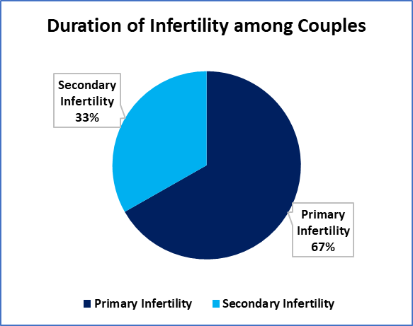
Figure 1: Distribution of Female Infertility Types
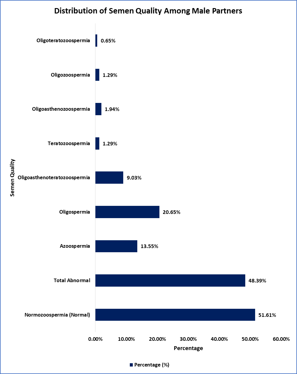
Figure 2: Distribution of Semen Quality Among Male Partners
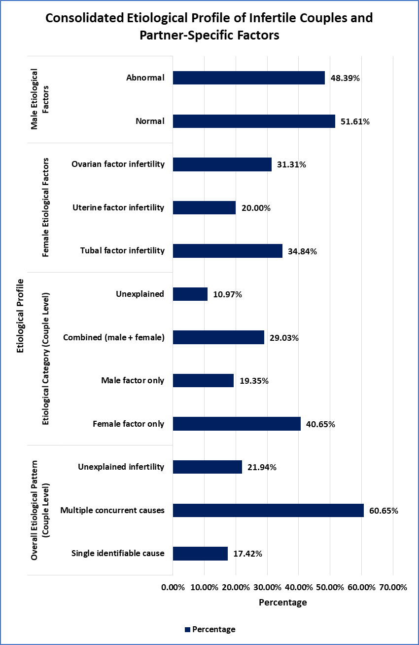
Figure 3: Consolidated Etiological Profile of Infertile Couples and Partner-Specific Factors
DISCUSSION:
Infertility is a multifactorial reproductive health condition requiring comprehensive evaluation of both partners. The present tertiary care hospital-based study assessed the etiological profile of infertility using clinical examination, imaging, endoscopic procedures, and semen analysis, providing an integrated overview of female, male, combined, and unexplained infertility factors in North Indian couples.
In the current cohort, primary infertility (66.77%) was approximately twice as common as secondary infertility (33.23%), indicating that most women presented without any prior conception. This distribution is closely comparable to several clinic-based studies: Singh et al. reported a predominance of primary infertility (nearly two-thirds).4 Similarly, Taha et al. observed a higher proportion of primary than secondary infertility.5 In contrast, Pal et al., a community-based prevalence study, documented relatively greater representation of secondary infertility compared with primary in their screened population, which can be justified by fundamental differences in study setting: community screening captures women who have conceived previously but later develop tubal damage, post-abortal/postpartum infections, or other secondary causes, whereas tertiary infertility clinics often see a higher fraction of primary infertility due to earlier referral for “no conception since marriage.”6Zargar et al. highlighted substantial primary infertility in a community survey context but with a clinical evaluation subset, suggesting that both community prevalence and clinic attendance can show high primary infertility depending on local sociocultural patterns and the timing of care-seeking.7
Hysterosalpingography demonstrated normal uterine cavity in 73.33% and abnormal cavityin26.67% of evaluated women, while tubal patency was normal in53.33%, with unilateral block in 21.90%andbilateral block in 24.76%. These findings are strongly comparable to those reported by Singh et al., who documented unilateral and bilateral tubal block in a substantial proportion of infertile women, and align with Zargar et al., where tubal factors were among the most frequent female etiologies.4,8 In contrast, studies such as Masoumi et al. reported a higher emphasis on ovulatory disorders, which may reflect population differences or earlier-stage evaluation.9 The relatively high prevalence of bilateral tubal block in the present study can be justified by tertiary-care referral patterns, as women with suspected or previously treated pelvic infections are more likely to undergo HSG.
Hysteroscopic assessment in the current study revealed that 63.49% had both tubal ostia open, some have unilateral (26.98%) or bilateral ostial block (9.52%), showing intrauterine-tubal junction pathology that may not be fully seen on HSG alone. Intrauterine adhesions were detected in 26.98%, endometrial polyps in 15.87% and submucous fibroids in 23.81%. These results are in agreement with clinic-based etiological data published by Singh et al. and Taha et al., in which uterine factors were significant contributors to infertility, but those were not systematic hysteroscopic stratification.4,5 Conversely, community-based studies, includingPal et al. and Zargar et al. mentioned uterine factors in a more generalized way without direct visualization, which can be underestimated as intracavitary pathology.6,8 The increased rate of detection in the current study can be explained by the fact that the hysteroscopy was specifically used in women who had abnormal screening tests, which is why it has an added value of diagnostic data and therapeutic value in tertiary infertility cases.
Diagnostic laparoscopy showed tubal abnormalities affecting 36.51% (right) and 44.44% (left) tubes and tubal block in 52.38% of women (distal more common). Endometriosis was identified in 44.44%, including 15.87% with advanced (Stage III-IV) disease, and pelvic adhesions in 52.38%, reflecting extensive pelvic pathology. These results are rather similar to the focus on tubal and peritoneal factors described by Zargar et al. and Singh et al., where tubal disease constituted a major female etiology.4,8 In contrast, studies such as Masoumi et al. reported a larger percentage of ovulatory causes which could be due to earlier assessment or different population demographics.9 The increased prevalence of advanced disease in the current cohort is likely to be due to tertiary referral bias and late presentation, which supports laparoscopy as the gold standard to make a conclusive diagnosis of tuboperitoneal infertility.
Semen analysis in present study revealed that 48.39% of male partners had abnormal semen parameters, with oligospermia (20.65%) and azoospermia (13.55%) being the most frequent defects. This distribution is similar to that of male-factor contributions reported by Umar et al., Awodele et al., and Jehan et al., who all reported that about half to three-quarters of infertile men have at least one abnormal semen parameter, with oligospermia and azoospermia being the most common.10–12 The slightly reduced rate of abnormalities in the current study relative to the male-only infertility clinic cohorts can be explained by the couple-based design, in which men are tested in the presence of female partners as opposed to being pre-selected on the basis of the suspected male factor infertility. These results support the idea that male factors play significant roles, either individually or in combination, to infertility and emphasize the importance of regular semen analysis as an inseparable part of overall couple assessment.
The consolidated etiological assessment revealed that multiple concurrent causes (60.65%) were more common than single isolated factors (17.42%), while unexplained infertility accounted for 21.94% of couples. Female factors alone contributed to infertility in 40.65%, male factors alone in 19.35%, and combined male–female factors in 29.03% of cases. This trend is very similar to the etiologic distributions of Singh et al. and Zargar et al., in which mixed and multifactorial infertility was common, and is consistent with the findings of Hull et al., who noted that infertility is often caused by multiple defects and not by just one.4,8,13 Conversely, other studies with a male focus like Awodele et al. and Jehan et al. found the prevalence of apparent male-factor to be higher, as it should be since their study designs were based on male infertility clinics.11,12 The reduced percentage of unexplained infertility in the current study relative to certain previous studies can be explained by the wide application of high-technological diagnostic methods, such as TVS, HSG, hysteroscopy, and laparoscopy, which increase etiologic diagnosis. These results support the need to assess both partners as a whole and avoid the tendency to infertility in one partner without proper research.
CONCLUSION:
This hospital-based cross-sectional study evaluated the etiological profile of infertility among couples attending a tertiary-care fertility clinic. Female-factor infertility was the most common cause, followed by combined male–female factors, male-factor infertility, and unexplained infertility, highlighting the multifactorial nature of infertility. Ovulatory dysfunction, tubal pathology, and uterine abnormalities were major female etiological factors, while advanced investigations frequently detected endometriosis, pelvic adhesions, and tubal block.
Male-factor infertility also contributed substantially, with abnormal semen parameters such as oligospermia, azoospermia, and oligoasthenoteratozoospermia commonly observed. The high proportion of combined etiologies emphasizes the importance of comprehensive couple-based infertility evaluation.
REFERENCES:
1. WHO. Infertility. Fact Sheets. World Health Organization: Geneva; n.d. Available from: https://www.who.int/news-room/fact-sheets/detail/infertility [Last accessed: 1/13/2026].
2. Chandra A, Copen Ce, Stephen Eh. Infertility and impaired fecundity in the United States, 1982-2010: data from the National Survey of Family Growth. Natl Health Stat Rep 2013;67(2):12–18.
3. Ciacchini R, Piarulli A, Bottai B, et al. Initial Psychological Evaluation in Couples with Unexplained Infertility: Focusing on Gender Differences. Psychol Int 2025;7(3):63.
4. Singh K, Kumari R, Ranjan A, et al. Analysis of causes and clinical pattern of infertility in couples coming to a tertiary care centre in Bihar, India. Int J Reprod Contracept Obstet Gynecol 2017;6(6):2279.
5. Taha A, Rashid K. Etiology of infertility in couples attending maternity hospital in Erbil. Zanco J Med Sci 2013;17(1):322–330.
6. Pal M, Devgun P, Chalana H, et al. A study of prevalence and socio-demographic profile of infertile couples in field practice area of a tertiary care centre, Amritsar, Punjab, India. Int J Community Med Public Health 2016;1472–1476.
7. Zargar AH, Wani AI, Masoodi SR, et al. Epidemiologic and etiologic aspects of primary infertility in the Kashmir region of India. Fertil Steril 1997;68(4):637–643.
8. Zargar MH, Ahmad F, Lateef M, et al. Understanding Male Infertility for Promising ART. In: IntechOpen eBooks IntechOpen; 2021.
9. Masoumi SZ, Yavangi M. An epidemiologic survey on the causes of infertility in patients referred to infertility center in Fatemieh Hospital in Hamadan. n.d.
10. Umar AG, Panti AA, Mbakwe M, et al. The Pattern of Seminal Fluid Analysis among Male Partners Attending an Infertility Clinic in a Nigerian Tertiary Health Institution. Open J Obstet Gynecol 2020;10(07):957–967.
11. Awodele K, Adeyemo SC, Olabode ED, et al. Major Etiologies of Male Infertility Among Couples Attending Fertility Clinics in Osun State, Nigeria: Findings From a Mixed‐Method Study. Raza F. ed. Int J Clin Pract 2025;2025(1):7884777.
12. Jehan S, Khan A, Riaz M. Etiological spectrum and contributing factors of male infertility among Pakistani men. Pak J Med Sci 2025;41(12):3493–3497.
13. Hull MGR, Glazener CMA, Kelly NJ, et al. Population study of causes, treatment, and outcome of infertility. Br Med J 1985;291(6510):1693–1697.