Phenotypic Characterisation And Antifungal Susceptibility Testing Of Candida Species Isolated From Various Samples In A Tertiary Care Hospital.
- Manish Yadav , PG final Year Department of Microbiology Raipur institute of medical sciences Raipur (CG)
- Sanchita Nihal , Professor Department of Microbiology Raipur institute of medical sciences Raipur (CG)
- Vivek Kumar , Associate Professor Department of Microbiology Raipur institute of medical sciences Raipur (CG).
- Aakash Sharma , PG final Year Department of Microbiology Raipur institute of medical sciences Raipur (CG)
Article Information:
Abstract:
Background: Candida is a major genus of opportunistic pathogenic yeasts, and its epidemiological profile is currently shifting.Non-albicans Candida species and antifungal drug resistance have become increasingly prominent issues.Since distinct Candida species differ in virulence, intrinsic drug susceptibility, and biofilm-associated persistence capacity, accurate species identification and antifungal susceptibility testing are critically important. Research Objectives: Isolate and perform phenotypic characterization of Candida strains from various clinical specimens, summarize their distribution across demographic groups, hospital departments, and specimen types, evaluate their antifungal susceptibility and selected virulence factors, and compare the performance of traditional identification methods and the automated VITEK 2 system. Materials and Methods: This prospective study was conducted over 18 months at the Department of Microbiology, Raipur Medical College. A pre-set sample size of 410 cases was used, and 9 types of clinical specimens including blood and urine were enrolled. A total of 11 phenotypic assays such as Gram staining were applied, the disk diffusion method and VITEK 2 system were used for susceptibility testing, three categories of virulence factors were assessed, and descriptive statistical analysis was adopted. Results: Among the 410 test records included in this study, there were 260 males (63.41%) and 150 females (36.59%). The age group of 21~30 years had the largest number of cases, with 109 cases (26.59%). The proportions of samples submitted from outpatient departments, inpatient wards, and ICUs were 44.15%, 33.21%, and 22.64% in sequence. There were 145 records of Candida albicans and 265 records of non-albicans Candida, and urine and blood were the most common sample sources. Among the 145 isolated strains, Candida albicans accounted for the largest share. For virulence factor detection, protease was detected in 98 strains. The drug susceptibility rate data had a non-complementary issue that required traceability verification. Conclusion: Non-albicans Candida is the dominant type of isolated strains. Phenotypic identification combined with antifungal susceptibility testing still has clinical value, but rare strains and strains with uncertain classification must be verified before their test results are applied in clinical practice.
Keywords:
Article :
INTRODUCTION:
Candida is a group of symbiotic yeast that colonize the human oral cavity, gastrointestinal tract, skin, and urogenital tract. They only cause disease when the host’s defense capability, microbial ecology, or epithelial barrier is damaged, and can trigger a series of conditions ranging from local mucocutaneous lesions to candidemia and disseminated infections. Exposure to broad-spectrum antimicrobial agents, placement of various invasive catheters, ICU admission, major surgery, parenteral nutrition, malignant tumors, organ transplantation, and immunosuppressive conditions all greatly increase the risk of infection. For invasive candidiasis, delayed identification or inappropriate initial treatment will significantly worsen patient outcomes.
The World Health Organization (WHO) has classified Candida albicans and Candida auris as critical-priority pathogens, the highest priority level, and called for strengthening laboratory capacity, surveillance, and research, as well as expanding access to fair and affordable diagnostic and treatment options. Currently, the epidemiology of global Candida bloodstream infections is constantly changing, and drug resistance continues to rise. Local laboratory data are critical to optimizing antimicrobial stewardship and implementing effective infection prevention and control measures1[3].
For a long time, Candida albicans was the main pathogenic species of the Candida genus. It can complete colonization, invasion, and immune evasion through mechanisms such as adhesins, invasins, and biofilm formation [4,5]. In recent years, the proportion of clinically isolated non-albicans Candida species has kept rising. These species all have distinct characteristics: Candida tropicalis, Candida glabrata (with reduced azole susceptibility), Candida parapsilosis (linked to the transmission of in-hospital intravascular devices), and Candida krusei (naturally resistant to fluconazole) [6-8]. Furthermore, the multidrug-resistant Candida auris has highlighted the urgent need for accurate pathogen identification. Species identification is far from a trivial taxonomic matter; it directly affects the selection of clinical treatment regimens.
Our lab adopts a hierarchical fungal identification strategy. In resource-limited settings, initial screening is first performed using traditional methods, which include colony morphology observation on Sabouraud dextrose agar, Gram staining, and other related techniques. CHROMagar can achieve rapid species differentiation, but traditional methods are prone to misjudging rare closely related species, and their results are easily interfered with by multiple factors. The automated platform VITEK 2 can shorten turnaround time and cover most common yeast species, but abnormal results need to be cross-checked with mass spectrometry or sequencing [9-12]. Ultimately, a practical algorithm that integrates screening, confirmation, and drug susceptibility testing must be established.
Fungal drug susceptibility testing is central to managing drug resistance and pathogenic risks. Drug resistance can be divided into three categories: intrinsic natural resistance, resistance acquired during treatment, and resistance generated by prior selection pressure from antifungal exposure. With reference to the standardized broth microdilution method, the disk diffusion method and automated systems can only be applied if they meet general standards. Species names cannot cover all pathogenic characteristics; biofilms and various virulence factors can increase infection risks. In clinical settings, test results must be combined with clinical information. This study was adapted from a dissertation. A prospective study was conducted over 18 months in Raipur, which integrated multi-dimensional analysis and transparently disclosed inconsistencies in internal data.
Objectives
This study set clear primary and secondary research objectives, and states in advance the statistical limitations of this study. The primary objectives are: first, to isolate and classify Candida strains from clinical samples collected across multiple departments, and distinguish Candida albicans from non-albicans Candida species using traditional phenotypic methods; second, to organize and record data by sex, age, campus, and sample type, and establish the local epidemiological characteristics of Candida detection in a tertiary care setting.
The secondary objectivesinclude testing antifungal susceptibility and assessing phenotypic virulence traits, with species identification and drug susceptibility tests performed using both the automated VITEK 2 system and manual methods. Limited by the fact that the original thesis did not provide consistency data at the paired isolate level, this study only reports methodological findings in a descriptive manner, and did not calculate sensitivity, specificity, kappa values, or other indicators for categorical consistency.
MATERIALS AND METHODS:
Study Design, Setting, and Population: This 18-month prospective laboratory study was conducted in the Department of Microbiology at RIMS, Raipur, to screen for Candida isolates in specimens submitted from outpatient departments, inpatient wards, and the intensive care unit (ICU). The sample size was calculated using the formula n=4pq/e². With a assumed prevalence of 30% and an allowable error of 4.5 percentage points, the calculated sample size was 410. Demographic data and specimen records for all 410 cases were included, but only 145 isolated strains entered genotyping, phenotyping, and virulence testing. The denominator for each study stage was not forced to be unified, to avoid erroneous calibration that lacked support from raw original data.
Inclusion Criteria
All Candida strains isolated from specimens including blood, urine, bronchoalveolar lavage fluid (BAL), pus, swabs of wound exudate, high vaginal swabs, catheter tips, and umbilical cord tips were included. All isolates were required to be verified by a second specimen collected from the same patient, and have complete, classifiable records.
Exclusion Criteria
This study only included biological samples belonging to the Candida genus. All other non-Candida organisms, cultures that did not contain Candida, contaminated or inactive isolates, and records with insufficient information to support the analysis of pre-set characteristics were excluded, and were not eligible for species-specific analysis. The original doctoral dissertation did not provide an algorithm for deduplicating patient-level data, so this study does not assume that each record corresponds to a unique incident of disease in an individual patient.
Data Collection Process
Smears were stained using Gram stain. Microscopic examination detected Gram-positive oval budding yeast cells, which could be present with or without pseudohyphae. Suspected yeast isolates were inoculated onto Sabouraud dextrose agar containing chloramphenicol, and incubated at approximately 25°C for 48 to 72 hours. For the germ tube test, pooled human serum was used, and samples were incubated at 35–37°C for 2 to 3 hours. Cornmeal agar was used for microscopic examination of chlamydospores, blastospores, and pseudohyphae at 27–30°C. Putative species were identified based on colony color using HiMediaCHROMagar, with Candida albicans ATCC 10231 used as the quality control strain. Additional tests were conducted to assess metabolic patterns: carbohydrate assimilation testing on yeast nitrogen base agar, and fermentation testing in indicator broth with Durham tubes.
Phenotypic information was supplemented by the urease test, nitrate utilization test, and 45°C growth test. For identification using the VITEK 2 system, colonies from an overnight culture were resuspended in 0.45% sterile saline, and the McFarland turbidity was adjusted to approximately 2.0 using a DensiChek. After loading the identification card, the system automatically incubated the sample and read the results. For automated antimicrobial susceptibility testing, the inoculum turbidity was adjusted to a McFarland value of 1.8–2.2. For the manual Kirby-Bauer disk diffusion method, Mueller-Hinton agar containing 2% glucose and methylene blue was used. The tested drugs included fluconazole, voriconazole, amphotericin B, ketoconazole, itraconazole, miconazole, nystatin, and clotrimazole. The results of tests for phospholipase, protease, and hemolysin were also recorded. The original study only mentioned biofilms but did not provide a complete data set, so this study could not reconstruct the prevalence of biofilms.
Statistical Data Analysis
Categorical data are summarized with frequencies and percentages. Percentages were recalculated only when both the numerator and denominator were explicitly stated; otherwise, the numerical values reported in the original dissertations were retained. Due to the inaccessibility of patient-level data, uncertainty estimates, and complete paired observations for conventional testing and VITEK testing, this study did not replicate inferential statistical tests. We identified two inconsistencies in the original data: the specimen spreadsheet categorized 410 records into 145 Candida albicans isolates and 265 non-Candida albicans isolates, but the strain species spreadsheet recorded that only 59 of the 145 isolates were Candida albicans; in addition, the sum of susceptibility rates and resistance rates for some antimicrobial agents did not reach 100%. For these reasons, the antimicrobial susceptibility data are only reported as they were categorized in the source materials, and they may not be converted into count data until the main dataset is audited.
RESULTS:
A total of 410 records were included in this study for demographic analysis. Among these records, 260 cases were male, accounting for 63.41%, and 150 cases were female, accounting for 36.59%, with a sex ratio of approximately 1.73:1. The age group with the largest proportion was 21–30 years old, which included 109 cases (26.59%). This was followed by the 31–40 years old and 41–50 years old groups, each with 80 cases, both accounting for 19.51%. Of the 265 non-Candida albicans isolates recorded in the original paper, 117 were from outpatient settings, 88 from inpatient wards, and 60 from the intensive care unit (ICU). The corresponding counts of Candida albicans isolates across these settings were 66, 46, and 33, summing to 145 cases. The proportion of Candida albicans isolates was roughly similar across all care settings.
Table 1: Demographic and hospital-location distribution
|
Characteristic |
Category |
n |
% |
|
Sex |
Male |
260 |
63.41 |
|
Sex |
Female |
150 |
36.59 |
|
Age |
0–10 |
20 |
4.88 |
|
Age |
11–20 |
72 |
17.56 |
|
Age |
21–30 |
109 |
26.59 |
|
Age |
31–40 |
80 |
19.51 |
|
Age |
41–50 |
80 |
19.51 |
|
Age |
51–60 |
20 |
4.88 |
|
Age |
61–70 |
19 |
4.63 |
|
Age |
71–80 |
10 |
2.44 |
|
Location* |
ICU |
60 |
22.64 |
|
Location* |
IPD |
88 |
33.21 |
|
Location* |
OPD |
117 |
44.15 |
*Location percentages use the thesis denominator of 265; corresponding albicans-category counts were 33, 46 and 66.
The clinical fungal test samples included in this study are categorized into 8 types, ranked by their proportion as urine, blood, pus, airway-related samples, and others. The proportions of Candida albicans and non-Candida albicans strains vary across these sample types. Additionally, among the 145 independently isolated strains, Candida albicans accounts for 40.69%, while all non-Candida albicans strains together make up 59.31%, with the last three types being the dominant non-Candida albicans strains.
Table 2: Specimen-wise distribution reported in the thesis
|
Specimen |
Albicans |
NAC |
Total |
|
Blood |
37 |
56 |
93 |
|
BAL |
15 |
23 |
38 |
|
Pus |
22 |
13 |
35 |
|
Urine |
18 |
89 |
107 |
|
HVS |
11 |
14 |
25 |
|
Body fluid |
3 |
5 |
8 |
|
Eye discharge |
0 |
0 |
0 |
|
Ear discharge |
2 |
0 |
2 |
|
Nail |
2 |
5 |
7 |
|
Catheter tip |
5 |
12 |
17 |
|
Umbilical tip |
5 |
12 |
17 |
|
Endotracheal tube |
7 |
24 |
31 |
|
Others |
18 |
12 |
30 |
|
Total |
145 |
265 |
410 |
Table 3: Species distribution in the separately reported 145-isolate set
|
Species |
n |
% |
|
C. albicans |
59 |
40.69 |
|
C. tropicalis |
38 |
26.21 |
|
C. glabrata |
18 |
12.41 |
|
C. parapsilosis |
14 |
9.66 |
|
C. krusei |
5 |
3.45 |
|
C. rugosa |
4 |
2.76 |
|
C. haemulonii |
3 |
2.07 |
|
C. guilliermondii |
2 |
1.38 |
|
C. famata |
1 |
0.69 |
|
C. lusitaniae |
1 |
0.69 |
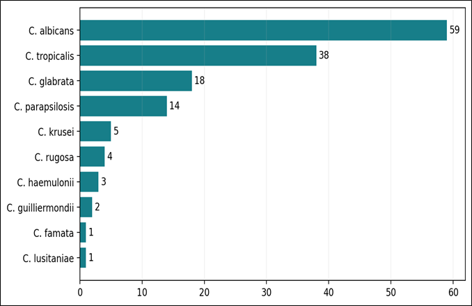
Figure 1: Distribution of Candida species (n=145)
The 145 fungal samples included in this study completed three types of tests: the consistency of phenotypic identification met the standard, and the biochemical tests complied with national standards; in the drug susceptibility test, fluconazole had a susceptibility rate of 89% but presented a contradiction with a reported drug resistance rate of 60%; the detection rate of virulence factors ranged from 60.69% to 67.59%.
Table 4: Antifungal susceptibility values reproduced from the thesis
|
Agent |
Sensitivity |
Resistance |
|
Fluconazole |
89% |
60% |
|
Voriconazole |
47% |
52% |
|
Amphotericin B |
62% |
35% |
|
Ketoconazole |
86% |
24% |
|
Itraconazole |
45% |
21% |
|
Miconazole |
30% |
12% |
|
Nystatin |
95% |
15% |
|
Clotrimazole |
97% |
11% |
Note: categories are non-complementary and require verification; they must not be interpreted as mutually exclusive outcomes.
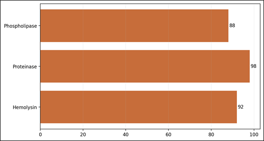
Figure 2: Activity of selected virulence factors among 145 isolates
DISCUSSION:
This study confirms that Candida species are widely distributed in clinical settings, and the clinical significance of non-albicans Candida species deserves attention. Among the 145 strains isolated in this study, Candida albicans was the strain with the highest detection rate among single species, but the total proportion of non-albicans Candida species reached 59.31%. The top three non-albicans Candida species with the highest detection rates were Candida tropicalis, Candida glabrata, and Candida parapsilosis. This trend is consistent with the conclusion from previous studies that the Candida population is shifting from being dominated by Candida albicans to a heterogeneous distribution. A study from a tertiary care institution in India also recorded the high proportion of non-albicans Candida species, while noting differences across regions and institutions. This study did not set clinical judgment variables, and only reported the strain detection patterns, so it cannot represent the incidence of invasive candidiasis.
The germ tube test, CHROMagar, and cornmeal agar show excellent apparent performance and can be integrated into stepwise fungal identification workflows. In this study, 144 out of 145 samples tested positive for the germ tube test, and all samples presented characteristic chromogenic phenotypes, meaning these methods have value for initial screening. While conventional and automated systems can identify most common yeast species, previous research has confirmed their limitations in detecting rare species. The four rare Candida species identified in this study were difficult to distinguish due to overlapping biochemical characteristics and incomplete system databases. Cases presenting abnormal phenotypes require rechecking with updated MALDI-TOF or sequencing. This study did not match manual and VITEK system results, so consistency could not be calculated. Future research needs to collect complete relevant data to measure consistency and other indicators.
Although existing conclusions on drug susceptibility and virulence have clinical relevance, their interpretation and application still require caution. The original doctoral thesis proposed that clotrimazole and nystatin have high activity, and there is heterogeneity in response to systemic azole drugs. However, its drug susceptibility data contain logical contradictions, which may stem from transcription errors, inconsistent statistical denominators, the existence of intermediate drug susceptibility categories, and mixed use of research methods. Furthermore, the data failed to meet the standard that drug susceptibility breakpoints must match the specific bacterial species, drug, and detection method used. Unaudited data must never be used to guide clinical treatment. The virulence data in the original thesis are internally consistent: about two-thirds of the strains express three types of virulence factors. Combined with published studies, the expression levels of these factors vary. The activity of these factors alone cannot be used to determine the severity of an infection; instead, five types of indicators must be combined to optimize the value of their interpretation.
The authors of this paper propose that all demographic and service area distribution data must be interpreted in combination with contextual settings to avoid misinterpretation when separated from their background. The observed findings of a high proportion of male cases and a concentration of cases among working-age adults cannot be directly interpreted as evidence of differences in biological susceptibility; these patterns must be linked to background factors such as care-seeking patterns and occupational exposure. Similarly, the fact that outpatient cases account for the largest proportion cannot be misinterpreted to mean that candidiasis in outpatient settings is more severe than cases in intensive care units (ICUs). Due to the lack of denominator data on the number of patients tested and culture samples collected across different departments, it is impossible to calculate setting-specific prevalence rates. For future surveillance, it is necessary to collect denominator data such as the number of hospital admissions and device-days to calculate standardized infection rates. It is also essential to categorize cases into three groups: community-onset cases, healthcare-associated onset cases, and ICU-acquired onset cases, to ensure that data can be compared across time and locations.
From an infection stewardship perspective, the core principle for the diagnosis and treatment of Candida infections is to combine diagnostic accuracy with therapeutic restraint. Detection of this fungus in non-sterile sites does not require automatic initiation of antifungal therapy, while detection of yeast in the bloodstream or the onset of invasive syndromes demands urgent intervention. Rapid species identification and validated drug susceptibility testing can identify intrinsic resistance and avoid unnecessary azole drug exposure, respectively. Routine surveillance must be stratified by fungal species and specimen type, and duplicate isolates must be excluded. This study provides a foundation for such surveillance, and the development of a standardized database with audit trails can further enhance the value of surveillance efforts.
Limitations of the Study
The single-center fungal drug susceptibility study we assessed has multifaceted methodological flaws. First, its single-center design severely limits the generalizability of its results. The study uses inconsistent terminology, has no documented patient deduplication process, shows obvious mismatches between its core specimen dataset and isolate species profile, and has non-complementary values for susceptibility rates and resistance rates. No molecular identification was performed, drug susceptibility records are not standardized, the new echinocandin class of drugs was not included, and no susceptibility breakpoints are specified. All core clinical information, such as comorbidities and prior treatments, is missing. Its descriptive design cannot support causal inference, and it lacks paired data and quality control results. The study also failed to address missing data, did not adopt blinding, and a full audit of the raw data and laboratory worksheets must be completed before submission.
CONCLUSION:
This 18-month study tracked the population heterogeneity of Candida strains across clinical samples from hospitals in different locations. Among the 145 isolated strains, Candida albicans accounted for the largest single proportion, while non-albicans Candida species collectively made up the majority. Urine and blood were the main sample sources. Commonly used identification methods showed stable consistency, and most strains carried pathogenic enzyme activities. It is recommended that hospitals implement routine species-level identification of Candida in clinical settings.
The core prerequisite for clinical translation is having reliable denominator standards and standardized interpretation of antimicrobial susceptibility test (AST) results. Currently, the percentages of antifungal activity reported in research studies are inconsistent, making it impossible to derive the prevalence of drug resistance or the therapeutic advantages of specific treatments. Three revisions must be completed before manuscript submission: identify unique patients and isolates in the main dataset; clarify the quantitative association between the 410 and 145 strains; reconstruct AST results using corresponding standards and species-specific denominators, and add MALDI-TOF or sequencing verification for rare fungal species. After these revisions, a practical local baseline for antimicrobial stewardship can be established. Long-term integration of multi-dimensional data for monitoring is needed to better guide targeted therapy and reduce selection pressure on antifungal agents.
Building on findings from previous studies, the authors of this paper propose an implementation framework centered on a hierarchical diagnostic strategy for Candida infections. This framework breaks down the technical process into four layers following the logical sequence from initial screening to definitive diagnosis, while also specifying four supporting requirements: first, drug susceptibility testing must be prioritized for isolates from invasive bloodstream infections, recurrent or refractory infections, infections in patients with relevant exposure histories, and infections caused by hard-to-predict Candida species; second, laboratories must distinguish between Candida colonization and active infection, and immediately report high-risk cases such as candidemia to clinical teams; third, clinical treatment must be coordinated with concurrent device assessment and infection source control, and these measures must not be managed as separate, disconnected tasks.
Medical institutions can generate cumulative Candida antibiotic resistance profiles following unified standards. Before generating these profiles, duplicate isolates must be excluded. Meanwhile, data should be stratified by strain type, department, and infection source type to prevent key resistance issues from being masked by aggregated data. Infection control teams can use these data to respond to clustered infections and implement prevention and control measures. The dataset used in this study can also support the development of a long-term monitoring system covering fields including diagnosis and antimicrobial stewardship.
REFERENCES:
1. World Health Organization. WHO fungal priority pathogens list to guide research, development and public health action. Geneva: WHO; 2022.
2. Kotey FC, Dayie NTKD, Tetteh-Uarcoo PB, Donkor ES. Candida bloodstream infections: changes in epidemiology and increase in drug resistance. Infect Drug Resist. 2021;14:11786337211026927.
3. Kullberg BJ, Arendrup MC. Invasive candidiasis. N Engl J Med. 2015;373(15):1445-56.
4. Mayer FL, Wilson D, Hube B. Candida albicans pathogenicity mechanisms. Virulence. 2013;4(2):119-28.
5. Lopes JP, Lionakis MS. Pathogenesis and virulence of Candida albicans. Virulence. 2022;13(1):89-121.
6. Deorukhkar SC, Saini S, Mathew S. Non-albicans Candida infection: an emerging threat. Interdiscip Perspect Infect Dis. 2014;2014:615958.
7. Silva S, Negri M, Henriques M, Oliveira R, Williams DW, Azeredo J. Candida glabrata, Candida parapsilosis and Candida tropicalis. FEMS Microbiol Rev. 2012;36(2):288-305.
8. Jamiu AT, Albertyn J, Sebolai OM, Pohl CH. Update on Candida krusei. Med Mycol. 2021;59(1):14-30.
9. Freydière AM, Guinet R, Boiron P. Yeast identification in the clinical microbiology laboratory. Med Mycol. 2001;39(1):9-33.
10. Sood P, Mishra B, Dogra V, Mandal A. Comparison of Vitek Yeast Biochemical Card with conventional methods. Indian J Pathol Microbiol. 2000;43(2):143-5.
11. Jain N, Mathur P, Misra MC, Behera B, Xess I, Sharma SP. Rapid identification of yeast isolates from clinical specimens. J Lab Physicians. 2012;4(1):30-4.
12. Kaur R, Dhakad MS, Goyal R, Kumar R. Identification and antifungal susceptibility testing of Candida species. J Glob Infect Dis. 2016;8(4):139-46.
13. Pfaller MA, Diekema DJ, Procop GW, Rinaldi MG. Multicenter comparison of VITEK 2 with CLSI broth microdilution. J Clin Microbiol. 2007;45(11):3522-8.
14. Bourgeois N, Dehandschoewercker L, Bertout S, et al. Antifungal susceptibility of 205 Candida spp. J Clin Microbiol. 2010;48(1):154-61.
15. Mohandas V, Ballal M. Distribution of Candida species and their virulence. J Glob Infect Dis. 2011;3(1):4-8.
16. Gökçe G, Cerikçioğlu N, Yağci A. Acid proteinase, phospholipase, and biofilm production. Mycopathologia. 2007;164(6):265-9.
17. Chakrabarti A, Sood P, Rudramurthy SM, et al. ICU-acquired candidemia in India. Intensive Care Med. 2015;41(2):285-95.
18. Dharwad S, Saldanha DRM. Species identification of Candida isolates and susceptibility patterns. J Clin Diagn Res. 2011;5(6):1177-81.
19. Achkar JM, Fries BC. Candida infections of the genitourinary tract. Clin Microbiol Rev. 2010;23(2):253-73.
20. Udayalaxmi, Jacob S, D’Souza D. Virulence factors of albicans and non-albicans Candida. J Clin Diagn Res. 2014;8(11):DC15-7.