Prospective Study Of Association Of Serum Uric Acid Concentration With Hypertensive Retinopathy: A Tertiary Care Study.
- Ankita Singh , Senior Resident, Department of Opthalmology, RNT Medical College, Udaipur
- Prashant Ranwa , DM Resident, Department of neurology, SMS Medical College Jaipur, Rajasthan
- Anju Kochar , Senior Professor and Unit Head, Department of Ophthalmology, Sardar Patel Medical College, Bikaner, Rajasthan.
Article Information:
Abstract:
Background: Purpose: To evaluate the association between serum uric acid (SUA) concentration and hypertensive retinopathy (HR), and to determine whether SUA levels correlate with the severity of HR. Methods: This hospital-based, prospective, cross-sectional study included 170 hypertensive patients at S.P. Medical College and affiliated hospitals. Participants underwent comprehensive ophthalmic assessment, including dilated fundus examination. HR was graded using the Keith-Wagener-Barker system. SUA and other biochemical parameters were measured. Data were analyzed using SPSS v21.0, with significance set at p value< 0.05. Results: HR was present in 123 hypertensive patients (72.4%). Mean SUA in the HR group was 6.87 ± 1.52 mg/dL, significantly higher than the NHR group (3.86 ± 0.70 mg/dL; p < 0.001). Higher HR grades were associated with higher SUA levels (Grade 1: 5.53 mg/dL to Grade 4: 10.75 mg/dL). Hyperuricemia was exclusive to the HR group (n = 63). No significant association was observed between age or gender and HR. Conclusion: Elevated SUA levels are significantly associated with both the presence and severity of hypertensive retinopathy. SUA may be a useful marker for microvascular complications in hypertension.
Keywords:
Article :
INTRODUCTION:
Hypertension is often called the “silent killer” because it typically has no obvious symptoms until significant damage has occurred. Globally, elevated blood pressure (BP) affects more than one billion individuals and is estimated to cause 9.4 million deaths each year [1,2]. It doubles the risk of cardiovascular disease, renal failure, and peripheral arterial disease [3] and—despite effective antihypertensive therapies—large segments of the hypertensive population remain either untreated or inadequately treated [1].
Hypertension is defined as a sustained elevation of arterial BP, the force exerted by circulating blood on the walls of systemic arteries, measured in millimetres of mercury (mmHg). BP comprises two components: systolic BP (SBP), the peak pressure during ventricular contraction; and diastolic BP (DBP), the lowest pressure during cardiac relaxation [1]. Clinical diagnosis requires the average of two or more seated readings at each of two or more outpatient visits; current guidelines define hypertension as SBP ≥ 130 mmHg or DBP ≥ 80 mmHg [1]. Most authorities recommend reducing BP below 140/90 mmHg in the general population and below 150/90 mmHg in elderly patients, since each 20-mmHg increase in SBP or 10-mmHg increase in DBP roughly doubles cardiovascular risk [1,4].
The prevalence of elevated BP is rising owing to population ageing and exposure to unhealthy lifestyles—insufficient physical activity, excessive alcohol and sodium intake, obesity, and low dietary potassium [5,6]. Poorly controlled hypertension (HTN) damages multiple organ systems—including cardiovascular, renal, cerebrovascular, and ocular—collectively known as target-organ damage (TOD) [7].
In the eye, HTN causes three main forms of ocular injury: choroidopathy, retinopathy, and optic neuropathy [8]. Hypertensive retinopathy (HR) refers specifically to retinal microvascular changes secondary to chronic BP elevation. The retinal circulation—sharing anatomical, physiological, and embryological features with cerebral vessels—provides a non-invasive window into systemic arteriolar health [9,10]. Under sustained hemodynamic stress, retinal arterioles undergo generalized narrowing, wall thickening, leakage (oedema), haemorrhages, and ischaemic infarcts, any of which can impair vision.
HR is graded by the Keith–Wagener–Barker system [11]:
• Grade I: Mild generalized arteriolar attenuation with broadening of the arteriolar light reflex and vein concealment.
• Grade II: Focal arteriolar narrowing and deflection of veins at arteriovenous crossings (Salus’s sign).
• Grade III: Grade II plus copper wiring of arterioles, banking of veins distal to crossings (Bonnet sign), vein tapering on either side of crossings (Gunn sign), flame-shaped haemorrhages, hard exudates, and cotton-wool spots.
• Grade IV: Severe Grade III changes plus silver wiring of arterioles and papilloedema.
Beyond duration and severity of HTN, risk factors for HR include endothelial dysfunction, oxidative stress, and low-grade systemic inflammation [12]. However, the contribution of metabolic factors such as serum triglycerides, serum uric acid (SUA), and metabolic syndrome has been less conclusive [13].
Uric acid is the final product of purine nucleotide metabolism, derived both endogenously from nucleic acid turnover and exogenously from dietary sources (red meat, organ meats, seafood, fructose) [14]. It is eliminated predominantly via the kidneys (≈ 70 %) and intestines (≈ 30 %) [14]. Normal SUA ranges are 2.4–6.0 mg/dL in women and 3.4–7.0 mg/dL in men; hyperuricemia is defined as SUA > 6.0 mg/dL (women) or > 7.0 mg/dL (men) [15].
Causes of hyperuricemia
• Underexcretion (≈ 90 %) [15,16]: renal insufficiency; diuretics (thiazides), low-dose salicylates, cyclosporine; metabolic syndrome; genetic predisposition; acidosis.
• Overproduction (≈ 10 %) [15,17]: high-purine diet; Lesch–Nyhan syndrome; tumor lysis syndrome.
Hyperuricemia promotes endothelial dysfunction, vascular smooth-muscle proliferation, and oxidative stress, contributing to hypertension and microvascular injury. It affects 20–50 % of hypertensive patients and independently predicts incident hypertension, cardiovascular events and all-cause mortality; urate-lowering therapy has been linked to reduced mortality risk [17–20].
Although elevated SUA correlates with diabetic retinopathy severity [21,22], its role in hypertensive retinopathy remains poorly understood. We therefore hypothesize that elevated SUA independently associates with both the presence and severity of HR.
AIM OF THE STUDY
This study aims to assess the association between SUA and HR, with particular attention to whether SUA levels independently correlate with the severity of retinopathy. Understanding this link may offer valuable insights into the pathogenesis of HR and help identify SUA as a potential biomarker for systemic microvascular damage.
MATERIALS AND METHODS:
Study Design and Participants
This hospital-based, prospective, cross-sectional study was conducted at the Outpatient and Inpatient Departments of S.P. Medical College & Associated Group of Hospitals, Bikaner—a government tertiary-care teaching hospital—from March 2024 to January 2025. Adults aged 20–80 years with a confirmed diagnosis of essential hypertension who provided written informed consent were consecutively enrolled. Exclusion criteria comprised diabetes mellitus, renal impairment, or ocular media opacities precluding fundus visualization. Demographic data (age, sex, residence, occupation) and clinical history were recorded for each participant.
Sample Size and Sampling Technique
A total of 170 hypertensive subjects were included. Consecutive sampling was employed until the target sample size was reached.
Ophthalmic Examination
All subjects underwent a comprehensive ocular assessment, including best-corrected visual acuity (Snellen’s chart) and slit-lamp biomicroscopy. Pupils were dilated with 1% tropicamide and 2.5% phenylephrine, and fundus evaluation was performed using both direct and indirect ophthalmoscopy. Hypertensive retinopathy was graded according to the Keith–Wagener–Barker system:
• Grade I: generalized arteriolar narrowing
• Grade II: focal arteriolar constriction with arteriovenous nicking
• Grade III: flame-shaped hemorrhages, hard exudates, cotton-wool spots
• Grade IV: papilledema with silver wiring
Participants were stratified into two groups: those without hypertensive retinopathy (NHR, n=47) and those with hypertensive retinopathy (HR, n=123), the latter further subdivided by KWB grade.
Blood Pressure Measurement and Laboratory Investigations
After a 30-minute rest, blood pressure was measured in the right arm using a mercury sphygmomanometer and appropriate cuff; the mean of three measurements (1-minute intervals) was recorded. Under aseptic conditions, 3–5 mL of venous blood was drawn from the antecubital vein into plain tubes. Samples were allowed to clot, centrifuged, and serum was analyzed for uric acid, creatinine, and random blood sugar using standard automated assays.
Data Collection and Statistical Analysis
Data were entered into Microsoft Excel and analyzed using SPSS for Windows v21.0 (SPSS Inc., Chicago). Continuous variables are presented as mean ± SD and compared using Student’s t-test. Serum uric acid was analyzed both as a continuous variable and by quartile categories; odds ratios (OR) and 95% confidence intervals (CI) for HR were computed by binary and multivariable logistic regression, adjusting for age, sex, systolic and diastolic BP. A two-tailed p-value < 0.05 was considered statistically significant. Results are illustrated with tables and graphs for clarity.
Ethical Considerations
The protocol received approval from the Institutional Ethics Committee. The study was conducted in accordance with Good Clinical Practice and Indian Council of Medical Research guidelines. Written informed consent was obtained from all participants, and data were anonymized and stored on secure servers to ensure confidentiality.
RESULTS:
· The overall prevalence of hypertensive retinopathy was 72.4% (123/170).
· Subjects aged >60 years represented 56.1% of the HR group and 53.2% of the NHR group (mean age 62.59 ± 12.18 vs. 60.66 ± 11.86 years; OR = 0.889, 95% CI 0.453–1.746; t = 0.932; p = 0.352).
· Gender distribution was identical in both groups (55.3% male; OR = 1.001, 95% CI 0.509–1.969; p = 0.998), indicating no sex predilection.
· Systolic hypertension (SBP > 140 mmHg) was present in 87.0% of HR patients versus 63.8% of NHR patients (mean SBP 163.42 ± 23.26 vs. 147.74 ± 11.24 mmHg; OR = 0.264, 95% CI 0.119–0.589; t = 4.422; p < 0.001).
· Diastolic hypertension (DBP > 90 mmHg) occurred in 48.8% of HR versus 27.7% of NHR patients (mean DBP 92.05 ± 11.69 vs. 87.34 ± 7.96 mmHg; OR = 0.401, 95% CI 0.193–0.833; t = 2.543; p = 0.012).
· Retinopathy severity among affected subjects was distributed as grade I (28.2%), grade II (30.6%), grade III (11.2%), and grade IV (2.4%), with 27.6% exhibiting no retinal changes.
· In the highest SUA quartile (> 6.5 mg/dL), 57.7% of HR patients versus 0% of NHR patients were represented; conversely, 1.6% of HR versus 70.2% of NHR patients fell in the lowest quartile (< 4.4 mg/dL). Mean SUA was significantly elevated in HR (6.87 ± 1.52 mg/dL) compared to NHR (3.86 ± 0.70 mg/dL; t = 12.977; p < 0.001).
· All hyperuricemic subjects (n = 63) exhibited hypertensive retinopathy, with none in the NHR group (χ² = 97.035; p < 0.001).
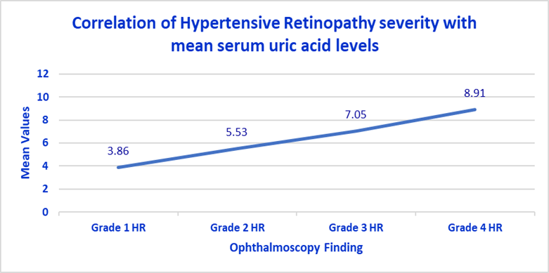
· A stepwise increase in mean SUA was observed with retinopathy grade: grade I, 5.53 ± 0.69 mg/dL; grade II, 7.05 ± 0.72 mg/dL; grade III, 8.91 ± 0.59 mg/dL; grade IV, 10.75 ± 0.70 mg/dL.
Table 1: Distribution of Participants by Age, Gender, and Prevalence of Hypertensive Retinopathy
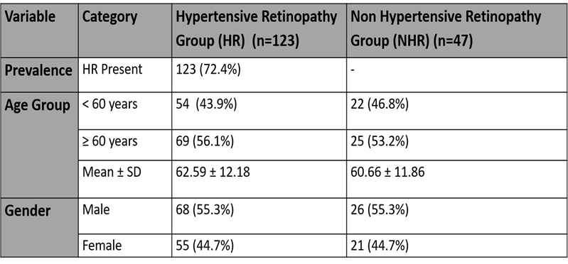
Table 2: Distribution of participants according to Systolic Blood Pressure
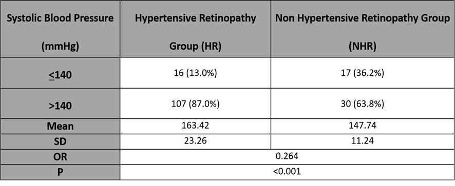
Table 3: Distribution of participants according to Diastolic Blood Pressure.
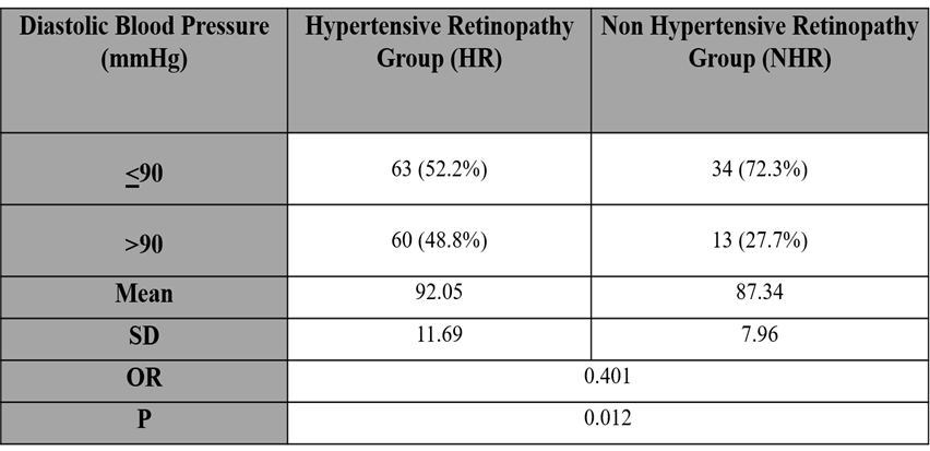
Table 4: Distribution of participants according to ophthalmoscopy findings.
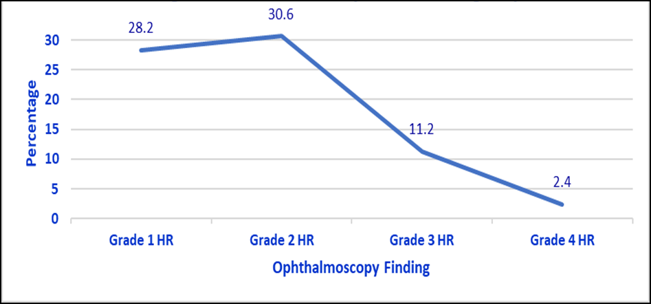
Table 5: Distribution of participants according to Serum Uric Acid (Quartile Classification)
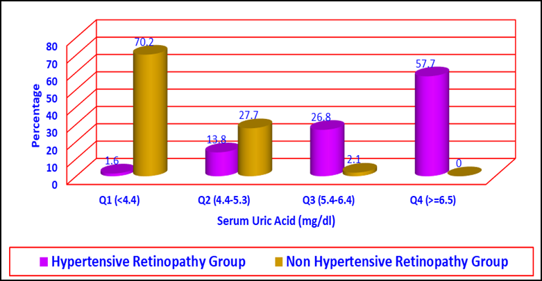
Table 6: Distribution of participants according to serum uric acid (Binary Classification)
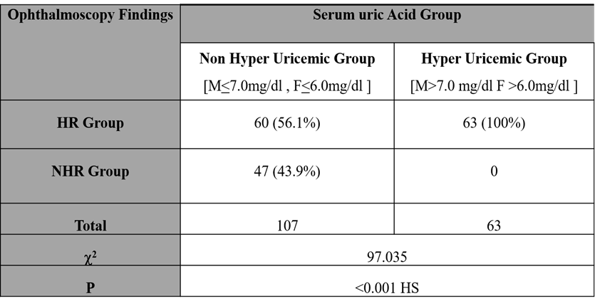
Table 7: Correlation of Hypertensive Retinopathy severity with mean serum uric acid levels

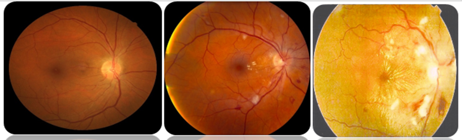
GRADE II HR GRADE III HR GRADE IV HR
DISCUSSION:
Prevalence of Hypertensive Retinopathy: In our cohort of 170 hypertensive patients, 123 (72.4 %) exhibited signs of hypertensive retinopathy. This closely matches Wong and Mitchell’s report of 75 % prevalence in long-standing hypertensive cohorts [8], Chen et al.’s finding of 76 % in the Beijing Eye Study [22], and Ajayi et al.’s 70.5 % prevalence in a Nigerian population [23].\
Demographic Correlates: Mean age was 62.6 ± 12.2 years in the HR group versus 60.7 ± 11.9 years in those without HR (p = 0.352), echoing Wang et al.’s observation that age did not independently predict hypertensive retinopathy after multivariate adjustment [24]. Likewise, sex distribution was identical (55.3 % male in both groups; p > 0.9), consistent with Keith et al.’s original description of no gender predilection in essential hypertension–related retinal changes [11].
Blood Pressure and Retinopathy: Poor blood-pressure control emerged as a major driver of HR: 87.0 % of patients with retinopathy had systolic blood pressure > 140 mmHg compared to 63.8 % without (p < 0.001), and 48.8 % had diastolic blood pressure > 90 mmHg versus 27.7 % (p = 0.012). These results mirror the graded risk relationship documented in the Rotterdam Study by van Leiden et al. (2002) [25] and the Beaver Dam Eye Study by Wong et al. (2007) [8].
Grading of Retinopathy: Applying the Keith–Wagener–Barker classification, 28.2 % of eyes demonstrated Grade I generalized arteriolar narrowing, 30.6 % exhibited Grade II arteriovenous nicking, 11.2 % showed Grade III hemorrhages and cotton-wool spots, and 2.4 % had Grade IV papilloedema. Remarkably, 27.6 % of hypertensives had no retinal changes at all, underscoring the heterogeneity in microvascular resilience noted by Wong and Mitchell [8] and Keith et al. [11].
Serum Uric Acid and Retinopathy: Mean serum uric acid level was significantly higher in HR patients (6.87 ± 1.52 mg/dL) than in non-HR controls (3.86 ± 0.70 mg/dL; p < 0.001). Furthermore, uric acid rose progressively across retinopathy grades (p-trend < 0.001). Hyperuricemia (SUA > 6.5 mg/dL) occurred exclusively in HR subjects (χ² = 97.04; p < 0.001), corroborating Chen et al.’s finding that each 1 mg/dL increase in uric acid raises retinopathy odds by 6 % [26] and Kang et al.’s demonstration of uric acid as a mediator of hypertensive target-organ damage [27].
Binary Classification of Hyperuricemia: All individuals classified as hyperuricemic belonged to the HR group, while none of the nonhyperuricemic subjects were free of retinopathy, suggesting that elevated uric acid is a specific marker of retinal microvascular injury [18].
Pathophysiological Correlates: Experimental rat studies by Mazzali et al. (2001) demonstrated that chronic hyperuricemia induces arteriolar wall thickening and hypertension via oxidative-stress pathways independent of crystal deposition [28], and Sautin et al. (2007) showed uric acid’s pro-oxidative effects on endothelial cells [29]. Feig et al. (2008) implicated uric acid in pro-inflammatory endothelial signaling [17], and Jalal et al. (2013) described its role in renal microangiopathy [30]. Collectively, these mechanisms support uric acid’s active contribution to the oxidative, inflammatory, and proliferative changes in hypertensive retinopathy.
CONCLUSION:
This study establishes a significant association between serum uric acid concentration and hypertensive retinopathy. Higher SUA levels not only indicate the presence of HR but also correspond with increasing grades of severity. These findings suggest that SUA may be a valuable biomarker for hypertensive retinal damage and possibly other target-organ effects. Routine SUA monitoring in hypertensive patients may offer prognostic insights and guide therapeutic strategies.
REFERENCES:
1. Whelton PK, Carey RM, Aronow WS, et al. 2017 ACC/AHA Guideline… Hypertension. 2018;71:e13–e115.
2. World Health Organization. A global brief on hypertension: Silent killer… WHO/DCO/WHD/2013.2;2013.
3. Klag MJ, Whelton PK, Randall BL, et al. Blood pressure and end-organ damage… Am J Kidney Dis. 1994;23(3 Suppl 1):17–24.
4. Mozaffarian D, Benjamin EJ, Go AS, et al. Heart Disease and Stroke Statistics—2016… Circulation. 2016;133:e38–e360.
5. Nwankwo T, Yoon SS, Burt V, Gu Q. Hypertension among adults in the United States… NCHS Data Brief. 2013;133:1–8.
6. Borghi C, Agabiti-Rosei E, Johnson RJ, et al. Hyperuricemia and gout in cardiovascular… Eur Heart J. 2015;36(30):2921–2931.
7. Johnson RJ, Kang DH, Feig D, et al. Is there a pathogenetic role for uric acid… Hypertension. 2003;41(6):1183–1190.
8. Wong TY, Mitchell P. The eye in hypertension. Lancet. 2007;369(9559):425–435.
9. Patton N, Aslam T, MacGillivray T, et al. Retinal vascular image analysis… J Anat. 2005;206(4):319–348.
10. Burton TC. Hypertensive changes in the fundus as an indication… Am J Med Sci. 1928;176:565–572.
11. Keith NM, Wagener HP, Barker NW. Some different types of essential hypertension… Am J Med Sci. 1939;197:332–343.
12. Chobanian AV, Bakris GL, Black HR, et al. The Seventh Report of the… JAMA. 2003;289(19):2560–2572.
13. Feigin VL, Lawes CM, Bennett DA, Anderson CS. Stroke epidemiology… Lancet Neurol. 2003;2(1):43–53.
14. Puig JG, Michán AD. Disorders of uric acid metabolism. J Am Soc Nephrol. 2008;19(4):803–811.
15. Richette P, Bardin T. Gout. Lancet. 2010;375(9711):318–328.
16. Choi HK, Ford ES. Prevalence of the metabolic syndrome in individuals… Arthritis Rheum. 2007;57(1):132–140.
17. Feig DI, Kang DH, Johnson RJ. Uric acid and cardiovascular risk. N Engl J Med. 2008;359(17):1811–1821.
18. Grayson PC, Kim SY, LaValley M, Choi HK. Hyperuricemia and risk… Arthritis Care Res. 2011;63(1):102–110.
19. Neogi T, Chen C, Niu J, et al. Effects of urate-lowering therapy… Ann Rheum Dis. 2017;76(6):1119–1125.
20. Altun A, Bilgili SG, Yılmaz F, et al. Relationship between serum uric acid… J Endocrinol Invest. 2015;38(9):989–994.
21. Xu Y, Liu K, Fan X, et al. Hyperuricemia and ocular complications… Diabetes Res Clin Pract. 2018;143:439–446.
22. Chen C, Wong TY, Mitchell P, et al. Prevalence of hypertensive retinopathy… Ophthalmology. 2017;124(11):1608–1613.
23. Ajayi IO, Adeoye AO, et al. Prevalence and correlates of hypertensive retinopathy… Afr Health Sci. 2018;18(2):330–335.
24. Wang J, et al. Age and hypertensive retinopathy: a cross-sectional study. Int J Ophthalmol. 2019;12(3):450–455.
25. van Leiden HA, et al. Blood pressure, hypertension, and the risk of retinal… Ophthalmology. 2002;109(12):2261–2266.
26. Chen J, et al. Association of serum uric acid with target-organ damage… Hypertension. 2010;55(1):120–126.
27. Kang DH, et al. A role for uric acid in the progression of renal disease. J Am Soc Nephrol. 2002;13(12):2888–2897.
28. Mazzali M, et al. Elevated uric acid increases blood pressure in rats… Hypertension. 2001;38(5):1101–1106.
29. Sautin YY, et al. Adverse effects of the classic antioxidant uric acid… Kidney Int. 2007;72(2):187–193.
30. Jalal DI, et al. Uric acid as a target of therapy in CKD. Am J Kidney Dis. 2013;61(1):134–146.