Prevalence of AmpC β-lactamases and Comparison by Two Phenotypic Methods for the Detection of AmpC β-lactamases among the Isolates of Enterobacterales in a Tertiary Care Hospital.
- M Banusri , Post Graduate, Department of Microbiology, Annapoorana Medical College & Hospitals.
- A Shirleymaryon , Post Graduate, Department of Microbiology, Annapoorana Medical College & Hospitals.
- N Shanmuga Vadivoo , Professor and HOD, Department of Microbiology, Annapoorana Medical College & Hospitals
- K Sudha , Professor, Department of Microbiology, Annapoorana Medical College & Hospitals.
- B Usha , Professor & Dean, Department of Microbiology, Annapoorana Medical College & Hospitals.
Article Information:
Abstract:
Background: AmpC β-lactamases are Ambler class C enzymes that confer resistance to a wide range of β-lactam antibiotics, including Cephamycins and extended-spectrum cephalosporins. AmpC β-lactamases are detected by screening with cefoxitin in routine antibiotic susceptibility testing in microbiology laboratories. Following screening, confirmatory methods are needed for its detection. Effective antibiotic treatment and infection control procedures are seriously threatened by the rising incidence of Enterobacterales that produce AmpC β-lactamases. Therefore, early and precise identification is crucial to directing the right course of therapy and preventing spread. Aim: To determine the prevalence of AmpC β-lactamase among clinical isolates of Enterobacterales and to compare the performance of Boronic acid inhibitor assay and Disk approximation test for phenotypic detection. Methods: A prospective, observational study was conducted over six months in the Department of Microbiology. A total of 100 fermentative Gram-negative isolates obtained from various clinical specimens were included. Cefoxitin (30 µg) disc diffusion was used for screening of AmpC-producing Enterobacterales. The disk approximation test and the boric acid inhibitor assay were used for confirmatory testing on isolates exhibiting Cefoxitin resistance (zone diameter <18 mm). Frequency distribution and percentage were used to observe the data. Results: Among 100 isolates, 66 (66%) were resistant to cefoxitin and considered screen-positive for AmpC production. The predominant organism isolated was Escherichia coli (63%), followed by Klebsiella pneumoniae (29%), with urine being the most frequently received specimen (45%) from which the isolates were obtained. All 66 cefoxitin-resistant isolates were positive by the Boronic acid inhibitor assay (100%), whereas the Disk approximation test detected 58 (87.9%) isolates. The preferred treatment options are Carbapenems, and the susceptibility to Imipenem was 81%, and Meropenem was 87%. Among Aminoglycosides, Amikacin showed a susceptibility of 63% and Gentamicin showed susceptibility of 59%. Nitrofurantoin showed susceptible rate of 93% respectively. Conclusion: The prevalence of AmpC β-lactamase in this study was 66%. Boronic acid inhibitor assay demonstrated better detection when compared to Disk approximation test and is recommended for routine laboratory use.
Keywords:
Article :
INTRODUCTION:
Antimicrobial resistance among Gram-negative bacilli has emerged as a major global health concern. Over the past few decades, there has been a significant increase in resistance to β-lactam antibiotics, which remain the cornerstone of therapy for many bacterial infections.(1). Gram-negative bacteria have developed a number of mechanisms against β-lactam antibiotics, which interfere with bacterial cell wall synthesis by binding to PBPs of their cell walls. The synthesis of β-lactamase enzymes is a prevalent mechanism of resistance in Gram-negative bacteria, which hydrolyze the β-lactam ring and make the antibiotic ineffective.
(2). The additional mechanisms that confer resistance include reduced drug permeability by either loss of porin channels, efflux pump mediated extrusion of antibiotics followed by modification of PBPs. The β-lactamases includes Extended Spectrum β-lactamases (ESBLs), AmpC β-lactamases, and Carbapenemases. While many microbiology labs now routinely detect ESBL and screen for AmpC β-lactamases by Cefoxitin disc diffusion, the lack of defined detection protocols to confirm AmpC β-lactamases makes it difficult in routine practices. (3).
Ambler class C and Bush Jacoby group I categorization schemes include AmpC β-lactamases. These enzymes provide resistance to a wide range of β-lactam antibiotics, such as
monobactams like aztreonam, cephamycins like cefoxitin, and narrow and broad-spectrum cephalosporins. Clavulanic acid and other β-lactamase inhibitors do not effectively block AmpC enzymes, in contrast to ESBLs.(4).
Three forms of AmpC resistance mechanisms may be distinguished: plasmid-mediated production, non-inducible chromosomal overexpression brought on by promoter alterations, and inducible chromosomal expression. TheSPICE organisms, such as Serratia marcescens, Pseudomonas aeruginosa, Indole-positive Proteus species / Acinetobacter, Citrobacter freundii, and Enterobacter cloacae, have inducible chromosomal AmpC genes. Because of regulatory alterations, organisms such as Escherichia coli develop non-inducible chromosomal resistance. However, because plasmid-mediated AmpC enzymes may spread horizontally between bacterial species, such as Escherichia coli and Klebsiella pneumoniae, they are especially concerning.(5)
There are fewer treatment options available because plasmid-mediated AmpC enzymes are typically linked to multidrug resistance and are expressed at high levels. Clinical isolates with plasmid-mediated AmpC genes may seem susceptible to third-generation cephalosporins initially, but they may subsequently become resistant during treatment, which might result in treatment failure.(6). Escherichia coli and Klebsiella pneumoniae, which belong to the Enterobacterales family, are among the most common bacteria responsible for both hospital-acquired and community-acquired infections. In recent years, there has been a global increase in strains of these bacteria that produce AmpC β-lactamase, although the rate of increase varies across different regions. Studies from India have reported a significant prevalence of plasmid mediated AmpC-producing isolates, highlighting their growing role in antimicrobial resistance and the challenges they pose for effective treatment.(7).
AmpC production is detected by phenotypic procedures such as disk approximation tests, three-dimensional extract tests, inhibitor-based assays utilizing boronic acid, and genotypically by PCR.(5). Molecular techniques are very specific, yet they are expensive and difficult for everyday application in laboratories with limited settings. On the other hand, phenotypic techniques are easy to perforn, affordable, and appropriate for regular laboratory use. In the boronic acid inhibitor-based test, inhibition of AmpC enzymes by boronic acid leads to an increase in the zone of inhibition around the cefoxitin disk with boronic acid when compared with cefoxitin alone. The disk approximation test detects inducible AmpC production by demonstrating a characteristic blunting or flattening of the inhibition zone between ceftazidime and an inducer drug such as cefoxitin or imipenem. Since the sensitivity and specificity of these phenotypic methods vary, comparative evaluation of different testing approaches is important for accurate detection of AmpC-producing isolates.(8).
As the Clinical and Laboratory Standards Institute (CLSI) has not yet recommended a standardized method for the detection of AmpC β-lactamases, microbiology laboratories must evaluate and adopt reliable diagnostic approaches suitable for their settings. Therefore, studies comparing simple, cost-effective, and practical phenotypic methods are essential. The present study was undertaken to determine the prevalence of AmpC-producing Enterobacterales isolates in a tertiary care hospital and to compare the performance of two commonly used phenotypic methods—the disk approximation test and the boronic acid inhibitor-based assay—for routine laboratory detection in our tertiary care hospital.
MATERIALS AND METHODS:
Study Design and Setting
This prospective observational study was conducted in the Department of Microbiology of a tertiary care teaching hospital over a period of six months after obtaining approval from the Institutional Ethics Committee. The study aimed to determine the prevalence of AmpC β-lactamase among Enterobacterales and to compare two phenotypic detection methods.
Study Population and Sample Size
A total of 100 consecutive non-duplicate fermentative Gram-negative isolates recovered from various clinical specimens during the study period were included. The specimens comprised urine, pus swabs, pus aspirates, tissue bits, sputum, and other respiratory samples, blood, and other body fluids. Only pathogenic isolates were considered for the study excluding colonizers or contaminants.
Inclusion Criteria
1. Pathogenic fermentative Gram-negative bacilli isolated from patient specimens.
2. Isolates belonging to the order Enterobacterales.
3. Non-duplicate isolates obtained from patients of all age groups and both sexes.
Exclusion Criteria
1. Non-fermentative Gram-negative bacilli such as Pseudomonas spp. and Acinetobacter spp.
2. Gram-positive organisms isolated from clinical samples.
3. Duplicate isolates from the same patient during the same infection episode.
4. Colonizers, contaminants, or isolates with insignificant growth
Identification of Isolates
Specimens were processed according to standard microbiological guidelines (Bailey & Scott’s Diagnostic Microbiology, Murray’s Medical Microbiology, and the Clinical Microbiology Procedures Handbook). Identification of isolates was performed based on colony morphology, Gram staining, routine, basic/ standard biochemical tests - indole, citrate, urease, triple sugar iron test, mannitol motility medium, nitrate reduction test, methyl red & Voges-Proskauer tests. Organisms identified included Escherichia coli, Klebsiella pneumoniae, Klebsiella aerogenes, and Citrobacter koseri.
Antimicrobial Susceptibility Testing
Antibiotic susceptibility testing (AST) was performed using the Kirby–Bauer disc diffusion method on Mueller–Hinton agar in accordance with Clinical and Laboratory Standards Institute (CLSI 2023-2024) guidelines. A 0.5 McFarland standard suspension was prepared for each isolate, and lawn cultures were performed on Mueller-Hinton agar. Antibiotic discs were applied and the plates were incubated at 37 °C. Antibiotics tested included Ampicillin, Cefotaxime, Ceftazidime, Cefoxitin, Piperacillin–Tazobactam, Ciprofloxacin, Cotrimoxazole, Tetracycline/ Doxycycline, Gentamicin, Amikacin, Imipenem, Meropenem, and Nitrofurantoin (for urinary isolates).
Screening and Confirmatory Tests for AmpC
All isolates were screened using cefoxitin (30 µg) disc diffusion. Isolates showing a zone diameter <18 mm were considered screen-positive for AmpC production.
Confirmatory testing was performed using:
Boronic Acid Inhibitor Assay
An increase in zone diameter of ≥5 mm with cefoxitin plus phenylboronic acid compared to cefoxitin alone was interpreted as positive.
Disk Approximation Test
Blunting or flattening of the ceftazidime inhibition zone toward inducing agents (cefoxitin or imipenem) was considered indicative of inducible AmpC production.
Statistical Analysis
Data were entered into Microsoft Excel and analyzed using descriptive statistics. Results were expressed as frequency and percentage.
IEC Approval – the following are the IEC approval details: AMCH/IEC/Proc. No. 65/2024, Dated- 16/10/2024..
RESULTS:
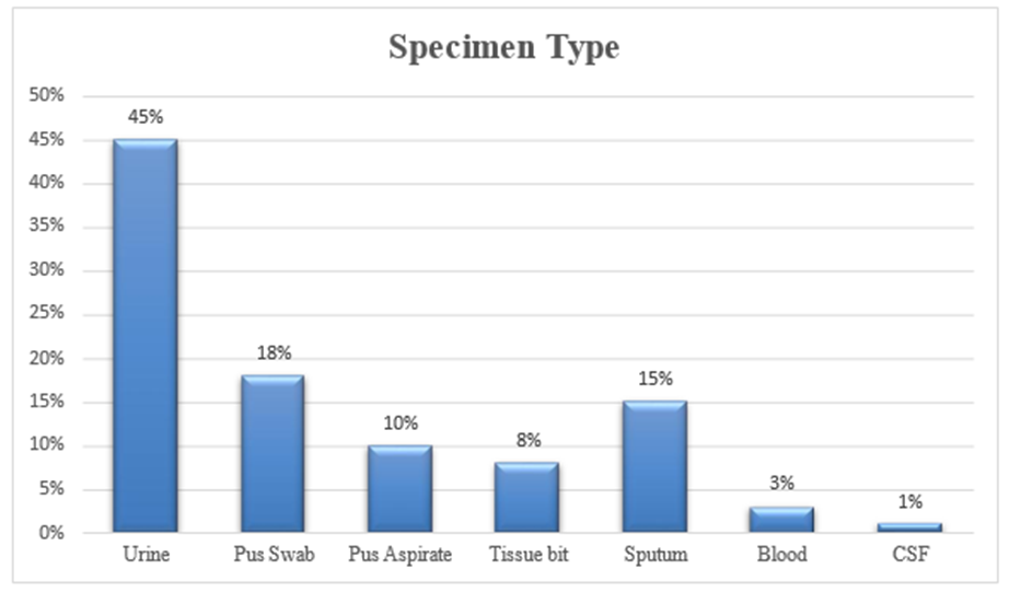
Chart 1: Distribution of fermentative Gram-negative Enterobacterales isolated from different clinical specimens (n = 100)
Chart 1 shows the distribution of clinical specimens from which the isolates were obtained. Out of 100 samples, urine constituted the majority with 45% (n=45), indicating urinary tract infections as the most common source of Enterobacterales isolates at our tertiary care centre. This was followed by pus swabs (18%), pus aspirates accounted for 10%, while tissue bits contributed 8%. Sputum samples constituted 15%). Blood cultures represented 3% of isolates, and cerebrospinal fluid (CSF) constituted 1%. The predominance of urinary samples highlights the significant burden of Gram-negative infections in the urinary tract.
Table 1. Organism-wise distribution of fermentative Gram-negative Enterobacterales (n = 100)
|
Organism |
Frequency |
Percentage (%) |
|
Escherichia coli |
63 |
63% |
|
Klebsiella pneumoniae |
29 |
29% |
|
Klebsiella aerogenes |
6 |
6% |
|
Citrobacter koseri |
2 |
2% |
Table 1 illustrates the distribution of isolated organisms. Escherichia coli was the predominant isolate, accounting for 63% (n=63) of cases. Klebsiella pneumoniae was the second most common organism at 29% (n=29). Klebsiella aerogenes comprised 6% (n=6), while Citrobacter koseri represented 2% (n=2). The high prevalence of E. coli reflects its well-established role as the leading pathogen in both community and hospital-acquired infections.
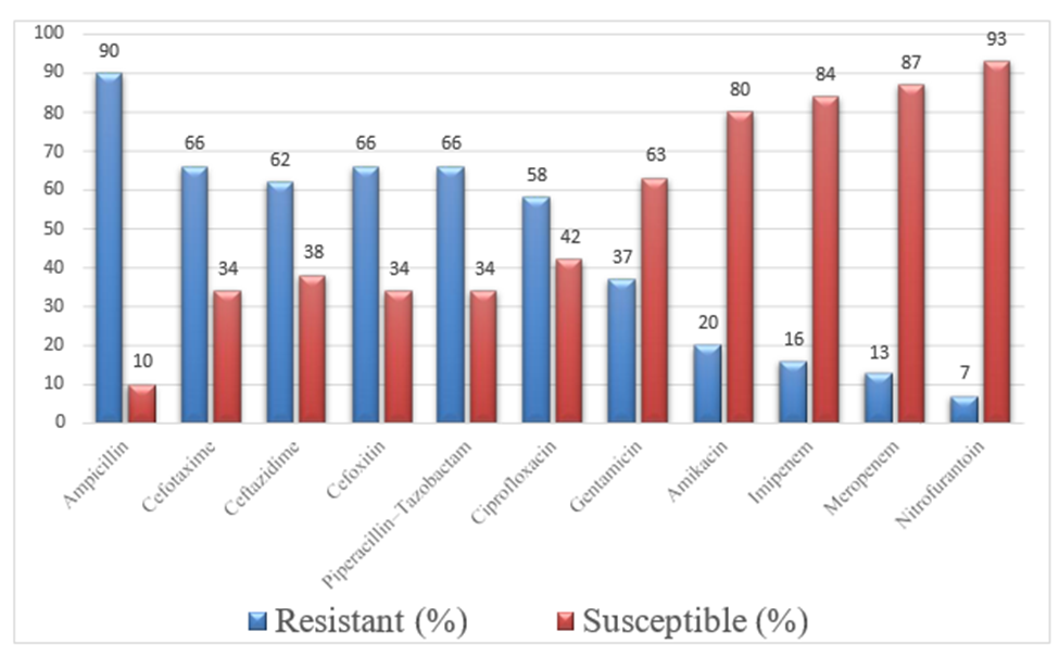
Chart 2: Antibiotic Susceptibility Pattern of fermentative Gram-negative Enterobacterales isolated (n = 100)
Chart 2 summarizes the antibiotic susceptibility pattern of all 100 isolates. Only Carbapenems showed more than 80% susceptibility rates- imipenem showing 81% and Meropenem showing 87%, indicating their continued effectiveness. Nitrofurantoin also showed a higher susceptibility of 93%, particularly among urinary isolates. Other antibiotic groups, such as Penicillins (Ampicillin 90%), Cephalosporins (Cefotaxime - 66%, Ceftazidime - 62%, Cefoxitin -66%), BL-BLIs (Piperacillin-tazobactam - 66%), Fluoroquinolones (Ciprofloxacin-58%) and Aminoglycosides (Gentamicin- 59% and Amikacin- 63%), showed less than 80% susceptibility rates, respectively.
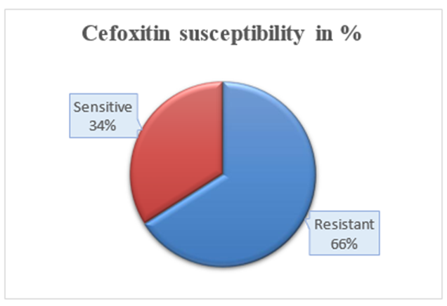
Chart 3: Distribution of Susceptibility Pattern of Cefoxitin by Disc Diffusion among Isolated Fermentative Gram-Negative Enterobacterales.
Chart 3 shows the susceptibility pattern of cefoxitin determined by the disc diffusion method. Among the 100 isolates tested, 66% (n=66) were resistant to cefoxitin, whereas 34% (n=34) were sensitive. The prevalence of screen-positive AmpC producers based on cefoxitin resistance was therefore 66%, indicating a substantial proportion of isolates requiring confirmatory testing.
Table 2: Distribution of Boronic Acid Inhibitor Assay among the Cefoxitin-resistant isolates (n = 66)
|
Result |
Frequency |
Percentage (%) |
|
Positive |
66 |
100% |
|
Negative |
0 |
0% |
Table2: Depicts the results of the Boronic acid inhibitor assay performed on the 66 cefoxitin-resistant isolates. All isolates (100%) tested positive for AmpC production using this method, with no negative results observed. This indicates excellent detection capability of the Boronic acid inhibitor assay in identifying AmpC producers.
Table 3: Disk Approximation Test among the Cefoxitin-resistant isolates (n = 66)
|
Result |
Frequency |
Percentage (%) |
|
Positive |
58 |
87.9% |
|
Negative |
8 |
12.1% |
Table 3 shows the results of the Disk approximation test conducted on the same 66 isolates. Out of these, 58 isolates (87.9%) were positive for AmpC production, while 8 isolates (12.1%) were negative. Compared to the Boronic acid assay, this method demonstrated slightly lower detection rates.
Table 4: Comparison of Cefoxitin screening with Two Phenotypic detection methods (n = 66)
|
Test Method |
No. of isolates tested |
Susceptible |
Resistant |
Positive |
Detection Rate |
|
Screening – Cefoxitin disk diffusion |
100 |
34 |
66 |
66 |
100% |
|
Detection – Boronic Acid Assay |
66 |
0 |
66 |
66 |
100% |
|
Detection – Disk Approximation Test |
66 |
8 |
58 |
58 |
87.9% |
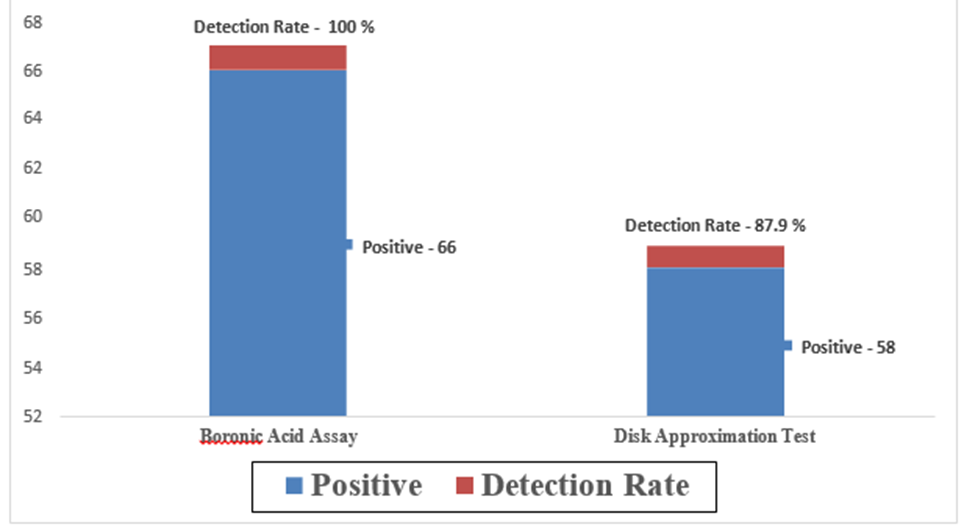
Chart 4: Comparison of Two Phenotypic Methods
Table 4 & Chart 4: compare the performance of the two phenotypic methods. The Boronic acid inhibitor assay detected AmpC production in all 66 isolates (100%), whereas the Disk approximation test detected AmpC production in 58 isolates (87.9%). This comparison demonstrates that the Boronic acid assay had superior detection efficiency in this study.
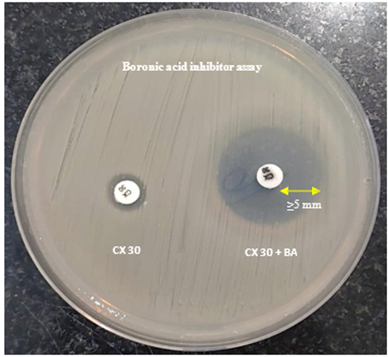
Figure 1: Boronic acid inhibitor assay showing a positive AmpC β-lactamase producer, demonstrated by an increase of ≥5 mm in the inhibition zone around the cefoxitin–boronic acid disc compared with the cefoxitin disc alone.
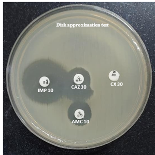
Figure 2: Positive disk approximation test for inducible AmpC β-lactamase production showing blunting of the ceftazidime inhibition zone adjacent to the AMC disc.
DISCUSSION:
AmpC β-lactamase production among Enterobacterales is an emerging concern due to its association with multidrug resistance and therapeutic failure.Our study compared two phenotypic detection techniques and assessed the frequency of AmpC β-lactamase production in 100 fermenting Enterobacterales.
As shown in Chart 1, in the present study, AmpC β-lactamase-producing Enterobacterales were most frequently isolated from urine specimens (45%; 45/100), followed by pus swabs (18%), sputum (15%), pus aspirates (10%), and tissue bits (8%). The predominance of urine specimens likely reflects the high burden of urinary tract infections (UTIs), which are predominantly caused by Gram-negative Enterobacterales. Similar findings have been reported in studies from India, where urine constituted the largest proportion of clinical samples processed for the isolation and identification of Gram-negative bacilli. Singhal S et al., Manoharan A et al. and Veeraraghavan B et al. (9–11) found that urine accounted for the majority of clinical specimens yielding Gram-negative bacilli, especially Klebsiella pneumoniae and Escherichia coli, with a significant prevalence of isolates producing AmpC β-lactamase. The frequent isolation of uropathogens from urine specimens emphasizes the importance of continuous surveillance of antimicrobial resistance mechanisms.
In the present study, as shown in Table 1, Escherichia coli (63%) was the predominant isolate, showing AmpC production, followed by Klebsiella pneumoniae (29%), Klebsiella aerogenes (6%), and Citrobacter koseri(2%). The predominance of Escherichia coli observed in this study is in concordance with a previous study by Gurung S et al., in which E. coli was the most frequently isolated uropathogen, accounting for 70.3% (52/74) of isolates, followed by K. pneumoniae, 29.7% (22/74), showing AmpC production (12). Similarly, Manoharan et al. reported that E. coli and Klebsiella spp. together accounted for the majority of plasmid-mediated AmpC β-lactamase-producing Enterobacterales isolated from five Indian tertiary care centres (11). Thus, E. coli demonstrates its continued role as a significant pathogen causing both community-acquired and healthcare-associated infections, particularly urinary tract infections. The antimicrobial susceptibility profile of the isolates, as shown in Chart 2, demonstrated high resistance to Ampicillin (90%), Cefoxitin (66%), Cefotaxime (66%) and Ceftazidime (62%). This resistance pattern is consistent with the study by Mohamudha Parveen et al., who observed resistance rates of 91.4% to Ampicillin, 67.2% to Cefoxitin, 69.4% to Cefotaxime, and 63.8% to Ceftazidime, among AmpC-producing Enterobacterales.(13) Similarly, Hemalatha et al. reported Cefoxitin resistance in 65% of AmpC-producing isolates (14), which is close to the 66% observed in the present study.
Carbapenems remained the most active agents in the present study, with Meropenem (87%) and Imipenem (81%) showing the highest susceptibility. Manoharan et al. reported similar higher susceptibility rates of 95% for Meropenem and 93% for Imipenem; also, recent IDSA guidance (2024) continues to recommend carbapenems as the preferred treatment for severe infections caused by AmpC-producing Enterobacterales. (11,15). Among the aminoglycosides, Amikacin (63%) showed better activity than Gentamicin (59%), although these rates were lower than those reported by Manoharan et al., who found susceptibility rates of 81% and 72%, respectively. Likewise, ciprofloxacin susceptibility (58%) in the present study was lower than the 68–75% reported in earlier Indian studies, suggesting a progressive increase in fluoroquinolone resistance among Enterobacterales.(11)
Nitrofurantoin demonstrated excellent activity (93%) against urinary isolates in the present study. This finding is consistent with recent surveillance studies, which have reported susceptibility rates of 85–95% among urinary Escherichia coli, supporting its continued use as a first-line agent for uncomplicated urinary tract infections.(11,15,16)
In the present study, 66% of the isolates were found to be resistant to Cefoxitin on routine screening by the disc diffusion method, indicating a high prevalence of potential AmpC producers, as shown inChart 3. A similar higher prevalence of AmpC was observed in a study by Das et al., which showed a prevalence of 62% of plasmid-mediated AmpC production among 71 isolates of Enterobacterales. (17). Also, the findings of our study are higher when compared to previous reports from India. Studies by A. Manoharan, Patricia E. Coudron, and S. Singhal have reported AmpC β-lactamase prevalence ranging from 20% to 50%. The comparatively higher prevalence observed in the present study could be explained by differences in study population, antimicrobial prescribing practices, local resistance epidemiology, and the variable use of cephalosporins in tertiary care settings(9,11,18)
As shown inTable 2, the Boronic acid inhibitor test showed a 100% detection rate for all 66 cefoxitin-resistant isolates. On the other hand, as shown in Table 3, the Disk approximation test identified AmpC synthesis in 58 cefoxitin-resistant isolates (87.9%), indicating the higher performance of the Boronic acid assay. The higher detection rate may be attributed to the specific inhibition of AmpC β-lactamase by phenylboronic acid, resulting in easier and more reliable interpretation. Our findings are comparable with those of Coudron PE, who reported a sensitivity of 97–100% for the Boronic acid assay, establishing it as a reliable phenotypic method for AmpC detection. (18). Similarly, Black et al. demonstrated that inhibitor-based assays had >95% sensitivity, whereas conventional disk-based methods showed lower sensitivities of 80–90%.(19). The findings of the present study further support the routine use of the Boronic acid inhibitor assay as a simple, cost-effective, and more sensitive phenotypic method for detecting AmpC β-lactamase-producing Enterobacterales, particularly in laboratories where molecular techniques are not readily available.
As shown in Table 4 & Chart 4,the comparison shows that the boronic acid inhibitor assay showed higher sensitivity than the disk approximation test for detecting AmpC production. Similar observations have been reported in previous studies by Coudron PE et al., which found boronic acid-based methods to be more sensitive and reliable (97- 100%), particularly for the identification of plasmid-mediated AmpC enzymes (18). Comparable observations were also reported by Singhal et al., who found that the Boronic acid inhibitor assay detected nearly all phenotypically confirmed AmpC-producing isolates (>95%). In contrast, the Disk Approximation Test missed a small proportion of isolates, resulting in a lower detection rate. The authors recommended the Boronic acid assay as the preferred phenotypic confirmatory test because of its ease of performance, reproducibility, and higher sensitivity.(9) The high level of concordance observed with the boronic acid test highlights its value as a reliable tool for routine use in clinical microbiology laboratories. Its higher sensitivity, simple methodology, and affordability make it a preferable option over inducible techniques such as disk approximation testing (18).
Overall, the present study demonstrates a high prevalence (66%) of AmpC β-lactamase-producing Enterobacterales, with Escherichia coli being the most frequently isolated organism. Among the phenotypic methods evaluated, the boronic acid inhibitor assay showed better detection rates than the disk approximation test, indicating its usefulness as a reliable detection method in routine laboratory practice, particularly in settings where PCR-based detection is unavailable. Also, the increasing resistance to third-generation cephalosporins observed among these isolates shows the ongoing threat of antimicrobial resistance. Early detection of AmpC producers, along with strict infection control practices, antimicrobial stewardship, and regular surveillance, is crucial to prevent the spread of these resistant organisms. Implementation of standardized detection methods in routine clinical laboratories can further improve patient management and control ofAmpC-producing pathogens.
CONCLUSION:
The present study demonstrates a high prevalence (66%) of AmpC β-lactamase production among Enterobacterales isolates in our tertiary care setting. Escherichia coli was the most often isolated pathogen showing AmpC β-lactamase production, and from urinary tract infection. The study findings highlight a high level of multidrug resistance among the isolates, particularly against third-generation cephalosporins and cephamycins. Although carbapenems remained the most effective antibiotics, their use should be carefully monitored to prevent the development of further resistance. Among the detection methods evaluated, the boronic acid inhibitor assay performed better than the disk approximation test and proved to be a simple, affordable, and reliable option for routine identification of AmpC-producing organisms. Early detection, appropriate antibiotic use, strict infection control practices, and continuous surveillance are essential to prevent the spread of these resistant pathogens.
LIMITATIONS OF THE STUDY
1. The study was only carried out at one tertiary care facility, which could have limited how broadly the findings can be applied.
2. Since the AmpC genes were not molecularly characterized, it was unable to distinguish between chromosomal and plasmid-mediated AmpC.
3. Molecular techniques were not used to assess the co-production of additional resistance mechanisms, such as ESBL or carbapenemases.
4. The relation between the clinical outcomes of the patient and AmpC production was not determined.
REFERENCES:
1. Kumari R, Saraogi I. Navigating Antibiotic Resistance in Gram-Negative Bacteria: Current Challenges and Emerging Therapeutic Strategies. ChemPhysChem. 2025;26(9):e202401057. doi:10.1002/cphc.202401057
2. Mora-Ochomogo M, Lohans CT. β-Lactam antibiotic targets and resistance mechanisms: from covalent inhibitors to substrates. RSC Med Chem. 2021 Oct 1;12(10):1623–39. doi:10.1039/d1md00200g
3. Oelschlaeger P. β-Lactamases: Sequence, Structure, Function, and Inhibition. Biomolecules. 2021 Jul;11(7):986. doi:10.3390/biom11070986
4. Philippon A, Arlet G, Labia R, Iorga BI. Class C β-Lactamases: Molecular Characteristics. Clin Microbiol Rev. 2022 Apr 18;35(3):e00150-21. doi:10.1128/cmr.00150-21
5. Tamma PD, Doi Y, Bonomo RA, Johnson JK, Simner PJ, Antibacterial Resistance Leadership Group. A Primer on AmpC β-Lactamases: Necessary Knowledge for an Increasingly Multidrug-resistant World. Clin Infect Dis. 2019 Oct 15;69(8):1446–55. doi:10.1093/cid/ciz173
6. Kaushik M, Kaushik A, Jain A, Chaudhary J, Gupta V. AmpC Inhibition: An Explicit Approach against Multi-Drug Resistance (MDR). Curr Top Med Chem. 2023 Aug 1;23(20):1919–27. doi:10.2174/1568026623666230504095005
7. Thakkar J, Shanoo A, Gupta S, Thakkar A, Thakkar J, Iv ABS, et al. The Pattern and Impact of Hospital-Acquired Infections and Its Outlook in India. Cureus. 2023 Nov 9;15. doi:10.7759/cureus.48583
8. Broekema NM, Van TT, Monson TA, Marshall SA, Warshauer DM. Comparison of Cefoxitin and Oxacillin Disk Diffusion Methods for Detection of mecA-Mediated Resistance in Staphylococcus aureus in a Large-Scale Study. J Clin Microbiol. 2009 Jan;47(1):217–9. doi:10.1128/jcm.01506-08
9. Singhal S, Mathur T, Khan S, Upadhyay D, Chugh S, Gaind R, et al. EVALUATION OF METHODS FOR AMPC β-LACTAMASE IN GRAM NEGATIVE CLINICAL ISOLATES FROM TERTIARY CARE HOSPITALS. Indian J Med Microbiol. 2005 Apr 1;23(2):120–doi:10.1016/S0255-0857(21)02652-9
10. Veeraraghavan B, Shankar C, Karunasree S, Kumari S, Ravi R, Ralph R. Carbapenem resistant Klebsiella pneumoniae isolated from bloodstream infection: Indian experience. Pathog Glob Health. 2017 Jul;111(5):240–6. doi:10.1080/20477724.2017.1340128 PubMed PMID: 28670975; PubMed Central PMCID: PMC5560201.
11. Manoharan A, Sugumar M, Kumar A, Jose H, Mathai D, Khilnani GC, et al. Phenotypic & molecular characterization of AmpC β-lactamases among Escherichia coli, Klebsiella spp. & Enterobacter spp. from five Indian Medical Centers. Indian J Med Res. 2012 Mar;135(3):359–64. PubMed PMID: 22561623; PubMed Central PMCID: PMC3361873.
12. Gurung S, Kafle S, Dhungel B, Adhikari N, Shrestha UT, Adhikari B, et al. <p>Detection of OXA-48 Gene in Carbapenem-Resistant <em>Escherichia coli</em> and <em>Klebsiella pneumoniae</em> from Urine Samples</p>. Infect Drug Resist. 2020 Jul 14;13:2311–21. doi:10.2147/IDR.S259967
13. Ovid [Internet]. [cited 2026 Jul 21]. Molecular description of plasmid-mediated AmpC... : Indian Journal of Medical Research. Available from: https://www.ovid.com/jnls/ijmr/fulltext/10.4103/0971-5916.93433~molecular-description-of-plasmid-mediated-ampc--lactamases
14. Sageerabanoo S, Malini A, Mangaiyarkarasi T, Hemalatha G. Phenotypic detection of extended spectrum β-lactamase and Amp-C β-lactamase producing clinical isolates in a Tertiary Care Hospital: A preliminary study. J Nat Sci Biol Med. 2015;6(2):383–7. doi:10.4103/0976-9668.160014 PubMed PMID: 26283835; PubMed Central PMCID: PMC4518415.
15. Tamma PD, Aitken SL, Bonomo RA, Mathers AJ, van Duin D, Clancy CJ. Infectious Diseases Society of America Guidance on the Treatment of Extended-Spectrum β-lactamase Producing Enterobacterales (ESBL-E), Carbapenem-Resistant Enterobacterales (CRE), and Pseudomonas aeruginosa with Difficult-to-Treat Resistance (DTR-P. aeruginosa). Clin Infect Dis Off Publ Infect Dis Soc Am. 2021 Apr 8;72(7):e169–83. doi:10.1093/cid/ciaa1478 PubMed PMID: 33106864.
16. Rodrígue z-Guerrero E, Callejas-Rodelas JC, Navarro-Marí JM, Gutiérrez-Fernández J. Systematic Review of Plasmid AmpC Type Resistances in Escherichia coli and Klebsiella pneumoniae and Preliminary Proposal of a Simplified Screening Method for ampC. Microorganisms. 2022 Mar 14;10(3):611. doi:10.3390/microorganisms10030611 PubMed PMID: 35336186; PubMed Central PMCID: PMC8954824.
17. Das D, Vohra P, Mane P, Shaozae CK. Extended spectrum, AmpC&metallo β-lactamases producing Escherichia coli in urinary isolates: A prospective study in north India. Indian J Med Res. 2025 Feb;161(2):167–73. doi:10.25259/IJMR_153_2024 PubMed PMID: 40257145; PubMed Central PMCID: PMC12010785.
18. Coudron PE. Inhibitor-based methods for detection of plasmid-mediated AmpC beta-lactamases in Klebsiella spp., Escherichia coli, and Proteus mirabilis. J Clin Microbiol. 2005 Aug;43(8):4163–7. doi:10.1128/JCM.43.8.4163-4167.2005 PubMed PMID: 16081966; PubMed Central PMCID: PMC1233913.
19. AmpC Disk Test for Detection of Plasmid-Mediated AmpC β-Lactamases in Enterobacteriaceae Lacking Chromosomal AmpC β-Lactamases | Journal of Clinical Microbiology [Internet]. [cited 2026 Jul 22]. Available from: https://journals.asm.org/doi/full/10.1128/jcm.43.7.3110-3113.2005.