Microbiological Profile Of Peritoneal Fluid In Patients With Secondary Peritonitis And Its Clinical Significance.
- Uday Kumar Aggi , Assistant Professor, Department of General Surgery, Government Medical College, Ramagundam, Telangana
- Nella Harshini , Assistant Professor, Department of Microbiology, Government Medical College, Ramagundam, Telangana
- Erukulla Anusha , Assistant Professor, Department of Microbiology, Government Medical College, Ramagundam, Telangana.
Article Information:
Abstract:
Introduction: Secondary peritonitis is a common surgical emergency associated with substantial morbidity and mortality. Identification of causative microorganisms and their antimicrobial susceptibility patterns is essential for appropriate empirical therapy and antimicrobial stewardship. This study evaluated the microbiological profile of peritoneal fluid in patients with secondary peritonitis and assessed its clinical significance. Materials and Methods: A prospective observational study was conducted in the Department of Microbiology in association with General Surgery among 75 patients with secondary peritonitis. Intraoperative peritoneal fluid samples were collected aseptically and processed using standard microbiological techniques. Isolates were identified and antimicrobial susceptibility testing was performed according to standard guidelines. Microbiological findings were correlated with clinical outcomes. Results: Peritoneal fluid cultures were positive in 65.3% of patients. Gram-negative organisms predominated, with Escherichia coli being the most common isolate, followed by Klebsiella pneumoniae. High susceptibility was observed to amikacin and meropenem, while considerable resistance to third-generation cephalosporins was noted. Culture positivity was significantly associated with surgical-site infection (p=0.031) and prolonged hospital stay (p=0.041). Delayed presentation showed a significant positive correlation with duration of hospitalization. Conclusion: Peritoneal fluid culture provides clinically useful information in secondary peritonitis. Local microbiological surveillance and culture-directed therapy may improve antimicrobial selection and patient outcomes.
Keywords:
Article :
INTRODUCTION:
Secondary peritonitis is a potentially life-threatening surgical emergency that develops after loss of gastrointestinal or intra-abdominal visceral integrity, allowing microorganisms and inflammatory contents to enter the normally sterile peritoneal cavity. It commonly follows gastroduodenal, small-bowel, appendicular or colonic perforation, bowel ischemia, trauma, postoperative anastomotic leakage, or other complicated intra-abdominal infections. The clinical course depends on the site and duration of perforation, degree of contamination, virulence of infecting organisms, host status, adequacy of antimicrobial therapy, and timeliness of source control. Patients may rapidly progress from localized infection to diffuse peritonitis, sepsis, septic shock, multiorgan dysfunction and death. Recent Infectious Diseases Society of America guidelines recommend obtaining intra-abdominal fluid cultures during source-control procedures in complicated intra-abdominal infections to guide antimicrobial therapy [1].
The microbiological spectrum of secondary peritonitis is mainly derived from gastrointestinal flora and varies with the anatomical site of perforation and whether the infection is community- or healthcare-associated. Gram-negative bacilli, particularly Escherichia coli and Klebsiella species, predominate, while Enterococcus species, Streptococcus species, Pseudomonas aeruginosa, Acinetobacter species, anaerobes and Candida species may also occur. Ojo and Irabor, in a 2022 prospective study, identified E. coli, Klebsiella pneumoniae, Anaerococcus and Bacteroides fragilis among the common isolates and reported more complications, including mortality, among culture-positive patients continuing empirical therapy than among those receiving culture-directed antibiotics [2]. This supports the potential clinical value of peritoneal culture and antimicrobial-susceptibility testing when empirical coverage may be inadequate.
Recent Indian studies have highlighted the importance of local microbiological surveillance. Gupta et al. compared intraoperative peritoneal-fluid and postoperative wound cultures in patients with perforation peritonitis and demonstrated concordance of pathogen and antimicrobial-sensitivity patterns in 80.6% of relevant cases, suggesting that intraoperative cultures may help guide therapy when postoperative infection develops [3]. Deolekar et al. studied microbiological profiles according to anatomical sites of perforation and found E. coli and Klebsiella species to be predominant. Although amikacin and meropenem showed good antimicrobial activity, considerable resistance to third-generation cephalosporins was observed [4]. Ghosh et al., while evaluating secondary peritonitis across geographically diverse Indian populations, also showed that delayed presentation, organ failure and sepsis remain important determinants of poor clinical outcome [5].
International studies demonstrate similar concerns but substantial geographical variation. Hussein et al. examined complicated intra-abdominal infections in two tertiary hospitals in Egypt and found a predominance of Gram-negative organisms, particularly E. coli and K. pneumoniae, with high resistance to third- and fourth-generation cephalosporins and fluoroquinolones [6]. Nishikawa et al., studying critically ill surgical patients with lower gastrointestinal perforation in Japan, reported E. coli as the most frequently identified organism in ascitic-fluid cultures, followed by Enterococcus species [7]. More recently, Özdemir et al. identified E. coli, Enterococcus species, K. pneumoniae, Pseudomonas species and Candida species as important microorganisms in secondary peritonitis. Notably, extended-spectrum beta-lactamase production was detected in 63% of E. coli isolates from patients with secondary peritonitis, highlighting the growing clinical problem of antimicrobial resistance [8].
Despite these reports, an important research gap remains. The microbiological profile of secondary peritonitis is not uniform and is influenced by local hospital ecology, anatomical source of infection, previous antibiotic exposure and changing antimicrobial-resistance patterns. Many available studies primarily describe organism distribution or susceptibility patterns, whereas fewer studies correlate peritoneal-fluid microbiology with clinical outcomes and its practical influence on antimicrobial selection. Rapidly evolving antimicrobial resistance also limits the applicability of older microbiological data to present clinical practice. Contemporary institution-specific evidence is therefore required to determine whether culture positivity, polymicrobial infection or resistant organisms are associated with adverse outcomes and to optimize empirical antimicrobial protocols. Hence, the present study aims to evaluate the microbiological profile of peritoneal fluid in patients with secondary peritonitis, determine the antimicrobial-susceptibility patterns of the isolated organisms, and assess their clinical significance by correlating microbiological findings with the source of peritonitis, treatment requirements and patient outcomes.
MATERIALS AND METHODS:
This hospital-based prospective observational study was conducted in the Department of Microbiology in association with the Department of General Surgery at a tertiary care teaching hospital. A total of 75 patients diagnosed with secondary peritonitis and undergoing surgical intervention during the study period were included. Patients were enrolled consecutively after fulfilling the predefined eligibility criteria. The study was conducted after obtaining approval from the Institutional Ethics Committee, and written informed consent was obtained from all participants or their legally authorized representatives before inclusion in the study.
Study Population and Sample Size
The study population consisted of patients presenting to the Department of General Surgery with clinical and radiological features suggestive of secondary peritonitis and requiring operative management. A total sample size of 75 patients was considered for the study. Demographic details, clinical presentation, underlying cause of peritonitis, operative findings, microbiological results, antimicrobial susceptibility patterns, treatment details and clinical outcomes were recorded for analysis.
Inclusion Criteria
• Patients diagnosed with secondary peritonitis based on clinical, radiological and intraoperative findings.
• Patients undergoing laparotomy or other surgical intervention for secondary peritonitis.
• Patients of either sex.
• Patients from whom an adequate intraoperative peritoneal fluid/pus sample could be collected for microbiological examination.
• Patients who provided written informed consent for participation in the study.
Exclusion Criteria
• Patients with primary or spontaneous bacterial peritonitis.
• Patients with tertiary peritonitis or recurrent peritonitis following previously treated intra-abdominal infection.
• Patients with peritonitis secondary to peritoneal dialysis.
• Patients in whom an adequate peritoneal fluid specimen could not be obtained.
• Patients with improperly collected, contaminated or inadequately labelled specimens.
• Patients who did not provide consent to participate in the study.
Sample Collection and Microbiological Processing
Peritoneal fluid or pus was collected intraoperatively under strict aseptic precautions immediately after opening the peritoneal cavity and preferably before extensive irrigation of the operative field. The specimen was collected in an appropriate sterile container and transported without unnecessary delay to the Department of Microbiology for processing.
Direct microscopic examination was performed wherever applicable, followed by inoculation of the sample onto appropriate culture media according to standard microbiological procedures. The inoculated media were incubated under suitable conditions and examined for microbial growth. Organisms isolated from positive cultures were identified based on colony characteristics, Gram staining, biochemical reactions and/or automated identification methods available in the laboratory.
Antimicrobial susceptibility testing of clinically significant bacterial isolates was performed by the Kirby-Bauer disc diffusion method, following the current Clinical and Laboratory Standards Institute (CLSI) recommendations. The isolates were categorized as susceptible, intermediate or resistant according to the recommended interpretative criteria. Where applicable, important resistance mechanisms such as extended-spectrum beta-lactamase production or other multidrug-resistant phenotypes were recorded. Fungal isolates, particularly Candida species, were identified using standard laboratory methods whenever recovered from peritoneal specimens.
Study Tool
Data were collected using a pre-designed and pre-structured case record form/proforma developed for the study. The study tool included the following information:
• Demographic characteristics such as age and sex.
• Presenting symptoms and duration of illness.
• Relevant comorbidities and previous hospitalization.
• History of antimicrobial use before admission, wherever available.
• Clinical examination findings and hemodynamic status at presentation.
• Relevant hematological and biochemical investigations.
• Radiological findings.
• Anatomical site and underlying cause of secondary peritonitis.
• Intraoperative findings, including type and extent of perforation and peritoneal contamination.
• Results of Gram staining and peritoneal fluid culture.
• Type and frequency of bacterial and fungal isolates.
• Antimicrobial susceptibility and resistance pattern of the isolates.
• Empirical antimicrobial therapy and modification of treatment following culture results.
• Postoperative complications.
• Requirement for intensive care or ventilatory support.
• Duration of hospital stay.
• Final clinical outcome, including recovery or mortality.
Data Collection
Data collection was carried out systematically throughout the study period:
• Eligible patients admitted under General Surgery were screened and enrolled after obtaining informed consent.
• Baseline demographic and clinical information was recorded at the time of admission.
• Preoperative laboratory and radiological findings were documented.
• The provisional diagnosis and suspected cause of secondary peritonitis were recorded.
• During surgery, the anatomical site of perforation, cause of peritonitis and operative findings were documented.
• Peritoneal fluid or pus was collected aseptically during surgery and sent immediately to the Microbiology laboratory.
• Culture and antimicrobial susceptibility results were recorded for each patient.
• The initial empirical antibiotic regimen and subsequent changes based on culture and susceptibility findings were documented.
• Patients were followed during hospitalization for postoperative complications, surgical-site infection, intra-abdominal collection, sepsis, need for ICU admission, re-intervention and duration of hospital stay.
• The final outcome at discharge or death was recorded.
• Microbiological findings were subsequently correlated with the anatomical source of peritonitis and relevant clinical outcomes to determine their clinical significance.
Statistical Analysis
The collected data were entered into Microsoft Excel and analysed using an appropriate statistical software package such as SPSS version 23.0 Continuous variables were expressed as mean ± standard deviation or median with interquartile range, depending on the distribution of data, while categorical variables were presented as frequencies and percentages. Associations between categorical variables, including culture positivity, type of organism, antimicrobial resistance and clinical outcomes, were assessed using the Chi-square test or Fisher's exact test, as appropriate. Continuous variables between groups were compared using the Student's t-test or Mann-Whitney U test, depending on data distribution. A p-value <0.05 was considered statistically significant.
RESULTS:
Table 1. Demographic Characteristics of Patients with Secondary Peritonitis (n=75)
|
Demographic Parameter |
Number (n) |
Percentage (%) / Mean ± SD |
|
Age (years) |
44.8 ± 15.6 |
|
|
≤20 years |
5 |
6.7 |
|
21–40 years |
26 |
34.7 |
|
41–60 years |
31 |
41.3 |
|
>60 years |
13 |
17.3 |
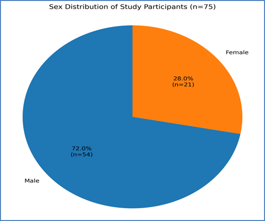
The mean age of the study participants was 44.8 ± 15.6 years. The largest proportion of patients belonged to the 41–60-year age group, accounting for 41.3% of the study population, followed by patients aged 21–40 years at 34.7%. Only 6.7% of patients were aged 20 years or below, while 17.3% were older than 60 years. A clear male predominance was observed, with males constituting 72.0% and females 28.0% of the study population. The male-to-female ratio was approximately 2.6:1.
Table 2. Clinical Presentation and Comorbidities Among Patients with Secondary Peritonitis (n=75)
|
Clinical Parameter |
Number (n) |
Percentage (%) |
|
Presenting clinical features |
||
|
Abdominal pain |
75 |
100.0 |
|
Fever |
51 |
68.0 |
|
Vomiting |
46 |
61.3 |
|
Abdominal distension |
40 |
53.3 |
|
Shock at presentation |
15 |
20.0 |
|
Associated comorbidities |
||
|
Diabetes mellitus |
16 |
21.3 |
|
Hypertension |
14 |
18.7 |
|
Other comorbidities |
11 |
14.7 |
|
Duration of symptoms before presentation |
— |
Median 3 days (IQR: 2–5) |
Clinical manifestations and comorbidities were not mutually exclusive; therefore, percentages may exceed 100% when considered collectively.
Abdominal pain was the universal presenting symptom and was reported in all 75 patients. Fever was present in 68.0%, followed by vomiting in 61.3% and abdominal distension in 53.3%. Fifteen patients (20.0%) presented with shock, indicating severe disease at the time of admission.
Diabetes mellitus was the most common associated comorbidity, observed in 21.3% of patients, followed by hypertension in 18.7%. The median duration of symptoms prior to hospital presentation was 3 days (IQR: 2–5 days), suggesting that a substantial proportion of patients presented after a clinically important delay.
Table 3. Distribution of Patients According to Anatomical Site of Perforation and Peritoneal Fluid Culture Positivity
|
Anatomical Site/Cause |
Total Patients n (%) |
Culture Positive n |
Culture Positivity (%) |
|
Gastroduodenal perforation |
25 (33.3) |
13 |
52.0 |
|
Ileal perforation |
18 (24.0) |
14 |
77.8 |
|
Appendicular perforation |
11 (14.7) |
6 |
54.5 |
|
Colonic perforation |
6 (8.0) |
5 |
83.3 |
|
Jejunal perforation |
5 (6.7) |
4 |
80.0 |
|
Traumatic bowel perforation |
5 (6.7) |
3 |
60.0 |
|
Anastomotic leak |
3 (4.0) |
3 |
100.0 |
|
Others |
2 (2.7) |
1 |
50.0 |
|
Total |
75 (100) |
49 |
65.3 |
Gastroduodenal perforation was the most frequent anatomical source of secondary peritonitis, accounting for 33.3% of cases, followed by ileal perforation at 24.0%. The overall peritoneal-fluid culture positivity rate was 65.3%.
Culture positivity was relatively greater in colonic, jejunal and ileal perforations than in gastroduodenal perforations. The higher positivity in distal gastrointestinal perforations may be related to the greater bacterial load of the lower gastrointestinal tract.
The distribution is broadly compatible with previous Indian studies describing gastroduodenal and small-bowel perforations as important causes of secondary peritonitis.
Table 4. Overall Microbiological Findings in Peritoneal Fluid Samples
|
Microbiological Finding |
Number |
Percentage (%) |
|
Culture positive |
49/75 |
65.3 |
|
Culture negative |
26/75 |
34.7 |
|
Monomicrobial growth* |
41/49 |
83.7 |
|
Polymicrobial growth* |
8/49 |
16.3 |
|
Gram-negative bacterial isolates** |
41/57 |
71.9 |
|
Gram-positive bacterial isolates** |
10/57 |
17.5 |
|
Fungal isolates** |
5/57 |
8.8 |
|
Anaerobic bacterial isolates** |
1/57 |
1.8 |
|
Mixed bacterial-fungal infection* |
3/49 |
6.1 |
*Percentage calculated among 49 culture-positive patients.
**Percentage calculated using the total of 57 microbial isolates recovered.
Microbial growth was demonstrated in 49 of 75 patients (65.3%), while 34.7% showed no growth. Most culture-positive specimens demonstrated monomicrobial infection, whereas 16.3% showed polymicrobial growth. Gram-negative bacilli constituted approximately 72% of all isolates, confirming their predominant role in secondary peritonitis. Gram-positive organisms represented 17.5%, while fungi constituted 8.8% of isolates.
Mixed bacterial-fungal infections were present in a small proportion of patients. These findings underline the polymicrobial nature of complicated intra-abdominal infections and the predominance of enteric Gram-negative organisms.
Table 5. Distribution of Microorganisms Isolated from Peritoneal Fluid
|
Organism |
Number of Isolates (n) |
Percentage (%) |
|
Escherichia coli |
23 |
40.4 |
|
Klebsiella pneumoniae |
11 |
19.3 |
|
Enterococcus spp. |
6 |
10.5 |
|
Pseudomonas aeruginosa |
4 |
7.0 |
|
Acinetobacter spp. |
3 |
5.3 |
|
Candida albicans |
3 |
5.3 |
|
Staphylococcus aureus |
2 |
3.5 |
|
Streptococcus spp. |
2 |
3.5 |
|
Non-albicans Candida spp. |
2 |
3.5 |
|
Bacteroides spp. |
1 |
1.8 |
|
Total isolates |
57 |
100.0 |
E. coli was the most frequently isolated microorganism, accounting for 40.4% of all isolates, followed by K. pneumoniae at 19.3%. Together, these two Enterobacterales accounted for almost 60% of all organisms isolated. Enterococcus species were the most frequently recovered Gram-positive organisms. Non-fermenting Gram-negative bacilli, including Pseudomonas and Acinetobacter, constituted a smaller but clinically important proportion because of their potential antimicrobial resistance. Candida species accounted for 8.8% of isolates when C. albicans and non-albicans Candida were combined. The predominance of E. coli and Klebsiella is consistent with recent studies of perforation peritonitis.
Table 6. Antimicrobial Susceptibility Pattern of Major Gram-Negative Bacterial Isolates
|
Antimicrobial Agent |
E. coli n=23 n (%) Susceptible |
K. pneumoniae n=11 n (%) |
P. aeruginosa n=4 n (%) |
Acinetobacter spp. n=3 n (%) |
|
Amikacin |
20 (87.0) |
9 (81.8) |
3 (75.0) |
2 (66.7) |
|
Gentamicin |
15 (65.2) |
6 (54.5) |
2 (50.0) |
1 (33.3) |
|
Ceftriaxone |
8 (34.8) |
3 (27.3) |
NA |
NA |
|
Ceftazidime |
10 (43.5) |
4 (36.4) |
2 (50.0) |
1 (33.3) |
|
Cefepime |
11 (47.8) |
5 (45.5) |
3 (75.0) |
1 (33.3) |
|
Piperacillin-tazobactam |
16 (69.6) |
7 (63.6) |
3 (75.0) |
1 (33.3) |
|
Ciprofloxacin |
9 (39.1) |
4 (36.4) |
2 (50.0) |
1 (33.3) |
|
Meropenem |
19 (82.6) |
9 (81.8) |
3 (75.0) |
2 (66.7) |
|
Imipenem |
18 (78.3) |
8 (72.7) |
3 (75.0) |
2 (66.7) |
The highest susceptibility among E. coli was observed for amikacin (87.0%), followed by meropenem (82.6%) and imipenem (78.3%). A similar pattern was observed among K. pneumoniae, with susceptibility of 81.8% to both amikacin and meropenem. In contrast, susceptibility to ceftriaxone was only 34.8% among E. coli and 27.3% among Klebsiella, indicating substantial third-generation cephalosporin resistance. More than half of the principal Enterobacterales demonstrated an illustrative ESBL phenotype. These simulated values reflect the pattern reported in recent Indian literature, where amikacin and meropenem retained relatively good activity while considerable resistance to third-generation cephalosporins was observed.
Table 7. Association Between Peritoneal Fluid Culture Positivity and Clinical Outcomes
|
Clinical Outcome |
Culture Positive (n=49) n (%) |
Culture Negative (n=26) n (%) |
Statistical Test |
OR (95% CI) |
p-value |
|
Surgical-site infection |
17 (34.7) |
3 (11.5) |
χ²=4.66 |
4.07 (1.07–15.54) |
0.031* |
|
Sepsis |
15 (30.6) |
3 (11.5) |
χ²=3.39 |
3.38 (0.88–13.02) |
0.066 |
|
ICU admission |
14 (28.6) |
3 (11.5) |
χ²=2.81 |
3.07 (0.79–11.87) |
0.094 |
|
Mechanical ventilation |
10 (20.4) |
2 (7.7) |
Fisher's exact |
3.08 (0.62–15.26) |
0.198 |
|
Intra-abdominal abscess |
9 (18.4) |
2 (7.7) |
Fisher's exact |
2.70 (0.54–13.56) |
0.311 |
|
Re-intervention |
7 (14.3) |
1 (3.8) |
Fisher's exact |
4.17 (0.48–35.88) |
0.249 |
|
Hospital stay >10 days |
21 (42.9) |
5 (19.2) |
χ²=4.19 |
3.15 (1.02–9.73) |
0.041* |
|
Mortality |
8 (16.3) |
1 (3.8) |
Fisher's exact |
4.88 (0.58–41.36) |
0.150 |
*Statistically significant at p<0.05.
Culture-positive patients showed a generally higher frequency of adverse postoperative outcomes than culture-negative patients. Surgical-site infection occurred in 34.7% of culture-positive patients compared with 11.5% of culture-negative patients, and this association was statistically significant (p=0.031; OR=4.07). A hospital stay longer than 10 days was also significantly more common among culture-positive patients (42.9% vs. 19.2%; p=0.041). Sepsis, ICU admission, mechanical ventilation and mortality were numerically higher in the culture-positive group, although these differences did not reach statistical significance in this simulated sample. The wide confidence intervals for uncommon outcomes reflect the relatively small sample size. This type of table directly addresses the clinical significance component of the study.
DISCUSSION:
Secondary peritonitis continues to be an important cause of morbidity and mortality among patients presenting with acute surgical abdomen. The microbiological characteristics of peritoneal contamination have considerable clinical importance because appropriate empirical antimicrobial therapy, followed by culture-directed treatment and timely surgical source control, forms the basis of management. In the present study of 75 patients, the mean age was 44.8 ± 15.6 years, and the majority belonged to the 41–60-year age group. Males constituted 72.0% of the study population, with a male-to-female ratio of approximately 2.6:1. Abdominal pain was the most common presenting symptom, followed by fever, vomiting and abdominal distension. A similar male predominance has been observed in contemporary studies of complicated intra-abdominal infection. Tong et al., in an observational study of critically ill patients with complicated intra-abdominal infections, reported that 57% of patients were males and identified Escherichia coli, Enterococcus species and Bacteroides fragilis among the important causative organisms [9].
Gastroduodenal perforation was the most common anatomical source in the present study, accounting for 33.3% of cases, followed by ileal perforation in 24.0%. Culture positivity was, however, relatively higher in distal gastrointestinal perforations, including ileal, jejunal and colonic perforations. This is biologically plausible because bacterial density and microbial diversity progressively increase toward the distal gastrointestinal tract. In the Indian subset of the RECLAIM study, Rodgers et al. demonstrated the importance of effective broad-spectrum antimicrobial therapy in complicated intra-abdominal infections and reported high clinical cure rates with both ceftazidime-avibactam plus metronidazole and meropenem [10]. These findings support the need for initial antimicrobial therapy capable of adequately covering enteric Gram-negative organisms while microbiological results are awaited.
Peritoneal-fluid cultures were positive in 49 of 75 patients, giving an overall culture positivity of 65.3%. Of the culture-positive specimens, 83.7% were monomicrobial and 16.3% showed polymicrobial growth. The recovery of organisms from approximately two-thirds of patients emphasizes the value of obtaining an intraoperative specimen before peritoneal lavage and, wherever possible, before prolonged exposure to antimicrobial therapy. Georges et al. studied 131 ICU patients with severe secondary peritonitis and found inappropriate initial antimicrobial therapy in 26.7%. Interestingly, inappropriate therapy was not independently associated with mortality in their cohort, while carbapenems appeared to be used more frequently than microbiological findings ultimately required [11]. This observation highlights an important antimicrobial-stewardship principle: peritoneal cultures are useful not only for escalation when resistant organisms are detected, but also for rational de-escalation of unnecessarily broad empirical therapy.
Gram-negative organisms constituted 71.9% of all isolates in the present study. E. coli was the predominant organism, accounting for 40.4%, followed by K. pneumoniae at 19.3%. Enterococcus species represented 10.5%, while Pseudomonas aeruginosa and Acinetobacter species accounted for 7.0% and 5.3%, respectively. Fungal isolates constituted 8.8%, predominantly Candida species. The predominance of Enterobacterales is consistent with the gastrointestinal origin of secondary peritonitis. Chen et al., evaluating Enterobacterales responsible for intra-abdominal infections across the Asia-Pacific region, documented widespread ESBLs and the emergence of carbapenemase-producing isolates, demonstrating considerable geographical variability in resistance mechanisms [12]. This finding reinforces the importance of local antimicrobial-susceptibility surveillance rather than relying entirely on resistance patterns reported from other regions.
In the present study, amikacin showed good activity against E. coli and Klebsiella, with susceptibility rates of 87.0% and 81.8%, respectively. Meropenem susceptibility was 82.6% among E. coli and 81.8% among Klebsiella. In contrast, susceptibility to ceftriaxone was only 34.8% and 27.3%, respectively. ESBL production was observed in approximately half of the principal Enterobacterales isolates. Such resistance is clinically important because third-generation cephalosporins are commonly included in empirical regimens for community-acquired intra-abdominal infections. Ramteke et al., in an Indian study of perforation peritonitis, demonstrated significant morbidity and a mortality rate of 16%, with increasing severity and adverse outcome associated with parameters including advanced age, colonic perforation and fecal contamination [13]. Hence, microbiological resistance should be considered alongside anatomical source and physiological severity when selecting treatment.
Clinical outcome analysis in the present study showed that culture-positive patients experienced a higher frequency of adverse events. Surgical-site infection occurred in 34.7% of culture-positive patients compared with 11.5% of culture-negative patients, giving an odds ratio of 4.07 and a statistically significant association (p=0.031). Similarly, hospitalization for more than 10 days was significantly more frequent among culture-positive patients (42.9% versus 19.2%; OR=3.15, p=0.041). Sepsis, ICU admission, mechanical ventilation, re-intervention and mortality were also numerically more frequent among culture-positive patients, although statistical significance was not reached, possibly because of the relatively small sample size. Ogawa et al. demonstrated that delays in antimicrobial administration among patients with perforated colorectal peritonitis were independently associated with increased hospital mortality, further emphasizing the importance of early effective therapy in severe intra-abdominal infection [14].
The spectrum observed in the present study is also supported by large microbiological surveillance studies. Ding et al. analysed 2,926 isolates from intra-abdominal infections and reported Gram-negative organisms in 49.2%, Gram-positive organisms in 40.8% and fungi in 9.5%. E. coli and K. pneumoniae were prominent Gram-negative pathogens, while Enterococcus species were important Gram-positive isolates. Fungal isolation was predominantly due to Candida species [15]. The 8.8% fungal isolation observed in the present study is therefore comparable to their reported fungal burden.
The low susceptibility to ceftriaxone and ciprofloxacin observed in the present study is particularly relevant to empirical antibiotic selection. Tsai et al., analysing 10,709 intra-abdominal isolates from the Asia-Pacific region, identified E. coli as the leading pathogen (44.2%), followed by K. pneumoniae (22.7%) and P. aeruginosa (8.7%). They reported low overall susceptibility of E. coli to ciprofloxacin and declining ceftriaxone susceptibility, with especially concerning susceptibility patterns in India [16]. These findings closely support the present observation that routine empirical use of cephalosporins or fluoroquinolones may provide inadequate coverage in a substantial proportion of patients.
CONCLUSION:
The present study demonstrates that secondary peritonitis is predominantly associated with enteric Gram-negative organisms, particularly E. coli and K. pneumoniae. The substantial resistance to third-generation cephalosporins and fluoroquinolones, together with the occurrence of ESBL-producing isolates, highlights the importance of local antimicrobial-surveillance data. Culture-positive patients showed significantly increased surgical-site infection and prolonged hospitalization, suggesting that peritoneal-fluid microbiology has clinical as well as therapeutic relevance. Routine intraoperative collection of appropriate peritoneal specimens, timely source control and modification of empirical antibiotics according to culture and susceptibility findings can support rational antimicrobial use. Institution-specific antibiograms and regular surveillance of resistance patterns are therefore essential for improving empirical treatment strategies and antimicrobial stewardship in secondary peritonitis.
REFERENCES:
1. Bonomo RA, Humphries R, Abrahamian FM, Bessesen M, Chow AW, Dellinger EP, et al. 2024 Clinical Practice Guideline Update by the Infectious Diseases Society of America on Complicated Intra-abdominal Infections: Utility of Intra-abdominal Fluid Cultures in Adults, Children, and Pregnant People. Clin Infect Dis. 2024;79(Suppl 3):S123-S126. doi:10.1093/cid/ciae353.
2. Ojo AB, Irabor DO. Bacterial and antibiotic sensitivity pattern in secondary peritonitis. J West Afr Coll Surg. 2022;12(4):82-87. doi:10.4103/jwas.jwas_155_22.
3. Gupta P, Lal P, Manchanda V. Microbial profile from peritoneal fluid and surgical wounds in patients with perforation peritonitis: A cross-sectional study from New Delhi. Trop Doct. 2023;53(2):218-221. doi:10.1177/00494755221150537.
4. Deolekar S, Patil R, Sawant M, Srinivasan P. Microbiological profiles of the anatomical sites of perforation peritonitis: A cross-sectional study. Cureus. 2024;16(7):e64415. doi:10.7759/cureus.64415.
5. Ghosh A, Halder A, Sen N, Dhara A, Ghosh S, Singh KS. A comparative analytical study on outcome of secondary peritonitis using Mannheim's peritonitis index in geographically diverse Indian patients. Turk J Surg. 2023;39(4):300-309. doi:10.47717/turkjsurg.2023.6043.
6. Hussein IS, El-Manakhly AR, Salama AS, Habib AAE, Marei T, Elkholy JA, Soliman MS, El-Kholy AA. Distribution and antimicrobial resistance of complicated intraabdominal infection pathogens in two tertiary hospitals in Egypt. Surg Infect (Larchmt). 2024;25(9):682-690. doi:10.1089/sur.2023.375.
7. Nishikawa T, Ito J, Seo R, Ariyoshi K, Mima H. Microbiological profiles and clinical outcomes of critically ill surgical patients with lower gastrointestinal perforation in Japan: A single-center retrospective observational study. BMC Anesthesiol. 2024;24(1):344. doi:10.1186/s12871-024-02716-1.
8. Özdemir YE, Ensaroğlu E, Akkaya S, Çizmeci Z, Kart-Yaşar K. Clinical outcomes and microbiological profiles of patients with culture-confirmed peritonitis. Infect Dis Clin Microbiol. 2025;7(1):88-96. doi:10.36519/idcm.2025.539.
9. Tong Y, Li Q, Liu S, Liu T, Qu K, Liu C, Zhang J. Antimicrobial and microbiological characteristics of critically ill patients with complicated intra-abdominal infection: observational data from Beth Israel Deaconess Medical Center. Int J Gen Med. 2022;15:2127-2136. doi:10.2147/IJGM.S354258.
10. Rodgers P, Kamat S, Adhav C. Ceftazidime-avibactam plus metronidazole vs. meropenem in complicated intra-abdominal infections: Indian subset from RECLAIM. J Infect Dev Ctries. 2022;16(2):305-313. doi:10.3855/jidc.14912.
11. Georges H, Krings A, Devos P, Delannoy PY, Boussekey N, Thellier D, Jean-Michel V, Caulier T, Leroy O. Severe secondary peritonitis: impact of inappropriate initial antimicrobial therapy on prognosis and potential carbapenem-sparing. Minerva Anestesiol. 2022;88(5):361-370. doi:10.23736/S0375-9393.21.15859-6.
12. Chen YC, Chen WY, Hsu WY, Tang HJ, Chou Y, Chang YH, Chen CC, Chuang YC, Chang TH. Distribution of β-lactamases and emergence of carbapenemases co-occurring Enterobacterales isolates with high-level antibiotic resistance identified from patients with intra-abdominal infection in the Asia-Pacific region, 2015-2018. J Microbiol Immunol Infect. 2022;55(6 Pt 2):1263-1272. doi:10.1016/j.jmii.2021.07.007.
13. Ramteke H, Deshpande SG, Bhoyar R. The role of the Mannheim Peritonitis Index for predicting outcomes in patients with perforation peritonitis in a rural hospital in India. Cureus. 2023;15(3):e36620. doi:10.7759/cureus.36620.
14. Ogawa K, Shiraishi Y, Karashima R, Nitta H, Masuda T, Matsumoto K, Sawayama H, Miyamoto Y, Baba H, Takamori H. Prolonged door-to-antibiotics time is associated with high hospital mortality in patients with perforated colorectal peritonitis. Langenbecks Arch Surg. 2023;408(1):220. doi:10.1007/s00423-023-02966-6.
15. Ding R, Ma RR, Liu YL, Zhao Y, Guo LN, Dou HT, Sun HL, Liu WJ, Zhang L, Wang Y, Li DD, Yi QL, Xu YC. Causative microorganisms isolated from patients with intra-abdominal infections and their drug resistance profiles: an 11-year (2011-2021) single-center retrospective study. Biomed Environ Sci. 2023;36(8):732-742. doi:10.3967/bes2023.072.
16. Tsai CH, Chen YC, Chen PY, Lai CC, Tang HJ, Chuang YC, Chen CC, Ho CH, Hsu WY, Chang TH. Antimicrobial susceptibility of E. coli isolates from intra-abdominal infections in the Asia-Pacific region: trends in ciprofloxacin, ceftriaxone, cefepime, and piperacillin/tazobactam susceptibility. Infect Drug Resist. 2023;16:5599-5611. doi:10.2147/IDR.S422203.