A Prospective Study Of Changes In Intraocular Pressure And Central Corneal Thickness After Congenital Cataract Surgery.
- Jayeeta Mazumder , Assistant Professor, MBBS, MS, Department Of Ophthalmology, Santiniketan Medical College.Gobindapur. Muluk.Bolpur. West Bengal 731240.
- Mahesh Chattopadhyay , Assistant Professor, MBBS, MS, Department Of Ophthalmology, Santiniketan Medical College.Gobindapur. Muluk.Bolpur. West Bengal 731240.
Article Information:
Abstract:
Introduction: Congenital cataract surgery is essential for preventing visual impairment in children; however, postoperative changes in intraocular pressure (IOP) and central corneal thickness (CCT) may influence ocular health and the interpretation of IOP measurements. Regular monitoring of these parameters is therefore important for early detection of postoperative ocular hypertension and glaucoma. Aims: To evaluate the changes in CCT and IOP following congenital cataract surgery and to assess their variation according to age, gender, and postoperative lens status. Materials and methods: A prospective observational study was conducted among 50 children diagnosed with congenital cataract who underwent cataract surgery and attended the Ophthalmology OPD of the Regional Institute of Ophthalmology, Medical College and Hospital, Kolkata, over an 18-month period. CCT and IOP were assessed preoperatively and at 1, 6, and 12 months postoperatively. The parameters were compared across different age groups, genders, and postoperative lens statuses. Result: Mean CCT increased progressively during follow-up from the preoperative assessment through 1, 6, and 12 months, with the overall change being statistically significant (p < 0.0001). IOP showed a slight initial reduction at 1 month followed by a progressive increase at 6 and 12 months, also demonstrating a statistically significant overall change (p < 0.0001). Younger children showed greater postoperative increases in CCT and IOP, particularly at 12 months. Gender-related differences were limited, with a significant difference in CCT at 6 months but no significant difference in IOP. Aphakic eyes demonstrated significantly higher CCT and IOP at 12 months than pseudophakic eyes (p < 0.001). Conclusion: Congenital cataract surgery is associated with significant longitudinal changes in CCT and IOP. Younger children and aphakic eyes require particular attention during follow-up. Assessment of CCT along with IOP may improve postoperative monitoring and facilitate early identification of ocular hypertension and glaucoma.
Keywords:
Article :
INTRODUCTION:
Congenital cataract refers to lens opacity present at birth or developing during the early period of life. It is an important cause of preventable childhood visual impairment because an optically significant cataract can interfere with normal visual development and lead to deprivation amblyopia. Early recognition and appropriate management are therefore essential for achieving satisfactory visual rehabilitation. The timing of surgery is particularly important in congenital cataract because prolonged visual deprivation may result in irreversible abnormalities of visual development. Surgical management is generally recommended when the cataract is visually significant, followed by appropriate optical correction and amblyopia management.[1][2]
Congenital cataract surgery has evolved considerably, with modern techniques allowing removal of the opaque lens and restoration of the visual axis through aphakic correction or primary intraocular lens implantation in appropriately selected children. However, surgery in the developing eye is associated with several short- and long-term considerations. The immature eye undergoes continuous anatomical growth and changes in ocular dimensions, while the postoperative eye may develop alterations involving the anterior segment, cornea and intraocular pressure. Consequently, successful cataract extraction does not eliminate the need for prolonged postoperative surveillance.[3]
One of the important complications following congenital cataract surgery is secondary glaucoma or postoperative ocular hypertension. The risk may persist for several years after surgery, making regular assessment of intraocular pressure (IOP) an important component of postoperative follow-up. The reported incidence of secondary glaucoma varies considerably between studies depending on patient age, duration of follow-up, type of cataract, surgical technique and whether an intraocular lens was implanted. Recent evidence indicates that secondary glaucoma remains a clinically relevant postoperative complication, with the risk influenced by factors including age at surgery and postoperative lens status.[4]˒[5] Early identification of abnormal IOP is important because sustained elevation may result in optic nerve damage and progressive visual impairment.
Assessment of IOP in children following congenital cataract surgery can, however, be influenced by changes in the corneal structure. Central corneal thickness (CCT) is an important parameter because the thickness and biomechanical properties of the cornea can affect tonometric estimation of IOP. A thicker cornea may result in an overestimation of the measured IOP, whereas a thinner cornea may have the opposite effect. Therefore, an apparently elevated IOP in a postoperative child should be interpreted in conjunction with CCT rather than in isolation.[6]
Several studies have demonstrated that CCT may increase following congenital cataract surgery, particularly in eyes that remain aphakic. Postoperative thickening of the central cornea has been demonstrated at different periods of follow-up, and the magnitude of change may vary according to the type of surgery and age at the time of surgery. In prospective longitudinal observations, significant increases in CCT have been reported after surgery, with greater changes observed in younger children and aphakic eyes.[7] These findings are clinically important because postoperative changes in CCT may influence the interpretation of IOP measurements and, consequently, the assessment of glaucoma risk.
The relationship between CCT and IOP is particularly relevant in children undergoing congenital cataract surgery. Evidence from prospective studies has shown that both parameters may change after surgery, and a positive association between changes in CCT and IOP has been observed during follow-up. In children undergoing primary intraocular lens implantation, postoperative increases in CCT have also been accompanied by changes in IOP, emphasizing the importance of assessing these two parameters together.[8] Thus, serial measurement of IOP and CCT may provide a more appropriate assessment of postoperative ocular status than measurement of IOP alone.
Therefore, the present prospective study is planned to evaluate the changes in intraocular pressure and central corneal thickness following congenital cataract surgery and to assess the relationship between these parameters during postoperative follow-up. A better understanding of these changes may contribute to more accurate interpretation of IOP, timely identification of children at risk of postoperative ocular hypertension or glaucoma, and improved long-term postoperative care.
MATERIALS AND METHODS:
Study design: Prospective observational study.
Place of study: OPD at Regional Institute Of Ophthalmology (RIO), Medical College and Hospital, Kolkata (RIO)
Period of study: 18 months
Study Population: Patients diagnosed with congenital cataract who undergo cataract surgery and attend the Ophthalmology OPD at Regional Institute of Ophthalmology (RIO), Medical College and Hospital, Kolkata, during the study period.
Sample size: 50
Inclusion Criteria:
• Children diagnosed with congenital cataract requiring surgical management.
• Children undergoing congenital cataract surgery at the study centre during the study period.
• Age appropriate for pediatric congenital cataract surgery and postoperative follow-up.
• Patients whose parents/legally authorized guardians provide written informed consent for participation.
• Patients available for regular postoperative follow-up and IOP and CCT assessment.
Exclusion Criteria:
• Cataract secondary to ocular trauma, uveitis, or other acquired ocular conditions.
• Pre-existing glaucoma or ocular hypertension before cataract surgery.
• Associated major ocular abnormalities or congenital anterior segment anomalies likely to affect IOP or CCT.
• Previous intraocular surgery in the study eye.
• Patients with systemic or neurological conditions that significantly affect ocular development or postoperative assessment.
• Patients who fail to attend the scheduled postoperative follow-up visits or in whom reliable IOP/CCT measurements cannot be obtained.
Statistical Analysis: For statistical analysis, data were initially entered into a Microsoft Excel spreadsheet and then analyzed using SPSS (version 27.0; SPSS Inc., Chicago, IL, USA) and GraphPad Prism (version 5). Numerical variables were summarized using means and standard deviations, while Data were entered into Excel and analyzed using SPSS and GraphPad Prism. Numerical variables were summarized using means and standard deviations, while categorical variables were described with counts and percentages. Two-sample t-tests were used to compare independent groups, while paired t-tests accounted for correlations in paired data. Chi-square tests (including Fisher’s exact test for small sample sizes) were used for categorical data comparisons. P-values ≤ 0.05 were considered statistically significant.
RESULTS:
Table 1. Baseline characteristics of study participants
|
Variable |
Category |
n (%) |
|
Age group (months) |
≤20 |
16 (32.0) |
|
21–30 |
17 (34.0) |
|
|
31–40 |
12 (24.0) |
|
|
>40 |
5 (10.0) |
|
|
Gender |
Female |
18 (36.0) |
|
Male |
32 (64.0) |
|
|
Postoperative status |
Aphakia |
16 (32.0) |
|
Pseudophakia |
34 (68.0) |
|
|
Total |
50 (100.0) |
|
Table 2. Changes in CCT and IOP during follow-up
|
Parameter |
Follow-up |
Mean ± SD |
p-value |
|
CCT (µm) |
Preoperative |
553.56 ± 29.30 |
<0.0001 |
|
1 month |
556.88 ± 28.52 |
||
|
6 months |
569.30 ± 34.05 |
||
|
12 months |
580.36 ± 35.84 |
||
|
IOP (mmHg) |
Preoperative |
12.38 ± 2.05 |
<0.0001 |
|
1 month |
11.88 ± 1.76 |
||
|
6 months |
15.12 ± 2.37 |
||
|
12 months |
16.24 ± 3.24 |
Table 3. Preoperative CCT and IOP according to age group
|
Age group (months) |
CCT (µm), Mean ± SD |
p-value |
IOP (mmHg), Mean ± SD |
p-value |
|
≤20 |
563.31 ± 10.14 |
0.0831 |
13.00 ± 2.97 |
0.3551 |
|
21–30 |
557.94 ± 38.34 |
12.29 ± 1.16 |
||
|
31–40 |
544.00 ± 27.22 |
11.58 ± 1.73 |
||
|
>40 |
530.40 ± 28.38 |
12.60 ± 1.14 |
Table 4. Postoperative CCT and IOP according to age group
|
Follow-up |
Age group |
CCT (µm), Mean ± SD |
p-value |
IOP (mmHg), Mean ± SD |
p-value |
|
1 month |
≤20 |
571.69 ± 6.96 |
0.0235 |
12.31 ± 2.57 |
0.6637 |
|
21–30 |
556.88 ± 36.31 |
11.59 ± 1.06 |
|||
|
31–40 |
546.58 ± 26.10 |
11.67 ± 1.30 |
|||
|
>40 |
534.20 ± 28.18 |
12.00 ± 1.58 |
|||
|
6 months |
≤20 |
585.44 ± 10.54 |
0.0596 |
15.81 ± 3.35 |
0.556 |
|
21–30 |
569.71 ± 45.85 |
14.65 ± 1.54 |
|||
|
31–40 |
555.08 ± 26.78 |
14.92 ± 2.11 |
|||
|
>40 |
550.40 ± 36.14 |
15.00 ± 1.41 |
|||
|
12 months |
≤20 |
608.00 ± 12.44 |
0.0001 |
19.06 ± 3.89 |
0.0001 |
|
21–30 |
578.00 ± 40.40 |
15.06 ± 2.01 |
|||
|
31–40 |
561.42 ± 27.62 |
14.67 ± 1.56 |
|||
|
>40 |
545.40 ± 29.12 |
15.00 ± 1.41 |
Table 5. Postoperative CCT and IOP according to gender
|
Follow-up |
Gender |
CCT (µm), Mean ± SD |
p-value |
IOP (mmHg), Mean ± SD |
p-value |
|
1 month |
Female |
547.22 ± 21.95 |
0.0721 |
11.50 ± 1.47 |
1.1507 |
|
Male |
562.31 ± 30.60 |
12.09 ± 1.89 |
|||
|
6 months |
Female |
554.83 ± 23.72 |
0.0226 |
15.06 ± 1.70 |
0.8871 |
|
Male |
577.44 ± 36.52 |
15.16 ± 2.70 |
|||
|
12 months |
Female |
568.44 ± 30.17 |
0.0776 |
15.83 ± 1.82 |
0.5116 |
|
Male |
587.06 ± 37.45 |
16.47 ± 3.83 |
Table 6. Association between postoperative status and CCT and IOP at 12 months
|
Postoperative status |
CCT (µm), Mean ± SD |
IOP (mmHg), Mean ± SD |
P-value |
|
Aphakia |
608.00 ± 12.43 |
19.06 ± 3.89 |
<0.001 |
|
Pseudophakia |
567.35 ± 35.95 |
14.91 ± 1.74 |
<0.001 |
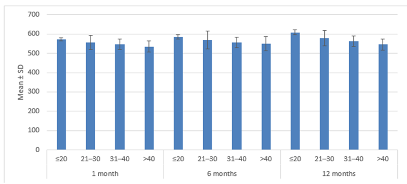
Figure 1. Postoperative CCT according to age group
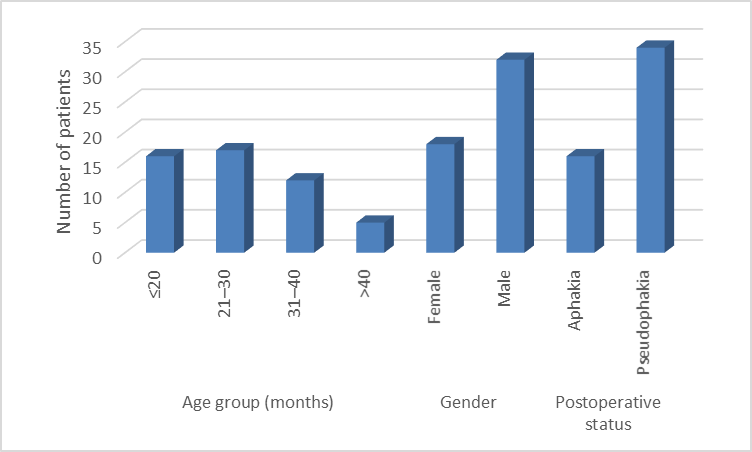
Figure 2. Baseline characteristics of study participants
Table 1 shows the baseline demographic and postoperative characteristics of the 50 study participants. The majority of participants were in the 21–30 months age group (34.0%), followed by ≤20 months (32.0%), 31–40 months (24.0%), and >40 months (10.0%). The mean age was 51.00 ± 36.17 months. Males constituted the majority of the study population (64.0%), while females accounted for 36.0%. Regarding postoperative status, 68.0% of participants were pseudophakic and 32.0% were aphakic.
Table 2 demonstrates the longitudinal changes in CCT and IOP following congenital cataract surgery. Mean CCT increased progressively from 553.56 ± 29.30 µm preoperatively to 556.88 ± 28.52 µm at 1 month, 569.30 ± 34.05 µm at 6 months, and 580.36 ± 35.84 µm at 12 months. This overall change was statistically significant (p < 0.0001). Similarly, mean IOP was 12.38 ± 2.05 mmHg preoperatively, decreased slightly to 11.88 ± 1.76 mmHg at 1 month, and subsequently increased to 15.12 ± 2.37 mmHg at 6 months and 16.24 ± 3.24 mmHg at 12 months. The change in IOP over follow-up was also statistically significant (p < 0.0001).
Table 3 evaluates preoperative CCT and IOP according to age group. The highest mean preoperative CCT was observed among children aged ≤20 months (563.31 ± 10.14 µm), while the lowest was observed in the 41–50 months group (530.40 ± 28.38 µm). However, the difference in preoperative CCT between age groups was not statistically significant (p = 0.0831). Similarly, mean preoperative IOP ranged from 11.58 ± 1.73 mmHg in the 31–40 months group to 13.00 ± 2.97 mmHg in the ≤20 months group, with no statistically significant difference between age groups (p = 0.3551).
Table 4 demonstrates postoperative CCT and IOP according to age group at different follow-up intervals. At 1 month, mean CCT was highest in the ≤20 months group (571.69 ± 6.96 µm) and lowest in the >40 months group (534.20 ± 28.18 µm), with a statistically significant difference (p = 0.0235). At 6 months, the highest CCT was again observed in the ≤20 months group (585.44 ± 10.54 µm), while the lowest was observed in the >40 months group (550.40 ± 36.14 µm); however, the difference was not statistically significant (p = 0.0596). At 12 months, CCT increased further, reaching 608.00 ± 12.44 µm in the ≤20 months group compared with 545.40 ± 29.12 µm in the >40 months group, with a highly significant difference (p = 0.0001). In contrast, IOP did not differ significantly between age groups at 1 month (p = 0.6637) or 6 months (p = 0.5560). At 12 months, however, a significant age-related difference was observed (p = 0.0001), with the highest IOP in the ≤20 months group (19.06 ± 3.89 mmHg) and lower values in the older groups. These findings suggest that younger children demonstrated greater postoperative increases in both CCT and IOP, particularly by 12 months.
Table 5 presents postoperative CCT and IOP according to gender. At 1 month, mean CCT was 547.22 ± 21.95 µm among females and 562.31 ± 30.60 µm among males, with no statistically significant difference (p = 0.0721). At 6 months, however, males had significantly higher mean CCT than females (577.44 ± 36.52 µm vs. 554.83 ± 23.72 µm; p = 0.0226). By 12 months, mean CCT remained higher among males (587.06 ± 37.45 µm) than females (568.44 ± 30.17 µm), although the difference was not statistically significant (p = 0.0776). For IOP, there was no statistically significant difference between males and females at 6 months (15.16 ± 2.70 vs. 15.06 ± 1.70 mmHg; p = 0.8871) or at 12 months (16.47 ± 3.83 vs. 15.83 ± 1.82 mmHg; p = 0.5116).
Table 6 compares CCT and IOP at 12 months according to postoperative lens status. The mean CCT was substantially higher in aphakic eyes (608.00 ± 12.43 µm) than in pseudophakic eyes (567.35 ± 35.95 µm), and this difference was statistically significant (p < 0.001). Similarly, mean IOP at 12 months was considerably higher among aphakic eyes (19.06 ± 3.89 mmHg) compared with pseudophakic eyes (14.91 ± 1.74 mmHg), with a statistically significant difference (p < 0.001). The association between postoperative status and gender was also statistically significant (χ² = 5.64, p = 0.0175), with aphakia being more frequent among males.
DISCUSSION:
The present prospective study evaluated changes in central corneal thickness (CCT) and intraocular pressure (IOP) following congenital cataract surgery in 50 children. Mean CCT increased progressively from 553.56 ± 29.30 µm preoperatively to 556.88 ± 28.52 µm at 1 month, 569.30 ± 34.05 µm at 6 months, and 580.36 ± 35.84 µm at 12 months, with the overall change being statistically significant (p < 0.0001). Similarly, mean IOP was 12.38 ± 2.05 mmHg preoperatively, decreased slightly to 11.88 ± 1.76 mmHg at 1 month, and subsequently increased to 15.12 ± 2.37 mmHg at 6 months and 16.24 ± 3.24 mmHg at 12 months (p < 0.0001). Resende, Lupinacci, Árieta, Costa and Costa reported a significant postoperative increase in CCT from 556.24 ± 44.19 µm to 585.07 ± 56.45 µm following congenital cataract surgery, along with an increase in IOP from 12.05 ± 2.3 mmHg to 13.89 ± 2.96 mmHg. These findings are consistent with the progressive postoperative changes observed in the present study.[7]. The increase in CCT in the present study was particularly evident among younger children. At 1 month, mean CCT was 571.69 ± 6.96 µm in children aged ≤20 months compared with 534.20 ± 28.18 µm among those aged >40 months (p = 0.0235). By 12 months, CCT had increased to 608.00 ± 12.44 µm in the youngest group compared with 545.40 ± 29.12 µm in the oldest group (p = 0.0001). Lim, Muir, Duncan and Freedman demonstrated a significant increase in CCT after removal of childhood cataracts and reported that postoperative corneal thickening was greater in younger children.[9] Muir, Duncan, Enyedi, Wallace and Freedman also reported increased CCT in children with congenital cataract, particularly in aphakic eyes.[6] These observations support the present finding that age at surgery may influence postoperative corneal thickness changes.
Postoperative lens status was strongly associated with CCT in the present study. At 12 months, aphakic eyes had a mean CCT of 608.00 ± 12.43 µm, compared with 567.35 ± 35.95 µm in pseudophakic eyes (p < 0.001). Faramarzi, Javadi, Jabbarpoor Bonyadi and Yaseri similarly found significantly greater CCT in aphakic eyes than in pseudophakic eyes after congenital cataract surgery.[10] Chang, Kou, Zhang, Wang, Tian and Zhao also reported a substantially greater increase in CCT in aphakic eyes than in pseudophakic eyes during postoperative follow-up.[11] These findings suggest that aphakia is associated with more pronounced postoperative corneal thickening. The greater CCT observed in younger and aphakic eyes may be clinically important because corneal thickness can influence the interpretation of tonometric IOP measurements. The present study also demonstrated a significant increase in IOP during follow-up, particularly at 6 and 12 months. At 12 months, IOP was 19.06 ± 3.89 mmHg in aphakic eyes compared with 14.91 ± 1.74 mmHg in pseudophakic eyes (p < 0.001). Nilforushan, Ghasemi Falavarjani, Razeghinejad and Bakhtiari reported higher IOP in children after congenital cataract surgery compared with controls, emphasizing the importance of long-term surveillance for postoperative ocular hypertension and glaucoma.[12] Simon, O'Malley, Gandham, Ghaiy, Zobal-Ratner and Simmons also demonstrated increased CCT in aphakic and pseudophakic children and highlighted the relevance of CCT when interpreting IOP in these patients.[13] Sukhija and Kaur reported a postoperative increase in both CCT and IOP in children younger than 2 years undergoing congenital cataract surgery with IOL implantation, with a significant relationship between changes in CCT and IOP.[8].
CONCLUSION:
The present prospective study demonstrated significant postoperative changes in both central corneal thickness and intraocular pressure following congenital cataract surgery. Central corneal thickness showed a progressive increase during postoperative follow-up, while intraocular pressure also increased after the initial postoperative period. These changes were more pronounced among younger children and aphakic eyes. Aphakic eyes demonstrated greater postoperative central corneal thickness and intraocular pressure compared with pseudophakic eyes. The findings highlight the importance of assessing central corneal thickness alongside intraocular pressure, as changes in corneal thickness may influence the interpretation of tonometric measurements. Regular and long-term postoperative monitoring is therefore essential, particularly in younger children and aphakic eyes, for early identification of ocular hypertension and postoperative glaucoma and for ensuring better long-term visual outcomes.
REFERENCES:
1. Mohammadpour M, Shaabani A, Sahraian A, Momenaei B, Tayebi F, Bayat R, Mirshahi R. Updates on managements of pediatric cataract. Journal of Current Ophthalmology. 2019 Jun 1;31(2):118-26.
2. Lloyd IC, Ashworth J, Biswas S, Abadi RV. Advances in the management of congenital and infantile cataract. Eye. 2007 Oct;21(10):1301-9.
3. Lagrèze WA. The management of cataract in childhood. Klinische Monatsblatter fur Augenheilkunde. 2009 Jan 27;226(1):15-21.
4. Li L, Wang X, Liu C, Wang S, Wang X. Incidence rate of secondary glaucoma following congenital cataract surgery: an in-depth systematic review and meta-analysis. American Journal of Ophthalmology. 2024 Sep 1;265:176-88.
5. Wang J, Chen J, Chen W, Wang Q, Zhao L, Wang R, Liu Z, Chen H, Cao Q, Tan X, Lin Z. Incidence of and risk factors for suspected glaucoma and glaucoma after congenital and infantile cataract surgery: a longitudinal study in China. Journal of glaucoma. 2020 Jan 1;29(1):46-52.
6. Muir KW, Duncan L, Enyedi LB, Wallace DK, Freedman SF. Central corneal thickness: congenital cataracts and aphakia. American journal of ophthalmology. 2007 Oct 1;144(4):502-6.
7. Resende GM, Lupinacci ÁP, Arieta CE, Costa VP. Central corneal thickness and intraocular pressure in children undergoing congenital cataract surgery: a prospective, longitudinal study. British Journal of Ophthalmology. 2012 Sep;96(9):1190-4.
8. Sukhija J, Kaur S. Central corneal thickness and intraocular pressure changes after congenital cataract surgery with intraocular lens implantation in children younger than 2 years. Journal of Cataract & Refractive Surgery. 2017 May 1;43(5):662-6.
9. Lim Z, Muir KW, Duncan L, Freedman SF. Acquired central corneal thickness increase following removal of childhood cataracts. American journal of ophthalmology. 2011 Mar 1;151(3):434-41.
10. Faramarzi A, Javadi MA, Bonyadi MH, Yaseri M. Changes in central corneal thickness after congenital cataract surgery. Journal of Cataract & Refractive Surgery. 2010 Dec 1;36(12):2041-7.
11. Chang P, Kou J, Zhang B, Wang J, Tian T, Zhao YE. Postoperative changes in central corneal thickness and intraocular pressure in a prospective cohort of congenital cataract patients. Cornea. 2020 Nov 1;39(11):1359-65.
12. Nilforushan N, Falavarjani KG, Razeghinejad MR, Bakhtiari P. Cataract surgery for congenital cataract: endothelial cell characteristics, corneal thickness, and impact on intraocular pressure. Journal of American Association for Pediatric Ophthalmology and Strabismus. 2007 Apr 1;11(2):159-61.
13. Simon JW, O’Malley MR, Gandham SB, Ghaiy R, Zobal-Ratner J, Simmons ST. Central corneal thickness and glaucoma in aphakic and pseudophakic children. Journal of American Association for Pediatric Ophthalmology and Strabismus. 2005 Aug 1;9(4):326-9.