Morphological Study Of Origin Of Profunda Femoris Artery In Human Cadavers: A Descriptive Cadaveric Study.
- Lingaswamy Veeramalla , Associate Professor, Department of Anatomy, Neelima Institute of Medical Sciences, Hyderabad, Telangana, India.
- Mithil Potuganti , Professor, Department of Anatomy, CMR Institute of Medical Sciences, Hyderabad, Telangana, India.
- Uday Kumar , Professor & Head, Department of Anatomy, Neelima Institute of Medical Sciences, Hyderabad, Telangana, India.
- Pawar S. E. , Professor & Head, Department of Anatomy, Vikhe Patil Institute of Medical Sciences, Ahilyanagar, Maharashtra, India.
Article Information:
Abstract:
Background: The profunda femoris artery (PFA) is the principal deep arterial supply of the thigh and an important collateral channel in the lower limb. Variation in its level and surface of origin from the femoral artery has practical implications for femoral arterial access, vascular reconstruction, orthopaedic procedures, and interpretation of angiographic studies. Objectives: To document the morphological patterns of origin of the PFA in adult human cadaveric lower limbs and to describe clinically relevant variations in its level and direction of origin. Methods: A descriptive cadaveric study was performed in the Department of Anatomy, Vikhea Patil Institute of Medical Sciences,Ahilyanagar,Maharastra and Neelima Institute of Medical Sciences, Hyderabad, Telangana, from January 2023 to January 2026. A total of 102 lower limbs from adult formalin-fixed cadavers used for routine undergraduate and postgraduate dissection were examined. The femoral triangle was dissected according to standard Cunningham-based dissection principles. The PFA was exposed from its point of origin, and the level and surface of origin in relation to the femoral artery were recorded. Representative variants were photographed. Results: All 102 lower limbs were included in the morphological assessment. The documented material demonstrated variability in both the level and surface of PFA origin. Four representative configurations were identified photographically: higher origin from the lateral aspect, higher origin from the medial aspect, lower division with posterolateral origin, and lower division with posteromedial origin. These observations showed that the PFA does not follow a single fixed topographical pattern. Conclusion: The origin of the PFA shows clinically relevant morphological variability. Awareness of high, low, lateral, medial, posterolateral, and posteromedial configurations is important during femoral arterial puncture, catheter-based interventions, vascular surgery, and procedures around the proximal thigh.
Keywords:
Article :
INTRODUCTION:
The profunda femoris artery (PFA), also termed the deep femoral artery, is the largest branch of the femoral artery and forms the principal arterial supply to the deep structures of the thigh. It usually arises within the femoral triangle from the posterolateral or lateral aspect of the femoral artery and descends posteriorly between the adductor muscles. Through its medial and lateral circumflex femoral branches and perforating arteries, the PFA contributes substantially to the vascularity of the hip region, femur, adductor compartment, and posterior thigh. It also participates in an important collateral network when flow through the superficial femoral arterial pathway is compromised [1,2].
Despite a broadly consistent anatomical description in standard texts, the level, surface, and branching configuration of the PFA demonstrate considerable variability. Indian cadaveric investigations have documented differences in the distance of origin from the mid-inguinal point and in the relationship of the PFA with the medial and lateral circumflex femoral arteries [2,3]. A meta-analysis involving 2,502 lower limbs identified six patterns of PFA origin and reported substantial heterogeneity in both the point and level of separation from the femoral artery [4]. More recent cadaveric studies have similarly shown high, intermediate, and low origins, reinforcing that a fixed-distance assumption does not reliably predict the position of the PFA [5].
This variability has direct clinical relevance. The femoral artery is routinely used for diagnostic angiography, endovascular intervention, cardiopulmonary support, and vascular access. A high PFA origin shortens the segment of femoral artery available for safe puncture, whereas an unusual medial or posteromedial origin can alter the expected relationship of the artery to adjacent veins and nerves. Variants of the PFA and its circumflex branches are also important during hip surgery, fracture fixation, vascular reconstruction, and anterolateral thigh flap harvesting [1,6]. Computed tomography angiography has demonstrated that the PFA can arise from posterior, posterolateral, lateral, medial, anterolateral, and posteromedial surfaces of the femoral artery, confirming that the spectrum seen in cadaveric dissection also has relevance in living patients [7].
Contemporary anatomical investigations continue to report clinically meaningful variation in PFA morphology and its relationship to femoral access landmarks [5,7]. Recent cadaveric data from India have also emphasized variability in origin, branching pattern, and morphometry [8], while newer dissection studies from other populations demonstrate that the distribution of posterior and posterolateral origins differs between cohorts [9]. Current morphometric work further shows that the level of PFA origin changes the length of the femoral arterial segment available for vascular access [10]. These population-related and procedure-related differences support the need for regional cadaveric observations rather than reliance on a single classical pattern.
Accordingly, the present study was undertaken to examine the morphology of the origin of the PFA in adult human cadaveric lower limbs. The objectives were to document variations in the level and surface of origin of the PFA from the femoral artery, identify representative high- and low-origin configurations, and highlight their anatomical and clinical significance for procedures involving the femoral triangle and proximal thigh.
METHODOLOGY:
Study design and setting: This was a descriptive observational cadaveric study conducted in the Department of Anatomy, Vikhea Patil Institute of Medical Sciences, Ahilyanagar, Maharastra and Neelima Institute of Medical Sciences, Hyderabad, Telangana, India. The work was carried out over a period from January 2023 to January 2026 using adult formalin-fixed cadaveric lower limbs available for routine undergraduate and postgraduate teaching. Cadaveric dissection remains an appropriate method for evaluating the topographical relationships and branching variability of the femoral arterial system and has been used in several comparable anatomical investigations [2,3,5,8].
Study material and sampling: A total of 102 lower limbs constituted the study material. All available adult formalin-fixed lower limbs in which the femoral triangle and proximal femoral arterial segment could be adequately demonstrated during the study period were included in the morphological assessment. The available study record did not contain complete donor-level information regarding age, sex, or paired right-left distribution; therefore, analyses based on these variables were not undertaken. The sample represented the complete set of eligible teaching specimens examined during the stated period rather than a probability-based population sample.
Dissection procedure: Dissection was performed according to the standard steps described in Cunningham’s Manual of Practical Anatomy. The skin and superficial fascia of the anterior thigh were reflected, and the fascia lata was opened to expose the femoral triangle. The femoral sheath and surrounding connective tissue were carefully cleared without disturbing the arterial relationships. The femoral artery was identified below the inguinal region and traced distally. The profunda femoris artery was then exposed from its point of separation from the femoral artery and followed for a sufficient distance to establish its direction and topographical relationship. Care was taken to preserve the femoral vein, femoral nerve, circumflex vessels, and adjacent muscular landmarks during exposure. Comparable dissection-based studies have emphasized careful preservation of these relationships because variation in the PFA can influence both vascular and neural topography [1,5].
Morphological assessment: The primary observations were the surface of origin of the PFA from the femoral artery and the relative level of its origin. The surface was described according to the observed orientation, including lateral, medial, posterolateral, or posteromedial configurations. The level was documented morphologically as a higher or lower origin in the available records. Absolute distance measurements from the inguinal ligament or mid-inguinal point were not available in the supplied dataset and were therefore not reconstructed. Representative variations were photographed after adequate exposure and labelled using FA for femoral artery and PFA for profunda femoris artery.
Data handling and statistical analysis: Observations were compiled descriptively. The total number of lower limbs examined was expressed as a count and percentage of the study sample. Morphological configurations supported by the photographic record were summarized by category. Because specimen-wise frequencies for each individual origin pattern, donor sex, and laterality were not available in the source dataset, prevalence estimates and inferential statistical comparisons were not calculated.
RESULTS:
A total of 102 adult formalin-fixed lower limbs were examined and included in the morphological assessment, representing 100% of the available study sample. The primary analysis focused on the topographical origin of the PFA in relation to the femoral artery. The available records and cadaveric photographs demonstrated variation in both the relative level of origin and the surface from which the artery arose. The principal study characteristics and source-supported observations are summarized in Table 1.
Table 1. Study sample and source-supported morphological observations
|
Parameter |
Specific value |
Observation |
|
Lower limbs examined |
102 (100%) |
All available limbs included in the descriptive assessment |
|
Study period |
3 years |
2023-2026 |
|
Specimen type |
Adult formalin-fixed lower limbs |
Cadaveric teaching specimens |
|
Method of assessment |
Dissection |
Femoral triangle exposed using standard anatomical dissection |
|
Photographically documented configurations |
4 types |
Higher-lateral, higher-medial, lower-posterolateral, and lower-posteromedial |
|
Higher-origin configurations represented |
2 types |
Lateral and medial surfaces of the femoral artery |
|
Lower-origin configurations represented |
2 types |
Posterolateral and posteromedial surfaces of the femoral artery |
Note. The counts of higher- and lower-origin configurations refer to distinct morphological types represented in the photographic documentation, not their frequency among the 102 lower limbs.
The higher-origin morphology was represented by two distinct configurations. In one specimen, the PFA arose at a relatively high level from the lateral aspect of the femoral artery (Figure 1). A second representative specimen showed a relatively high origin from the medial aspect of the femoral artery (Figure 2). The medial configuration is particularly notable because it departs from the more frequently described lateral or posterolateral relationship. The two higher-origin patterns demonstrate that proximal separation of the PFA can occur from different surfaces of the parent artery.
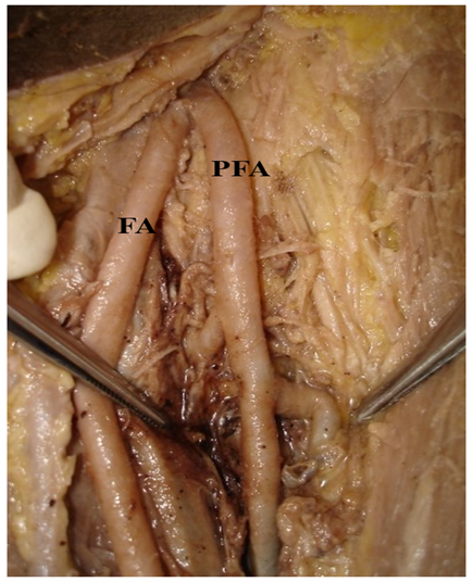
Figure 1. Higher origin of the profunda femoris artery from the lateral aspect of the femoral artery.
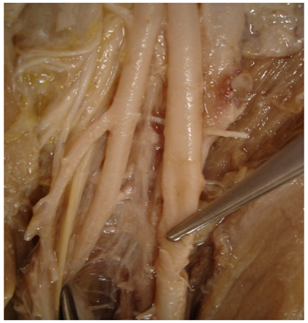
Figure 2. Higher origin of the profunda femoris artery from the medial aspect of the femoral artery.
The lower-origin morphology was likewise represented by two directional patterns. Figure 3 demonstrates a lower division in which the PFA arises from the posterolateral aspect of the femoral artery. Figure 4 shows a lower division with a posteromedial origin. Thus, the supplied material documents variation not only in proximal versus distal level but also around the circumference of the femoral artery. The four configurations and their defining features are presented in Table 2.
Table 2. Morphological configurations of profunda femoris artery origin documented in the study material
|
Configuration |
Relative level |
Surface of origin |
Morphological description |
|
Pattern 1 |
Higher |
Lateral |
PFA separates at a relatively high level from the lateral aspect of the femoral artery; illustrated in Figure 1. |
|
Pattern 2 |
Higher |
Medial |
PFA separates at a relatively high level from the medial aspect of the femoral artery; illustrated in Figure 2. |
|
Pattern 3 |
Lower |
Posterolateral |
Lower division with the PFA arising from the posterolateral aspect of the femoral artery; illustrated in Figure 3. |
|
Pattern 4 |
Lower |
Posteromedial |
Lower division with the PFA arising from the posteromedial aspect of the femoral artery; illustrated in Figure 4. |
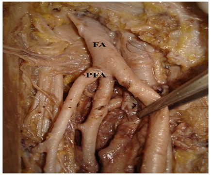
Figure 3. Lower division with posterolateral origin of the profunda femoris artery from the femoral artery.
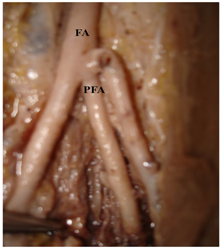
Figure 4. Lower division with posteromedial origin of the profunda femoris artery from the femoral artery.
Abbreviations: FA, femoral artery; PFA, profunda femoris artery.
Taken together, the descriptive findings demonstrate a broad topographical spectrum of PFA origin within the study material. The photographic record confirms higher and lower separation levels and lateral, medial, posterolateral, and posteromedial directional origins. Specimen-wise frequency data for the four patterns were not available in the supplied dataset; consequently, no prevalence percentages, right-left comparisons, sex-based analyses, or significance testing were presented.
DISCUSSION:
The present cadaveric study demonstrates that the origin of the profunda femoris artery is topographically variable and cannot be represented by a single constant pattern. In the 102 lower limbs examined, the available photographic documentation showed four representative configurations: a higher lateral origin, a higher medial origin, a lower posterolateral origin, and a lower posteromedial origin. These observations are important because they demonstrate variation in two separate anatomical dimensions: the level at which the PFA leaves the femoral artery and the surface from which it arises. Although specimen-level frequencies were not available for calculation of prevalence, the documented configurations correspond to patterns repeatedly described in anatomical literature. Nasr et al. reported the posterolateral aspect as the most frequent origin in their cadaveric series, with additional posterior, lateral, and posteromedial origins [1]. Manjappa and Prasanna also found posterolateral origin to be common in a South Indian population and emphasized variability in the distance from the mid-inguinal point [2]. Prakash et al. documented a median separation of 4.2 cm distal to the midpoint of the inguinal ligament in Indian cadavers and linked distal PFA origin with variant circumflex femoral branching [3]. At the evidence-synthesis level, Tomaszewski et al. identified six origin patterns across 2,502 lower limbs and calculated a pooled mean origin distance of 41.15 mm from the mid-inguinal point [4]. The spectrum illustrated in the current study is therefore anatomically plausible and consistent with the established heterogeneity of PFA origin.
The high-origin patterns observed in the present photographic material deserve particular clinical attention. Claassen et al. showed that high and intermediate deep femoral artery origins were common in their cadaveric series [5], while Rajani et al. reported a high origin in 21.21% of limbs and described rare medial and anterolateral configurations [11]. A medially arising PFA can alter its relationship to the femoral vein and can increase the risk of unexpected vascular injury during exposure or catheterization. Choy et al. further demonstrated that PFA variation can modify its relationship with the femoral nerve, emphasizing the importance of precise regional anatomy during nerve blocks and femoral procedures [12]. The lower posterolateral and posteromedial configurations documented here are also clinically relevant because distal separation changes the available length of the femoral arterial segment and the relationship of circumflex branches. Perera demonstrated that the level of deep femoral origin varies with the origin pattern of the circumflex femoral arteries [13]. Łabętowicz et al. subsequently proposed a detailed classification of circumflex femoral branching and showed substantial variation in the distance from the inguinal ligament to the deep femoral artery origin [14]. Recent imaging and cadaveric studies continue to confirm this variability [7-10]. Smółka et al. specifically showed that the femoral arterial access window varies markedly with PFA origin [10]. Taken together, the present observations reinforce the need for clinicians to anticipate non-classical PFA anatomy. Recognition of high, low, lateral, medial, posterolateral, and posteromedial origins can improve anatomical interpretation and reduce avoidable injury during femoral puncture, endovascular access, orthopaedic surgery, vascular reconstruction, and reconstructive flap procedures.
LIMITATIONS
This study has limitations. Donor-level age, sex, laterality, and paired-specimen information were not available in the supplied dataset. Absolute distances between the inguinal ligament and PFA origin were also unavailable. In addition, specimen-wise counts for each photographed origin pattern were not recorded, preventing calculation of variant prevalence or side-based statistical comparisons. Formalin fixation and the use of teaching cadavers can also influence tissue relationships during dissection.
CONCLUSION:
The profunda femoris artery demonstrates meaningful variation in both the level and surface of its origin from the femoral artery. In this cadaveric series of 102 lower limbs, representative higher lateral, higher medial, lower posterolateral, and lower posteromedial configurations were documented. These findings support the concept that the PFA should not be assumed to arise at a constant level or from a uniform surface. Detailed knowledge of these patterns is important for safe femoral arterial access, angiographic procedures, vascular reconstruction, orthopaedic surgery, and interventions involving the proximal thigh. Careful anatomical assessment and, where clinically appropriate, pre-procedural vascular imaging can reduce unexpected vessel injury associated with variant PFA anatomy during practice.
REFERENCES:
1. Nasr AY, Badawoud MH, Al-Hayani AA, Hussein AM. Origin of profunda femoris artery and its circumflex femoral branches: anatomical variations and clinical significance. Folia Morphol (Warsz). 2014;73(1):58-67. doi:10.5603/FM.2014.0008.
2. Manjappa T, Prasanna LC. Anatomical variations of the profunda femoris artery and its branches-a cadaveric study in South Indian population. Indian J Surg. 2014;76(4):288-292. doi:10.1007/s12262-012-0677-3.
3. Prakash, Kumari J, Bhardwaj AK, Jose BA, Yadav SK, Singh G. Variations in the origins of the profunda femoris, medial and lateral femoral circumflex arteries: a cadaver study in the Indian population. Rom J Morphol Embryol. 2010;51(1):167-170.
4. Tomaszewski KA, Henry BM, Vikse J, Pękala P, Roy J, Svensen M, et al. Variations in the origin of the deep femoral artery: a meta-analysis. Clin Anat. 2017;30(1):106-113. doi:10.1002/ca.22691.
5. Claassen H, Schmitt O, Schulze M, Wree A. Deep femoral artery: a new point of view based on cadaveric study. Ann Anat. 2021;237:151730. doi:10.1016/j.aanat.2021.151730.
6. Tzouma G, Kopanakis NA, Tsakotos G, Skandalakis PN, Filippou D. Anatomic variations of the deep femoral artery and its branches: clinical implications on anterolateral thigh harvesting. Cureus. 2020;12(4):e7867. doi:10.7759/cureus.7867.
7. Turan B, Tuncer MC, Ertuğrul ÖZ, Çetinçakmak MG. Morphological investigation of deep femoral artery variations using multidetector computed tomography angiography. Acta Chir Belg. 2023;123(5):509-516. doi:10.1080/00015458.2022.2092962.
8. Dhaminirithika AG, Rajilarajendran H, Kavinnilavan G, Indra P. Unveiling the secrets of the profunda femoris artery: a cadaveric journey with morphometric insights. Turk J Surg. 2025;41(3):294-299. doi:10.47717/turkjsurg.2025.6571.
9. de Villiers EL, Keet K. Anatomical variations of the deep femoral artery: a dissection-based study. Surg Radiol Anat. 2025;47(1):205. doi:10.1007/s00276-025-03702-z.
10. Smółka L, Strugała M, Pomianowski B, Dutka WB, Kursa K, Blady K, et al. Morphometric variability of the femoral artery access window in relation to profunda femoris artery origin: a cadaveric study with clinical implications for femoral vascular access. Folia Morphol (Warsz). 2026;85:e01726115. doi:10.5603/fm.112528.
11. Rajani SJ, Ravat MK, Rajani JK, Bhedi AN. Cadaveric study of profunda femoris artery with some unique variations. J Clin Diagn Res. 2015;9(5):AC01-AC03. doi:10.7860/JCDR/2015/13913.5876.
12. Choy KW, Kogilavani S, Norshalizah M, Rani S, Aspalilah A, Hamzi H, et al. Topographical anatomy of the profunda femoris artery and the femoral nerve: normal and abnormal relationships. Clin Ter. 2013;164(1):17-19. doi:10.7417/T.2013.1504.
13. Perera J. Anatomy of the origin of the deep femoral artery. Ceylon Med J. 1995;40(4):139-141.
14. Łabętowicz P, Olewnik Ł, Podgórski M, Majos M, Stefańczyk L, Topol M, et al. A morphological study of the medial and lateral femoral circumflex arteries: a proposed new classification. Folia Morphol (Warsz). 2019;78(4):738-745. doi:10.5603/FM.a2019.0033.