Bacteriological Profile and Antibiotic Susceptibility Patterns of Blood Culture Isolates in a Tertiary Care Centre in Central India.
- Madkey M V , Assistant Professor, Department of Microbiology, AIIMS, Raipur, Chhattisgarh
- Meshram V M , Associate Professor, Department of General Medicine, Govt. Medical College, Nagpur, Maharashtra
- Gedam D S , Associate Professor, Department of Microbiology, Indira Gandhi Govt, Medical College, Nagpur, Maharashtra
Article Information:
Abstract:
Background: Bloodstream infections (BSIs) are major causes of morbidity and mortality among hospitalized patients, particularly in critical care units. Increasing trends of drug resistance in blood culture isolates complicating treatment is serious concern all over the world. Objective: To determine the bacteriological profile and drug susceptibility patterns of blood culture isolates. Methodology: This retrospective cross-sectional study was conducted at Indira Gandhi Government Medical College, Nagpur from February 2023 to January 2024. Out of total 254 blood samples, 110 blood culture positive isolates were included in the study. Data regarding patient demographics, hospital wards, isolated organisms, and antimicrobial susceptibility patterns were analysed. Isolation and identification of causative organisms were done on the basis of standard microbiological protocol. Drug susceptibility patterns of organisms were assessed using the Kirby-Bauer Disc Diffusion method in accordance with Clinical and Laboratory Standards Institute (CLSI) 2022 guidelines. Results: Out of total 254 blood culture samples, blood culture were positive in 110 cases (43.30 %). Gram-negative bacteria were the predominant isolates, accounting for 61 (55.5%) cases, followed by Gram-positive bacteria in 46 (41.8%) and Candida albicans in 3 (2.7%). Staphylococcus aureus was the most common organism, isolated in 27 (24.54%) cases, followed by Klebsiella pneumoniae 18(16.36%), Escherichia coli 12(10.90%), and Acinetobacter spp. 12(10.90%) each. Extended Spectrum Beta Lactamase (ESBL) production was seen in 37.5 % of gram-negative isolates and methicillin resistance was observed in 29.62% of Staphylococcus aureus (MRSA) isolates. Conclusion: Gram-negative bacteria were the predominant blood culture isolates, while Staphylococcus aureus was the most frequent individual pathogen. Early diagnosis and detection of drug resistance patterns results are thus life-saving during patient management.
Keywords:
Article :
INTRODUCTION:
Bloodstream infections (BSIs) are one of the most severe infectious diseases seen in clinical practice and are a significant cause of morbidity and mortality globally [1]. They are caused by the presence of pathogenic microorganisms in the blood, which results in a general inflammatory reaction that can quickly escalate to septic shock, multiple organ dysfunction syndrome and death when left undetected and untreated [2]. Sepsis is estimated to impact millions of people every year and is one of the top contributors to preventable deaths, with the highest rates in neonates, elderly people, critically ill cases, and those with weakened immune systems [3]. Bloodstream infection burden is particularly high in low- and middle-income countries, especially due to delayed diagnosis, limited laboratory resources, and misuse of antibiotics which lead to poor clinical results [4]. Blood culture is gold standard for the diagnosis of bloodstream infections, since blood can be used to isolate and identify the causative microorganisms and to assess antimicrobial susceptibility [5]. With blood culture, the diagnosis is not just clinical, but microbiological as well, which allows antimicrobial therapy to be tailored rather than given as a blanket treatment for a range of bacteria. Early detection of pathogens can enhance patient survival as well as reduce unnecessary antibiotic usage, adverse drug reactions, hospital stay, and health care costs. Therefore, blood culture surveillance is now an integral part of hospital infection control and antimicrobial stewardship programs [2,6].
The range of microorganisms causing bloodstream infections is dependent on geographical location, health care environment, patient characteristics, and infection control efforts [7]. Gram-positive organisms like Staphylococcus aureus, coagulase-negative staphylococci and Enterococcus species still continue to be a major cause of bloodstream infections, especially in patients that have an intravascular catheter, a prosthetic device or long-term hospital stay. Concurrently, Gram-negative bacteria, such as Escherichia coli, Klebsiella pneumoniae, Acinetobacter baumannii and Pseudomonas aeruginosa are becoming the cause of serious nosocomial infections, particularly in the intensive care unit [8]. In addition, opportunistic fungal pathogens have become significant causes of bloodstream infection in critically ill, immunocompromised and intensive care patients, mainly species of Candida. The rapid development and spread of antimicrobial resistance is one of the most difficult aspects of the control of bloodstream infections. Bacterial resistance has been increasing due to the widespread and often inappropriate use of antibiotics in hospitals and in the community [9]. Multidrug-resistant (MDR) organisms, such as methicillin-resistant Staphylococcus aureus (MRSA), methicillin-resistant coagulase-negative staphylococci (MRCONS), extended-spectrum β-lactamase (ESBL)-producing Enterobacteriaceae, carbapenem-resistant Acinetobacter spp. and multidrug-resistant Pseudomonas aeruginosa are increasingly found all around the world. These organisms have been linked to a lack of therapeutic alternatives, more treatment failures, longer hospital stays, higher health care costs, and higher mortality rates [10]. Antimicrobial resistance (AMR) is one of the greatest public health risks of the 21st century. Antimicrobial resistance is a global health problem and an issue of major importance to the World Health Organization, which is working to combat the spread of resistance among the world's population. Resistance reduces the impact of standard antibiotics and delays clinical cure, leading to greater use and reliance on expensive reserve antimicrobial agents [11].
The purpose of this study is to generate new epidemiological evidence at the local level to inform clinicians of the most common bacterial pathogens responsible for Blood Stream Infections and whether they are susceptible to commonly-used antimicrobial agents, which will have a positive impact on patient outcome, Antimicrobial Stewardship Programs (AMSP) and infection prevention programs.
METHODOLOGY:
This retrospective cross-sectional study was conducted at Indira Gandhi Government Medical College, Nagpur from February 2023 to January 2024. The study included patients of any age and either sex whose blood cultures showed microbial growth during the study period. Out of total 254 blood samples 110 blood culture positive isolates were included in the final analysis. Blood culture isolates with available organism identification and antimicrobial susceptibility testing results were considered eligible for analysis. Blood cultures with no microbial growth, contaminated cultures and records with incomplete microbiological identification or susceptibility data were excluded from the study.
Blood Culture
Venous blood (approximately 10 mL) was collected aseptically using a disposable 18–20-gauge needle and a 10–20 mL syringe, taking care to avoid contamination of the venipuncture site.1 to 5 ml of blood was collected from the pediatric patients depending on their age and weight. The blood was immediately inoculated into conventional blood culture bottles containing Tryptic Soy Broth. The blood-to-broth ratio was maintained at approximately 1:10 to dilute any circulating antimicrobial agents below inhibitory levels [12].
Patients with clinical suspicion of septicemia were identified and their blood samples were collected in Tryptic Soy Broth. Then the broth was incubated overnight aerobically at 37°C. The sample was inoculated onto Blood Agar, MacConkey Agar and Sabouraud’s Dextrose Agar. Isolates were identified on the basis of colony morphology and standard biochemical tests. If there is no growth then the sample is sub-cultured on alternate days for 7 days before reporting it as sterile.
Antimicrobial Susceptibility Testing
Antimicrobial susceptibility testing was performed using commercially available antibiotic discs of appropriate content and potency (HiMedia Laboratories Pvt. Ltd., Mumbai, India) on Mueller–Hinton agar. Zone diameters were determined by the modified Kirby–Bauer disk diffusion method and resistance profiles were categorized as sensitive, intermediate, or resistant based on CLSI Guidelines 2022.
Quality Control
Escherichia coli ATCC® 25922, Pseudomonas aeruginosa ATCC® 27853 and Staphylococcus aureus ATCC® 25923 strains were used for quality control. ESBL was tested by applying the disks of Ceftazidime (30µg) and Ceftazidime + Clavulanic acid (30µg + 10µg) to the lawn culture of the test organism. If the zone of inhibition around Ceftazidime-Clavulanic acid is >5mm than the zone of inhibition around the Ceftazidime disk, then the test organism is said to be ESBL producer.
Data Collection
Data were retrieved from the microbiology laboratory records in a retrospective manner. Data on age, sex, hospital ward, date of specimen collection, isolated organism and antimicrobial susceptibility results were documented on a structured data collection sheet. Samples for blood culture were processed by conventional microbiological methods. Colony morphology, Gram stain and biochemical tests were used for identification of isolates. Antimicrobial susceptibility testing was done by the Kirby–Bauer disk diffusion method and results were interpreted based on Clinical and Laboratory Standards Institute guidelines 2022. The major study variables were type of isolated organism, Gram positive or Gram-negative culture, fungal isolate, antimicrobial susceptibility pattern, and the presence of multiple drug resistance.
Statistical Analysis
Data were analyzed and entered IBM SPSS Statistics version 28.0. The quantitative data like age were expressed as mean
± standard deviation and the categorical data like sex, ward of the hospital, organism type and appearance of resistance pattern to the antimicrobials were expressed as frequencies and percentages. Chi-Square test was used to determine the association between categorical variables. The criterion for statistical significance was a p-value of < 0.05.
RESULTS:
In the present study, blood culture samples from a total of 254 patients with the clinical diagnosis of septicemia were processed out of which 110 (43.30%) were positive. Among the culture positive samples, gender distribution was 67 (60.90%) males and 43 (39.1%) females. All the blood culture samples were processed by the conventional blood culture methods. Maximum numbers of samples were from patients in the age group of <1 year followed by 1-10 years. Most of the blood culture positive samples were obtained from patients admitted to the Neonatal Intensive Care Unit (NICU), accounting for 27 (24.5%) cases, followed by Paediatric wards with 23 (20.9 %) cases, the Dialysis Unit with 10 (9.1%) cases. (Table 1)
Table 1. Baseline Characteristics of Patients with Positive Blood Cultures (n = 110)
|
Variable |
Category |
n (%) |
|
Age (years) |
< 1 year 1-10 years 11-20 years 21-30 years 31-40 years 41-50 years 51 -60 years 61-70 years 71-80 years |
27 (24.55) 23 (20.90) 7 (6.36) 6 (5.46) 7 (6.36) 6 (5.45) 14 (12.73) 10 (9.09) 10 (9.09) |
|
Gender |
Male Female |
67 (60.90%) 43 (39.1%) |
|
Ward of admission |
NICU Paediatrics ward Dialysis Unit MICU PICU Other wards |
27 (24.5%) 23 (20.9%) 10 (9.1%) 7 (6.4%) 7 (6.4%) 36 (32.7%) |
In present study, Gram-negative bacilli were found to be the commonest isolates 61(55.5%). Gram-positive cocci were found in 46 (41.8%) of cases, while fungal isolates (Candida albicans) in 3 (2.7%) of cases as given in (Figure 1).
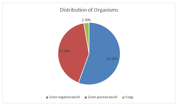
Figure 1: Distribution of Organisms
Among the 110 blood culture isolates, Staphylococcus aureus was the most frequently isolated organism, accounting for 27 (24.5%) cases In gram negative bacilli isolates were Klebsiella pneumoniae 18 (16.36%), Acinetobacter spp.12(10.90%), and Escherichia coli 12(10.90%) each. Non-fermenters accounted for 8 (7.3%) isolates, while Enterococcus faecalis and Pseudomonas aeruginosa were each isolated in 6 (5.5%) cases. MRCONS constituted 5 (4.5%) isolates, whereas CONS accounted for 3 (2.7%) cases.(Table 2)
Table 2. Distribution of Blood Culture Isolates (n = 110)
|
Organism |
n (%) |
|
Staphylococcus aureus |
27 (24.5) |
|
Klebsiella pneumoniae |
18 (16.36) |
|
Acinetobacter spp. |
12 (10.90) |
|
Escherichia coli |
12 (10.90) |
|
Citrobacter spp. |
10 (9.09) |
|
Non-fermenters |
8 (7.3) |
|
Enterococcus faecalis |
6 (5.5) |
|
Pseudomonas aeruginosa |
6 (5.5) |
|
MRCONS |
5 (4.5) |
|
CONS |
3 (2.7) |
|
Fungal isolates |
3(2.7) |
 |
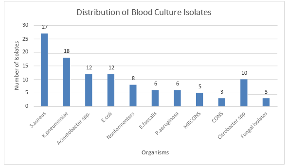
Figure 2: Distribution of Blood Culture Isolates (n=110)
All Gram-positive isolates were sensitive to vancomycin and linezolid. All the isolates of Staphylococcus aureus were resistant to penicillin (Table 3).
Table 3. Antibiotics Sensitivity Pattern for Gram Positive Bacteria.
|
Organisms |
Antibiotics Sensitivity (%) |
|||||||||
|
P |
Cx |
G |
Levo |
E |
CD |
Lz |
TEI |
VA |
HLG |
|
|
Staphylococcus aureus (n=27) |
0 |
70.38 |
37.04 |
74.07 |
62.96 |
62.96 |
100 |
- |
100 |
- |
|
CoNS (n=8) |
0 |
37.5 |
87.5 |
50 |
75 |
75 |
100 |
- |
100 |
- |
|
Enterococcus faecalis (n=6) |
0 |
- |
- |
83.33 |
66.66 |
- |
100 |
83.33 |
100 |
66.66 |
Klebsiella pneumoniae isolates in our study showed complete resistance to cefazolin. Among 12 isolates of E. coli, maximum sensitivity was seen to Amikacin (75%), Tobramycin (75%), and piperacillin-tazobactam (83.33%). Most of the isolates were resistant to ampicillin, 1st and 2nd Generation Cephalosporins. All the isolates of Citrobacter spp. were resistant to cefazolin, and cefoxitin but sensitive to cefotaxime, piperacillin tazobactam and aminoglycosides (Table 4).
Table 4: Antibiotics Sensitivity Pattern for Gram Negative Bacteria.
|
Organisms |
Antibiotics sensitivity (%) |
||||||||||
|
AMP |
CZ |
CAZ |
CXM |
CTX |
CPM |
PIT |
MRP |
AK |
GEN |
TOB |
|
|
Klebsiella pneumoniae (n=18) |
- |
0 |
38.88 |
33.33 |
50 |
72.22 |
94.44 |
94.44 |
55.55 |
44.44 |
88.8 |
|
E. coli (n=12) |
0 |
25 |
66.67 |
50 |
66.67 |
75 |
83.33 |
83.33 |
75 |
66.67 |
75 |
|
Citrobacter freundii (n=5) |
- |
- |
- |
- |
80 |
60 |
80 |
80 |
80 |
100 |
80 |
|
Citrobacter koserii (n=5) |
- |
0 |
0 |
0 |
60 |
60 |
80 |
80 |
80 |
80 |
80 |
In the present study, 37.5% Gram-negative isolates were ESBL producers. ESBL production among Gram-negative bacilli was predominantly shown by E. coli (20 %) followed by Klebsiella pneumoniae (17.5%) (Table 5)
Table 5: ESBL production among Enterobacterales isolates (n = 40)
|
Method of detection of ESBL |
K. pneumoniae n (%) |
E. coli n (%) |
Citrobacter spp. n (%) |
Total Enterobacterales |
|
Phenotypic Confirmatory Disk Diffusion Method |
7 (17.5) |
8 (20) |
- |
15 (37.5) |
The antimicrobial sensitivity of Pseudomonas aeruginosa, Acinetobacter baumannii, Acinetobacter lwoffi shows that 5 out of 6 isolates of Pseudomonas aeruginosa and most of the isolates of Acinetobacter baumannii were sensitive to meropenem. For Pseudomonas aeruginosa strains effective antibiotics were piperacillin-tazobactam (83.33%) and amikacin (66.66%). Most of the isolates of Pseudomonas aeruginosa showed resistance to ceftazidime. Most of the isolates of Acinetobacter baumannii and Acinetobacter lwoffii were sensitive to amikacin and piperacillin-tazobactam (Table 6).
Table 6: Antibiotics Sensitivity Pattern for Non-fermenters.
|
Organisms |
Antibiotics Sensitivity (%) |
|||||||||
|
CAZ |
CPM |
TOB |
GEN |
AK |
PIT |
MRP |
COT |
AT |
NET |
|
|
Pseudomonas aeruginosa (n=6) |
33.33 |
83.33 |
66.66 |
66.66 |
66.66 |
83.33 |
83.33 |
66.66 |
83.33 |
66.66 |
|
Acinetobacter baumannii (n=8) |
50 |
75 |
62.50 |
62.50 |
75 |
75 |
87.5 |
50 |
- |
- |
|
Acinetobacter lwoffii (n=4) |
50 |
75 |
50 |
50 |
75 |
75 |
75 |
75 |
- |
- |
DISCUSSION:
Bacteria that invade the bloodstream continue to be a major cause of morbidity and mortality in the world, especially in hospitalized and critically ill individuals. Correct identification of causative organisms and regular monitoring of antimicrobial susceptibility trends are important to provide guidance on empirical antimicrobial treatment and to optimize patient outcomes. Pathogens isolated from blood are critical responsibilities of the diagnostic microbiology laboratory.
In this study out of total 254 blood samples 110 positive blood culture samples were analyzed, with 67 (60.90%) from male and 43 (39.1%) from female patients. The majority of samples were from individuals aged <1 year, followed by those aged 1–10 years. These findings are consistent with previous studies, such as Mehta et al. [13], who reported 65% male and 35% female samples. Out of total 254 samples, blood cultures were positive in 110 (43.30%) cases. This positivity rate aligns closely with previous reports by Khanal et al. [14] (44%) and Sultana et al. [15] (49.28%). It is well established that prior antibiotic administration can reduce pathogen detection in blood cultures. Prior antibiotic intake can reduce the likelihood of detecting pathogens in blood cultures. Collecting multiple blood culture sets and ensuring adequate blood volume per bottle increase the chances of identifying bloodstream infections. Clinical factors such as patient age, fever intensity, and source of infection also affect positivity rates.
Consistent with other studies, Gram-negative bacteria predominated as causative agents accounting for 55.50% of isolates, followed by Gram-positive cocci at 41.80%, and fungi at 2.70%. This predominance of Gram-negative pathogens concurs with findings reported by Latif et al. [16] (72.1%), Garg et al. [17] (65.5%), and Prashanth et al. [18] (70.47%).
Among the Gram-negative isolates in our study, Klebsiella pneumoniae was the predominant pathogen, accounting for 16.36% of cases, followed by Escherichia coli (10.90%), Acinetobacter baumannii (10..90%). These findings are consistent with Mehdinejad et al. [19], who observed Klebsiella pneumoniae (33.5%) and E. coli (20.6%) as the leading Gram-negative pathogens. This pattern underscores the significant role of Klebsiella pneumoniae in bloodstream infections and highlights the need for targeted antimicrobial strategies against these prevalent organisms. In our study, Pseudomonas aeruginosa was 5.5%, similar findings were also reported by Arora et al. [20] (7.63%) and Qureshi M et al. [21] (10.7%). A high percentage of Pseudomonas aeruginosa isolates were reported by Garg et al. [17] (16%), Mehta et al. [13] (19.75%).
In our study, Acinetobacter spp. (10.90%) were isolated in positive blood culture samples. In contrast, a very high percentage (32%) of Acinetobacter spp. (mainly Acinetobacter lwoffii followed by Acinetobacter baumannii) were reported by Barati M et al. [22], Malini A et al. [23] Acinetobacter baumannii (73.3%), Acinetobacter lwoffii (5.8%) . Samanta et al. [24] who also found Acinetobacter spp. (66%).
Among Gram-positive isolates, Staphylococcus aureus (24.5%) was the commonest followed by Enterococcus faecalis (5.5%) and Staphylococcus epidermidis (CoNS) (2.7%). This was in accordance with other studies carried out by Mehta et al. [13], and Ayobola et al. [25] who reported Staphylococcus aureus as 13.66% and 14.6% respectively. The results of the cefoxitin disk diffusion test, Methicillin resistance was seen in 29.62% of isolates of Staphylococcus aureus.
In our study, Enterococcus faecalis was isolated in 5.5% of blood culture positive samples which were similar to Anbumani et al. [26] (4.16%) and Alam M. S et al. [27] (6.8%). Apart from Gram-positive and Gram-negative organisms, Candida albicans were isolated in 2.7% positive blood culture samples. Similar, findings were found by Qursheed Sultana et al. [15] (1.19%), and Jena et al. [28] found (3.1%).
In the present study, maximum isolates of Enterobacterales were sensitive to Meropenem followed by Amikacin, Tobramycin, and Piperacillin-tazobactam. High resistance showed to Ampicillin, 1st, 2nd generation cephalosporins. Arora et al. [20], Barati M et al. [,22] and Kumar S et al. [29] also reported the same antibiotic resistance pattern in Gram-negative isolates from BSIs.
In the present study, 37.5% of Gram-negative isolates were ESBL producers, among these ESBL production was highest in E. coli (20 %) followed by Klebsiella pneumoniae (17.5%) which is similar to the ESBL production reported by Arora et al. [20] 34.35% and Anathan et al. [30] 25.4% respectively among Klebsiella pneumoniae.
In our study, Gram-positive cocci were 100% sensitive to Vancomycin and Linezolid. A similar sensitivity pattern to Vancomycin and Linezolid was reported by Mehdinejad M et al. [19], in contrast Chinna D et al. [31] Sadar et al. [32] reported 2.4% resistance to Vancomycin. A high degree of Vancomycin resistance was reported by Pavani et al. [33] (33.3%). In the present study, two isolates of Candida albicans were sensitive to Fluconazole and Amphotericin B, one isolate was resistant to Itraconazole, which was comparable with the findings of Pal et al. [34] showed the use of long-term antibiotics and intravascular catheter use directly correlated with a high incidence of Candida infection.
There are some limitations in this study. First, it was a single centre retrospective study, and the results of this study may not be generalizable to other healthcare institutions that have different patient populations and a different microbial epidemiology. Secondly, the study focused only on positive blood culture isolates, and detailed clinical data including comorbidities, severity of illness, previous antibiotic use, length of hospital stay, presence of invasive devices, and outcomes of patients were not always available, which prevented an analysis of their impact on bloodstream infection and antimicrobial resistance. Even with these drawbacks, the study offers a lot of baseline information on the local bacteriological profile and the antimicrobial resistance scenario, which can be used to guide empirical antibiotic use, to develop hospital antibiograms and antimicrobial stewardship programmes.
CONCLUSION:
It is concluded that Gram-negative bacteria were the predominant causes of bloodstream infections, Staphylococcus aureus was the single most frequently isolated pathogen. A high proportion of multidrug-resistant organisms was observed, particularly in critical care units, including the Neonatal Intensive Care Unit, highlighting the growing challenge of antimicrobial resistance. These findings emphasize the importance of routine blood culture surveillance, periodic hospital-specific antibiogram development, strict infection prevention and control measures, and effective antimicrobial stewardship programs to optimize empirical antibiotic therapy and improve patient outcomes.
REFERENCES:
1. Datta, Sangeeta; Kumar, Avinash*; Chatterjee, Biswaroop. Microbiological profile and antimicrobial resistance patterns of bloodstream infections in a tertiary care hospital in eastern India. Current Medicine Research and Practice 15(4):p 145-150, Jul–Aug 2025. | DOI: 10.4103/cmrp.cmrp_162_24
2. Oyekale OT, Ojo BO, Olajide AT, Oyekale OI. Bacteriological profile and antibiogram of blood culture isolates from bloodstream infections in a rural tertiary hospital in Nigeria. Afr J Lab Med. 2022;11(1), a1807. https://doi.org/10.4102/ajlm.v11i1.1807
3. Fasih F, Baig S, Zameer S, Naseem S, Sharafat S. Bacteriological profile and drug resistance patterns of blood culture isolates: A five year audit from tertiary care hospital.Isra Med J. 2019; 11(6): 449-453.
4. Gohel K, Jojera A, Soni S, Gang S, Sabnis R, Desai M. Bacteriological profile and drug resistance patterns of blood culture isolates in a tertiary care nephrourology teaching institute. Biomed Res Int. 2014;2014:153747. doi: 10.1155/2014/153747. Epub 2014 Apr 7. PMID: 24804199; PMCID: PMC3997982.
5. Saavedra JC, Fonseca D, Abrahamyan A, Thekkur P, Timire C, Reyes J, Zachariah R, Agudelo LG. Bloodstream infections and antibiotic resistance at a regional hospital, Colombia, 2019-2021. Rev Panam Salud Publica. 2023 Apr 19;47:e18. doi: 10.26633/RPSP.2023.18. PMID: 37082533; PMCID: PMC10105591.
6. Oyekale OT, Ojo BO, Olajide AT, Oyekale OI. Bacteriological profile and antibiogram of blood culture isolates from bloodstream infections in a rural tertiary hospital in Nigeria. Afr J Lab Med. 2022 Aug 24;11(1):1807. doi: 10.4102/ajlm.v11i1.1807. PMID: 36091350; PMCID: PMC9453184.
7. Wang X, Zhao C, Li H, Chen H, Jin L, Wang Z, Liao K, Zeng J, Xu X, Jin Y, Su D, Liu W, Hu Z, Cao B, Chu Y, Zhang R, Luo Y, Hu B, Wang H. [Microbiological profiles of pathogens causing nosocomial bacteremia in 2011, 2013 and 2016]. Sheng Wu Gong Cheng Xue Bao. 2018 Aug 25;34(8):1205-1217. Chinese. doi: 10.13345/j.cjb.180192. PMID: 30152206.
8. Vega S, Dowzicky MJ. Antimicrobial susceptibility among Gram-positive and Gram-negative organisms collected from the Latin American region between 2004 and 2015 as part of the Tigecycline Evaluation and Surveillance Trial. Ann Clin Microbiol Antimicrob. 2017 Jul 12;16(1):50. doi: 10.1186/s12941-017-0222-0. PMID: 28701170; PMCID: PMC5508790.
9. Denisuik AJ, Garbutt LA, Golden AR, Adam HJ, Baxter M, Nichol KA, Lagacé-Wiens P, Walkty AJ, Karlowsky JA, Hoban DJ, Mulvey MR, Zhanel GG. Antimicrobial-resistant pathogens in Canadian ICUs: results of the CANWARD 2007 to 2016 study. J Antimicrob Chemother. 2019 Mar 1;74(3):645-653. doi: 10.1093/jac/dky477. PMID: 30500898.
10. Birru M, Woldemariam M, Manilal A, Aklilu A, Tsalla T, Mitiku A, Gezmu T. Bacterial profile, antimicrobial susceptibility patterns, and associated factors among bloodstream infection suspected patients attending Arba Minch General Hospital, Ethiopia. Sci Rep. 2021 Aug 5;11(1):15882. doi: 10.1038/s41598-021-95314-x. PMID: 34354138; PMCID: PMC8342484.
11. Birru, M., Woldemariam, M., Manilal, A. et al. Bacterial profile, antimicrobial susceptibility patterns, and associated factors among bloodstream infection suspected patients attending Arba Minch General Hospital, Ethiopia. Sci Rep 11, 15882 (2021). https://doi.org/10.1038/s41598-021-95314-x
12. Collee JG, Fraser AG, Marmion BP, Simmons A. Mackie and McCartney practical medical microbiology. 14th ed. Edinburgh: Churchill Livingstone; 1996.
13. Mehta M, Dutta P, Gupta V. Antimicrobial susceptibility pattern of blood isolates from a teaching hospital in north India. Japanese journal of infectious diseases. 2005 Jun;58(3):174–6
14. Khanal B, Harish BN, Sethuraman KR, Srinivasan S. Infective endocarditis: report of a prospective study in an Indian hospital. Trop Doct. 2002 Apr;32(2):83-5.
15. Sultana Q, Ansari H, Ansari MW. Bacteriological profile and antimicrobial susceptibility patterns of organisms responsible for blood stream infections. Indian J Microbiol Res. 2016;3(2):113-117.
16. Latif S, Anwar M, Ahmad I. bacterial pathogens responsible for blood stream infection (bsi) and pattern of drug resistance in a tertiary care hospital of lahore 2017.
17. Garg A, Anupurba S, Garg J, Goyal RK, Sen MR. Bacteriological profile and antimicrobial resistance of blood culture isolates from a university hospital. Journal of Indian Academy of Clinical Medicine. 2007 Apr;8(2):139-43.
18. Prashanth H, Dominic Saldanha R, Shenoy S, Baliga S. Predictors of mortality in adult sepsis. Int J Biol Med Res. 2011;2:856–861.
19. Mehdinejad M, Khosravi AD, Morvaridi A. Study of Prevalence and Antimicrobial Susceptibility Pattern of Bacteria Isolated from Blood Cultures. Journal of Biological Sciences. 2009;9:249-253.
20. Arora U, Devi P. Bacterial profile of blood stream infections and antibiotic resistance pattern of isolates. JK Sci. 2007;9(4):186-90.
21. Qureshi M, Aziz F. Prevalence of microbial isolates in blood culture and their antimicrobial susceptibility profile. Biomedia Zoll. 2011;27:136-9.
22. Barati M, Talebi Taher M, Abasi R, Mohammad Zadeh M, Barati M. Bacteriological Profile and Antimicrobial Resistance of Blood Culture Isolates. Arch Clin Infect Dis. 2009;4(2):87-95.
23. Malini A, Deepa EK, Gokul BN, Prasad SR. Nonfermenting gram-negative bacilli infections in a tertiary care hospital in Kolar, Karnataka. J Lab Physicians. 2009 Jul;1(2):62.
24. Samanta P, Gautam V, Thapar R, Ray P. Emerging resistance of non-fermenting gram negative bacilli in a tertiary care centre. Indian J Pathol Microbiol. 2011;54:666-7.
25. Ayobola ED, Egbule OS, Omonigho O. Study of prevalence and antimicrobial susceptibility of blood culture bacterial isolates. Malaysian J Microbiol. 2011 Jan 1;7(2):78-82.
26. Anbumani N, Kalyani J, Mallika M. Distribution and antimicrobial susceptibility of bacteria isolated from blood cultures of hospitalized patients in a tertiary care hospital. Indian J Practising Doctor. 2008;5:5-6.
27. Alam MS, Pillai PK, Kapur P, Pillai KK. Resistant patterns of bacteria isolated from bloodstream infections at a university hospital in Delhi. J Pharm Bioallied Sci. 2011 Oct;3(4):525-30.
28. Jena PP, Gur R, Duggal S, Kumar A, Rongpharpi SR. Microbiological Profile and Antibiogram of Blood Stream Isolates at a Referral Hospital in North Delhi: A One Year Study. International Journal of Biomedical Research. 2015;6(10):819-824.
29. Kumar S, Rizvi M, Vidhani S, Sharma VK. Changing face of septicaemia and increasing drug resistance in blood isolates. Indian J Pathol Microbiol. 2004 Jul;47(3):441-6.
30. Ananthan S, Subha A. Cefoxitin resistance mediated by loss of a porin in clinical strains of Klebsiella pneumoniae and Escherichia coli. Indian J Med Microbiol. 2005;23(1):20–23.
31. Chinna D, Sharma A, Gupta V, Singh S. Bacterial profile and antimicrobial susceptibility pattern of blood isolates from a tertiary care hospital in North India. Int J Pharm Res Bio-Sci. 2013;2(2):24–35.
32. Sader HS, Jones RN, Andrade-Baiocchi S, Biedenbach DJ; SENTRY Participants Group (Latin America). Four-year evaluation of frequency of occurrence and antimicrobial susceptibility patterns of bacteria from bloodstream infections in Latin American medical centers. Diagn Microbiol Infect Dis. 2002 Nov;44(3):273-80.
33. Pavani D, Kommula DM, Mudaliar DJ. Bacteriological Profile and Multidrug Resistance Patterns of Blood Culture Isolates in a Teaching Hospital in South India. National Journal of Integrated Research in Medicine. 2012;3(3):55–59.
34. Pal N, Sharma R, Rishi S, Vyas L. Optimum Time to Detection of Bacteria and Yeast Species with BACTEC 9120 Culture System from Blood and Sterile Body Fluids. J Lab Physicians. 2009 Jul;1(2):69-72.