Fine Needle Aspiration as a Diagnostic Modality for Lymphadenopathy with Utility of CBNAAT in Tuberculous Lymphadenopathy.
- Shruthi , Assistant Professor, Department of Pathology, Navodaya Medical College Hospital & Research Centre, Raichur, Karnataka
- A.S. Aanand , Professor & Head, Department of Pathology, Navodaya Medical College Hospital & Research Centre, Raichur, Karnataka
- Vignesh Sai P , PG Resident, Department of Pathology, Navodaya Medical College Hospital & Research Centre, Raichur, Karnataka.
Article Information:
Abstract:
Background: Lymphadenopathy involving the cervical, axillary, and inguinal regions is a common clinical presentation encountered in routine clinical practice. The etiology ranges from reactive inflammatory conditions and tuberculous lymphadenitis to metastatic malignancies and lymphomas. Fine needle aspiration cytology (FNAC) is a rapid, minimally invasive, and cost-effective diagnostic modality that provides a high degree of diagnostic accuracy in the evaluation of lymphadenopathy. In suspected tuberculous lymphadenopathy, ancillary investigations such as Ziehl–Neelsen (ZN) staining and Cartridge Based Nucleic Acid Amplification Test (CBNAAT) improve diagnostic accuracy and facilitate early detection of Mycobacterium tuberculosis along with rifampicin resistance. Aim: To evaluate the role of FNAC as a diagnostic modality for lymphadenopathy and to assess the utility of CBNAAT in suspected cases of tuberculous lymphadenopathy. Materials and Methods: This prospective cross-sectional study will be conducted in the Department of Pathology, Navodaya Medical College Hospital and Research Centre, Raichur, over a period of 1 year. Patients presenting with lymph node enlargement and fulfilling the inclusion criteria will undergo FNAC. Smears will be stained with Hematoxylin & Eosin (H&E), Papanicolaou (PAP), May–Grünwald–Giemsa (MGG), and Ziehl–Neelsen (ZN) stains. In clinically suspected cases of tuberculous lymphadenopathy, aspirated material will be subjected to CBNAAT for confirmation of tuberculosis. Diagnostic accuracy parameters of FNAC will be assessed using CBNAAT as the reference investigation. Results: A total of 37 patients with lymphadenopathy were evaluated. Reactive lymphadenopathy was the most common cytological diagnosis (24; 64.9%), followed by granulomatous lymphadenitis (7; 18.9%). AFB staining was positive in 5 cases (13.5%). CBNAAT was performed in 10 clinically and cytomorphologically suspected cases of tuberculous lymphadenitis, of which 2 (20.0%) were positive and 8 (80.0%) were negative. A significant association was observed between AFB staining and CBNAAT results (Fisher’s exact test, p=0.018). Conclusion: The study is expected to determine the diagnostic utility of FNAC in lymphadenopathy and evaluate the additional role of CBNAAT in diagnosing tuberculous lymphadenopathy, thereby facilitating early diagnosis and appropriate patient management.
Keywords:
Article :
INTRODUCTION:
Lymphadenopathy involving the cervical, axillary, and inguinal regions is a common clinical presentation encountered in day-to-day clinical practice. Enlarged lymph nodes have diverse etiologies ranging from benign reactive hyperplasia secondary to viral or bacterial infections to tuberculosis, lymphomas, and metastatic malignancies. Fine needle aspiration cytology (FNAC) is widely used as an initial diagnostic investigation because it is rapid, minimally invasive, economical, and provides a high degree of diagnostic accuracy in the evaluation of lymphadenopathy.[1,2] Tuberculosis (TB) remains one of the oldest infectious diseases and continues to be a major public health problem worldwide. India contributes nearly one-quarter of the global tuberculosis burden.
The disease burden is further compounded by HIV co-infection and the emergence of multidrug-resistant tuberculosis (MDR-TB), primarily due to treatment default and rifampicin resistance. Early diagnosis is therefore essential for prompt initiation of therapy and prevention of disease transmission.[3] Although conventional cytomorphological examination and Ziehl–Neelsen staining serve as useful initial diagnostic tools in resource-limited settings, their sensitivity is limited, particularly in paucibacillary lesions. Mycobacterial culture remains the gold standard; however, it requires several weeks for organism isolation and drug susceptibility testing, delaying definitive diagnosis and treatment.[3] The Cartridge Based Nucleic Acid Amplification Test (CBNAAT/GeneXpert MTB/RIF) is a rapid, automated, real-time polymerase chain reaction assay endorsed by the World Health Organization for the diagnosis of tuberculosis. Besides detecting Mycobacterium tuberculosis, CBNAAT simultaneously identifies rifampicin resistance and provides results within approximately two hours, making it particularly valuable in extrapulmonary tuberculosis and paucibacillary specimens.[4] The present study is therefore designed to evaluate the role of FNAC as a diagnostic modality for lymphadenopathy and to assess the additional utility of CBNAAT in clinically suspected tuberculous lymphadenopathy in a tertiary care hospital.
MATERIALS AND METHODS:
Study Design: This was a hospital-based prospective cross-sectional study
Study Period
The study was conducted over a period of one year, from JUNE 2025 to MAY 2026.
Study Setting
The study was carried out in the Department of Pathology, Navodaya Medical College Hospital and Research Centre, Raichur. Patients attending the outpatient and inpatient departments who were referred for lymph node fine-needle aspiration cytology (FNAC) during the study period were included in the study.
METHODOLOGY
After obtaining written informed consent, demographic details and relevant clinical information, including age, sex, site of lymph node enlargement, size and duration of swelling, were recorded. Fine-needle aspiration cytology (FNAC) was performed under strict aseptic precautions. The lesion was identified by palpation after disinfecting the overlying skin using a sterile spirit swab. The lymph node was stabilized between the thumb and index finger of the non-dominant hand, and aspiration was performed using the aspiration technique. In each case, four smears were prepared.
Two smears were immediately fixed in 95% ethanol and stained with Hematoxylin and Eosin (H&E) and Papanicolaou (PAP) stains. Two air-dried smears were stained with May–Grünwald–Giemsa (MGG) and Ziehl–Neelsen (ZN) stains. In clinically suspected cases of tuberculous lymphadenopathy, the remaining aspirated material was collected in a Falcon tube containing 1 mL of normal saline and transported with appropriate labeling to the Department of Tuberculosis and Chest Medicine for Cartridge-Based Nucleic Acid Amplification Test (CBNAAT). CBNAAT was performed using the GeneXpert platform according to the manufacturer's instructions. Sample reagent was added to the aspirated material in a 2:1 ratio and mixed thoroughly.
The mixture was incubated for 10 minutes at room temperature, shaken again, and incubated for an additional 5 minutes. Subsequently, 2 mL of the processed material was transferred into the GeneXpert cartridge for analysis. Following staining, H&E-, PAP-, and MGG-stained smears were examined for cytomorphological diagnosis. Ziehl–Neelsen-stained smears were examined under oil immersion for the demonstration of acid-fast bacilli. The cytological diagnosis was correlated with CBNAAT findings in all suspected cases of tuberculous lymphadenopathy.
INCLUSION CRITERIA
1. Patients providing informed written consent for participation in the study.
2. Patients presenting with lymph node enlargement referred for FNAC evaluation.
EXCLUSION CRITERIA
1. Patients who do not provide informed consent for participation in the study.
SAMPLE SIZE
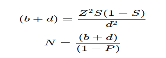
The sample size was calculated using the formula for estimation of specificity.
|
Parameter |
Value |
|
Expected specificity (S) |
0.80* |
|
Absolute precision (d) |
0.15 |
|
Confidence level (1−α) |
95% |
|
Z value |
1.96 |
|
Prevalence of MDR-TB among retreatment cases (P) |
0.15* |
|
Minimum sample size |
33 |
Substituting the above values into the formula, the calculated sample size was 33, which was rounded off to 35 subjects.¹
STATISTICAL ANALYSIS
The collected data will be entered into Microsoft Excel and analyzed using Statistical Package for Social Sciences (SPSS) version 20.0 (IBM SPSS Statistics; IBM Corp., Armonk, NY, USA). Descriptive statistics will be used to summarize the study variables. Continuous variables will be expressed as mean ± standard deviation or median with interquartile range (IQR) depending on the distribution of data, which will be assessed using appropriate statistical test.
ETHICAL CONSIDERATIONS
The study was conducted after obtaining approval from the Institutional Ethics Committee of Navodaya Medical College Hospital and Research Centre, Raichur, Karnataka. All procedures performed in this study involving human participants were in accordance with the ethical standards of the institutional committee and with the principles of the Helsinki Declaration (1975), as revised in 1983. Written informed consent was obtained from all participants prior to inclusion in the study. Patient confidentiality was maintained throughout, and no names, initials, or hospital numbers were used in the study records or illustrative material.
RESULTS:
A total of 37 patients presenting with lymphadenopathy who underwent fine needle aspiration cytology (FNAC) during the study period were included in the present study.
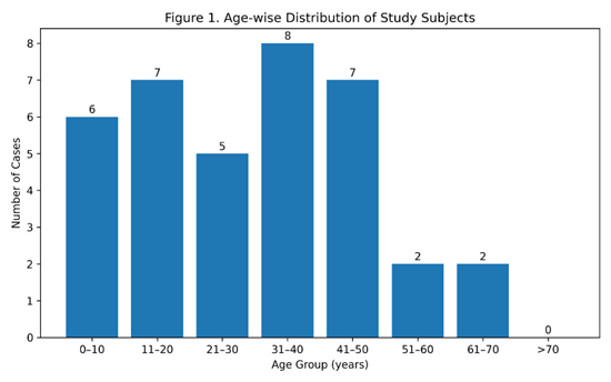
Figure 1. Age-wise distribution of the study population
The age of the study subjects ranged from 3 to 70 years. The highest number of patients belonged to the 31–40 years age group (8 cases; 21.6%), followed by the 11–20 years and 41–50 years age groups (7 cases each; 18.9%). Patients in the 0–10 years age group constituted 6 cases (16.2%), while those in the 21–30 years age group accounted for 5 cases (13.5%). The least represented age groups were 51–60 years and 61–70 years, with 2 cases (5.4%) each.( Figure 1)
Among the 37 study subjects, 27 (73.0%) were females and 10 (27.0%) were males, demonstrating a female predominance (Figure 2)
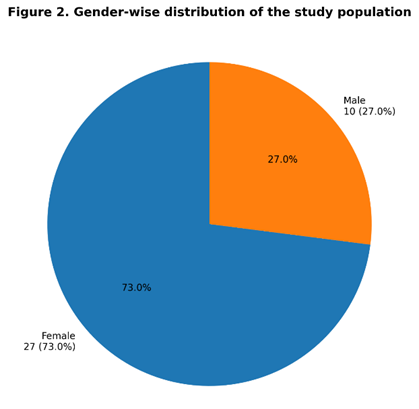
Figure 2. Gender-wise distribution of the study population
The cytomorphological spectrum of lymph node lesions is summarized in Table 1.
Reactive lymphadenopathy, including reactive lymphadenitis and chronic non-specific lymphadenitis, was the predominant diagnosis, accounting for 24 cases (64.9%), followed by granulomatous lymphadenitis in 7 cases (18.9%). Suppurative lymphadenitis constituted 3 cases (8.1%), while malignant lesions accounted for 2 cases (5.4%). One case (2.7%) yielded an inadequate aspirate and was categorized as unsatisfactory for definitive cytological diagnosis.
|
Cytomorphological diagnosis |
Number of cases (n) |
Percentage (%) |
|
Reactive lymphadenopathy* |
24 |
64.9 |
|
Granulomatous lymphadenitis |
7 |
18.9 |
|
Suppurative lymphadenitis |
3 |
8.1 |
|
Malignant lesions |
2 |
5.4 |
|
Unsatisfactory/inadequate aspirate |
1 |
2.7 |
|
Total |
37 |
100 |
*Includes reactive lymphadenopathy, reactive lymphadenitis and chronic non-specific lymphadenitis
Cytomorphological study of all 24 cases of Reactive lymphadenitis showed cellular smear with polymorphous lymphoid population composed of small lymphocytes, with admixed centrocytes, centroblasts and occasional immunoblasts. Background shows tingible-body macrophages and hemorrhage.
Granulomatous lymphadenitis constituted 7 cases, cytomorphologically all cases showed aggregates of epithelioid cells amoung scattered lymphocytes, with occasional multinucleated giant cells. Amoung 7 cases of granulomatous lymphadenitis, background caseous necrosis was seen in 5 cases (71.4%).
Suppurative lymphadenitis constituted 3 cases and cytomorphology of all cases showed, scattered numerous neutrophils admixed with macrophages, lymphocytes and occasional plasma cells in a background of abundant granular necrotic and inflammatory debris.
In the present study two malignant lesions were encountered, one case of medullary carcinoma of the thyroid metastatic to the lymph node and other case of metastatic carcinoma in the lymph node from a primary lung carcinoma.
Medullary carcinoma of the thyroid metastatic to the lymph node, cytological smear showed dispersed, loosely cohesive clusters of malignant cells. These tumor cells are polygonal, with moderate amounts of finely granular cytoplasm, nuclei are round to oval, showing moderate pleomorphism, hyperchromasia, and occasional eccentric nuclei. The background showed abundant finely granular material with hemorrhage. FNAC of Thyroid also showed similar cytomorphological features
Metastatic carcinoma in the lymph node from a primary lung carcinoma cytomorphology smears showed loose cohesive clusters and singly scattered malignant epithelial cells in a hemorrhagic and necrotic background. These tumor cells showed marked nuclear pleomorphism, enlarged hyperchromatic nuclei, with high N:C ratio, irregular nuclear contours, and prominent nucleoli with moderate amount of cytoplasm in a hemorrhagic background.
AFB staining was performed in all 37 cases. Of these, 5 cases (13.5%) were AFB positive, whereas 32 cases (86.5%) were AFB negative
A total of 10 cases were clinically and cytomorphologically suspected to represent tuberculous lymphadenitis, comprising 7 cases of granulomatous lymphadenitis and 3 cases of suppurative lymphadenitis. All these 10 cases were subjected to CBNAAT analysis.
Among the 10 suspected cases of tuberculous lymphadenitis, 2 cases (20.0%) were CBNAAT positive, 8 cases (80.0%) were CBNAAT negative
Among the 10 suspected cases of tuberculous lymphadenitis, 5 cases (50.0%) were AFB positive and 5 cases (50.0%) were AFB negative. Of the five AFB-positive cases, 2 cases (40.0%) were CBNAAT positive, while 3 cases (60.0%) were CBNAAT negative. All 5 AFB-negative cases were CBNAAT negative. No CBNAAT-positive case was identified among the AFB-negative cases.
Among the 10 cases of CBNAAT results, all 2 AFB-positive cases were CBNAAT positive, whereas all 5 AFB-negative cases were CBNAAT negative. A statistically significant association was observed between AFB staining and CBNAAT findings using Fisher's exact test (p = 0.018)
For descriptive assessment of diagnostic agreement, AFB staining was considered as the reference method among the 10 evaluable cases. CBNAAT demonstrated a sensitivity of 100.0%, specificity of 100.0%, positive predictive value of 100.0%, negative predictive value of 100.0%, and overall agreement of 100.0% relative to AFB staining.
The complete observed agreement between AFB staining and CBNAAT among cases with definitive molecular results indicates a strong association between the two diagnostic methods in the present study. However, the diagnostic agreement parameters were derived from 10 evaluable cases and represent agreement with AFB staining as the reference method within the present study population
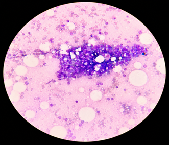
Image 1. Reactive lymphadenopathy (May–Grünwald–Giemsa stain, ×400) shows cellular smear with polymorphous lymphoid population composed of small lymphocytes, with admixed centrocytes, centroblasts and occasional immunoblasts. Background shows tangible body macrophages and hemorrhage.
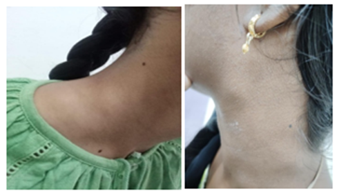
Image 2. Tubercular lymphadenopathy. Clinical picture showing multiple matted cervical lymph nodes of patient with extra pulmonary tuberculosis.
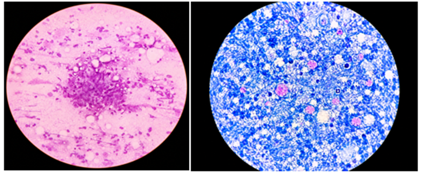
Image 3. A (H&E, 400X) Granulomatous lymphadenitis and B (Ziehl–Neelsen stain, ×1000, oil immersion). A) showing aggregates of epithelioid cell granuloma in necrotic background. B) showing numerous slender, beaded, bright red acid-fast bacilli against a blue necrotic background, confirming AFB positivity.
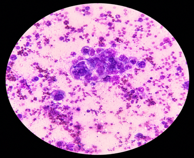
Image 4. Positive for malignancy (May–Grünwald–Giemsa stain, ×400). cytology smear showing clusters of atypical malignant cells exhibiting nuclear pleomorphism, hyperchromasia and increased nuclear-to-cytoplasmic ratio, consistent with positive for malignancy (metastatic carcinoma).
DISCUSSION:
Lymphadenopathy is a common clinical presentation with a wide range of etiologies, including reactive, inflammatory, tuberculous and malignant lesions. Fine needle aspiration cytology (FNAC) is a rapid, minimally invasive and cost-effective investigation that provides useful cytomorphological categorization of lymph node lesions. Hashmi et al.[2] and Chauhan and Behera[5] emphasized the usefulness of FNAC as an initial diagnostic modality in the evaluation of peripheral lymphadenopathy. In the present study, 37 cases were evaluated, with patients ranging from 3 to 70 years of age. Females predominated (73.0%).
Reactive lymphadenopathy was the most common cytological diagnosis, accounting for 24 cases (64.9%), followed by granulomatous lymphadenitis (18.9%), suppurative lymphadenitis (8.1%) and malignant lesions (5.4%). Ali et al.[1] reported granulomatous lymphadenopathy as the predominant cytological pattern, followed by reactive lymphadenopathy. The variation may be related to differences in study population, referral pattern and geographic distribution. Granulomatous lymphadenitis was identified in 7 cases, of which 5 showed caseous necrosis. AFB staining was positive in 5 of the 37 cases (13.5%). Among the 10 cases clinically and cytomorphologically suspected of tuberculous lymphadenitis, 5 (50.0%) were AFB positive. The relatively limited positivity of ZN staining may be attributed to the paucibacillary nature of extrapulmonary tuberculosis. Kumari et al.[6] and Dhote et al.[3] have highlighted the complementary role of molecular testing in suspected tuberculous lymphadenitis.CBNAAT was performed in 10 suspected cases.
In the present study, 2 cases were CBNAAT positive, 8 were negative. Patil et al.[7]reported a higher detection rate with CBNAAT than ZN staining in suspected tuberculous lymphadenitis, supporting the value of molecular testing as an adjunct to conventional cytology. Arpitha et al.[10]similarly demonstrated the complementary diagnostic utility of FNAC, ZN staining and GeneXpert in suspected tuberculous lymphadenopathy.
Complete observed agreement between AFB staining and CBNAAT was noted among cases with definitive molecular results, with a statistically significant association (Fisher's exact test, p=0.018). However, this finding should be interpreted cautiously because of the small number of evaluable cases and the absence of culture or another independent reference standard. The two insufficient CBNAAT samples also emphasize the importance of adequate aspiration and proper allocation of FNAC material for ancillary investigations.
The two malignant cases in the present study, comprising metastatic medullary carcinoma of the thyroid and metastatic lung carcinoma, further demonstrate the broad diagnostic utility of FNAC in lymphadenopathy. Vigliar et al.[11] also demonstrated the high diagnostic value of lymph node FNAC using standardized cytological classification.Overall, the present findings support FNAC as an effective first-line investigation for lymphadenopathy, with AFB staining and CBNAAT providing complementary information in suspected tuberculous lymphadenitis. The combined approach can facilitate early diagnosis and appropriate clinical management, particularly in extrapulmonary tuberculosis.
CONCLUSION:
FNAC is a rapid, minimally invasive and cost-effective first-line investigation for the evaluation of lymphadenopathy and provides useful cytomorphological diagnosis of a wide range of lymph node lesions. In suspected tuberculous lymphadenitis, the addition of Ziehl–Neelsen staining and CBNAAT provides complementary diagnostic information and improves the detection of tuberculosis, particularly in paucibacillary lesions. CBNAAT performed on FNAC material is therefore a useful adjunct to conventional cytology and AFB staining for the early diagnosis of tuberculous lymphadenitis. Overall, the combined approach of FNAC with appropriate ancillary investigations can facilitate early diagnosis and appropriate patient management.
REFERENCES:
1. Ali SR, Shilpa G, Hamza AM, Chaitra BE, Francis R. Role of fine needle aspiration cytology as a diagnostic tool in lymphadenopathy with utility of CBNAAT in tuberculous lymphadenopathy. IP Arch Cytol Histopathol Res. 2019;4(1):36-39. doi:10.18231/2456-9267.2019.0010.
2. Hashmi AA, Naz S, Ahmed O, Yaqeen SR, Irfan M, Kamal A, et al. Utility of fine needle aspiration cytology in the evaluation of lymphadenopathy. Cureus. 2020;12(12):e11990. doi:10.7759/cureus.11990.
3. Dhote AR, Gaikwad A, Datar S, Kowe B. Utility of cartridge based nucleic acid amplification test in diagnosis of tubercular lymph nodes on fine needle aspiration and its comparison with Ziehl-Neelsen staining and concentration bleach method. Panacea J Med Sci. 2022;12(1):61-66. doi:10.18231/j.pjms.2022.012.
4. Sushma SGS, Ingin RJ, Ahmed MM. Diagnosis of tubercular lymphadenopathy by cartridge-based nucleic acid amplification test (CBNAAT) and its correlation with fine needle aspiration cytology: an institution-based study. Int J Adv Res. 2023;12(11):68-75. doi:10.21474/IJAR01/19817.
5. Chauhan S, Behera SK. FNAC as a diagnostic tool in the evaluation of cytomorphological spectrum of peripheral lymphadenopathy along with histopathological agreement—an experience in a tertiary teaching hospital of Northern India. J Evid Based Med Healthc. 2020;7(48):2897-2902. doi:10.18410/jebmh/2020/601.
6. Kumari M, Khambra P, Panwar K, Yadav I. Rapid diagnosis of tubercular lymphadenopathy by cartridge-based nucleic acid amplification test (CBNAAT) and its correlation with Ziehl-Neelsen staining on fine needle aspiration cytology. Int J Health Sci Res. 2020;10(7):17-21.
7. Patil SB, Dhage SM, Umap PS, Ghorpade SV, Patharwat S. Cartridge based nucleic acid amplification test: a sensitive diagnostic tool for tuberculosis on fine needle aspirates samples. Int J Community Med Public Health. 2020;7(4):1511-1515. doi:10.18203/2394-6040.ijcmph20201465.
8. Onkari AS, Iravane JA. Comparative utility of GeneXpert assay over fine needle aspiration cytology in early diagnosis of tuberculous lymphadenitis. IOSR J Dent Med Sci. 2021;20(2):23-30. doi:10.9790/0853-2002012330.
9. Thakkar B, Patel VR, Panchal N, Amin B, Nakum C. Role of CBNAAT (cartridge based nucleic acid amplification test) in early diagnosis of tuberculous lymphadenopathy and drug resistant tuberculous lymphadenopathy with the help of fine needle aspiration cytology. Int J Health Clin Res. 2022;5(3):442-446.
10. Arpitha K, Ravish Kumar M, Sirasagi AK, Pattar PM. Comparison of fine needle aspiration cytology, Ziehl-Neelsen staining and GeneXpert methods in suspected cases of tubercular lymphadenopathy. Natl J Lab Med. 2021;10(3):PO26-PO29. doi:10.7860/NJLM/2021/47991:2498.
11. Vigliar E, Acanfora G, Iaccarino A, Mascolo M, Russo D, Scalia G, et al. A novel approach to classification and reporting of lymph node fine-needle cytology: application of the proposed Sydney system. Diagnostics (Basel). 2021;11(8):1314. doi:10.3390/diagnostics11081314.