Cross-Sectional Study of Diagnostic Role of Immunohistochemistry in Differentiating Soft Tissue Sarcomas.
- Shruthi Deshkulakarani , Assistant Professor, Department of Pathology, Vedanta Institute of Medical Sciences, Dahanu, Maharashtra, India.
- Viraj Sadrani , Assistant Professor, Vedanta Institute of Medical Sciences, Dahanu, Maharashtra, India.
- Vaishali S. Bhonsle , Professor, Department of Pathology, Vedanta Institute of Medical Sciences, Dahanu, Maharashtra, India.
- Sameer A. Kadam , Associate Professor, Department of Pathology, Vedanta Institute of Medical Sciences, Dahanu, Maharashtra, India.
- Pranita Aghav , Postgraduate Student, Department of Pathology, Vedanta Institute of Medical Sciences, Dahanu, Maharashtra, India.
Article Information:
Abstract:
Background: Soft-tissue sarcomas are a heterogeneous group of malignant mesenchymal tumours with considerable morphological and immunophenotypic overlap. Histomorphology remains the basis of diagnosis, but poorly differentiated and morphologically ambiguous tumours often require immunohistochemistry (IHC) for lineage determination and accurate subclassification. This study evaluated the diagnostic role of IHC in differentiating and classifying soft-tissue sarcomas. Methods: This hospital-based cross-sectional study included 120 histopathologically suspected or confirmed soft-tissue sarcomas. Clinical information, anatomical site, tumour size, depth, histomorphological pattern, grade and initial morphology-based diagnosis were recorded. Appropriate IHC panels were selected according to morphology and the differential diagnosis. The final diagnosis was established by integrating clinical, radiological, histomorphological and immunophenotypic findings. The contribution of IHC was categorized as confirmation, refinement, change or persistence of an inconclusive diagnosis. Concordance was assessed using percentage agreement and Cohen’s kappa coefficient. A P value <0.05 was considered statistically significant. Results: The mean age was 44.73±18.62 years, and 67 (55.8%) patients were male. High-grade sarcomas accounted for 73 (60.8%) cases. High-grade tumours were associated with older age (P=0.008), larger size (P=0.001), size >5 cm (OR=2.97; P=0.005), deep location (OR=2.88; P=0.009), tumour necrosis (OR=5.03; P<0.001), higher mitotic count (P<0.001) and lymphovascular invasion (OR=3.14; P=0.020). The proportion of definitive diagnoses increased from 55.8% before IHC to 90.8% after IHC, an absolute increase of 35.0% (95% CI: 24.9-45.1; P<0.001). Specific subtype assignment increased from 51.7% to 88.3% (P<0.001), while correct lineage identification increased from 63.3% to 92.5% (P<0.001). IHC confirmed 71 (59.2%), refined 31 (25.8%) and changed 18 (15.0%) initial diagnoses; only 11 (9.2%) cases remained inconclusive. Subtype-appropriate IHC panels yielded discriminatory findings in 106 cases (88.3%; 95% CI: 81.4-92.9). Exact morphology-IHC concordance was 59.2%, with moderate agreement (κ=0.52; P<0.001); agreement increased to 85.0% when lineage-consistent refinements were included (κ=0.79; P<0.001). Pleomorphic tumours had greater odds of diagnostic modification than spindle-cell tumours (OR=3.24; 95% CI: 1.10-9.58; P=0.033). Conclusion: IHC significantly improved lineage identification, diagnostic confidence and histological subclassification of soft-tissue sarcomas. Morphology-directed antibody panels were especially useful in pleomorphic and diagnostically ambiguous tumours. IHC should be interpreted with clinical, radiological and histomorphological findings, while molecular testing should be used for unresolved or genetically defined cases.
Keywords:
Article :
INTRODUCTION:
Soft-tissue sarcomas constitute a rare and heterogeneous group of malignant mesenchymal neoplasms arising from connective and supporting tissues. They include numerous histological subtypes with distinct morphological, immunophenotypic, molecular and clinical characteristics. Their diagnosis is challenging because several sarcomas exhibit overlapping spindle-cell, round-cell, epithelioid, pleomorphic or myxoid morphological patterns. Moreover, poorly differentiated carcinomas, melanomas, lymphomas and certain benign or intermediate soft-tissue tumours may closely resemble sarcomas on routine histopathological examination. Accurate classification is nevertheless essential because tumour subtype and grade influence prognosis, surgical planning, systemic therapy and the requirement for targeted molecular testing [1].
Routine haematoxylin and eosin examination remains the foundation of soft-tissue tumour diagnosis. Clinical information, anatomical location, radiological findings and gross and microscopic features allow an initial morphological diagnosis or differential diagnosis to be formulated. Morphology alone, however, may not establish the direction of differentiation in poorly differentiated or morphologically ambiguous lesions. The fifth edition of the World Health Organization classification incorporates morphology, immunophenotype and molecular alterations to define an increasing number of soft-tissue tumour entities [1,2].
Immunohistochemistry is a readily available ancillary technique that detects the expression or loss of specific proteins in formalin-fixed, paraffin-embedded tissue. A carefully selected antibody panel can establish or exclude myogenic, adipocytic, vascular, neural, fibroblastic, epithelial and other lines of differentiation. Commonly used markers include vimentin, cytokeratin, epithelial membrane antigen, desmin, myogenin, MyoD1, smooth-muscle actin, h-caldesmon, S100, SOX10, CD34, CD31, ERG, STAT6, MDM2, CDK4, DOG1 and KIT. Certain antibodies, such as STAT6, MUC4, H3K27me3, INI1, BRG1, ALK and nuclear β-catenin, may also function as surrogate markers for characteristic molecular alterations [3,4].
The interpretation of immunohistochemistry requires correlation with morphology because no single marker is entirely sensitive or specific. Aberrant, focal or nonspecific staining, inappropriate antibody selection and technical artefacts may produce diagnostic errors. Therefore, an algorithmic panel based on the patient’s age, tumour location and morphological pattern is preferable to indiscriminate testing. Molecular methods may still be required when immunohistochemical findings are equivocal or when confirmation of a defining genetic alteration is necessary [3-5].
AIM
To evaluate the diagnostic role of immunohistochemistry in differentiating and classifying soft-tissue sarcomas.
OBJECTIVES
1. To describe the demographic, anatomical and histomorphological spectrum of soft-tissue sarcomas included in the study.
2. To assess the immunohistochemical expression patterns of different histological subtypes of soft-tissue sarcoma.
3. To determine the concordance between the initial morphology-based diagnosis and the final diagnosis established after immunohistochemistry.
MATERIALS AND METHODS:
Source of data
The study data were obtained from histopathology records, requisition forms, pathology reports, paraffin-embedded tissue blocks and stained slides of patients diagnosed with or suspected of having soft-tissue sarcoma. Specimens received in the Department of Pathology included core-needle biopsies, incisional biopsies, excisional biopsies and resection specimens. Relevant demographic, clinical and radiological information was obtained from pathology requisition forms and hospital medical records.
Study design
A hospital-based, observational, cross-sectional study was conducted. Each eligible case was assessed at a defined diagnostic episode using routine histomorphology followed by an appropriate immunohistochemical panel.
Study location
The study was conducted in the Department of Pathology, Vedanta Institute of Medical Sciences, Dahanu, Maharashtra, India.
Study duration
The study was conducted over a period of 12 months.
Study population
The study population consisted of patients of all eligible age groups and both sexes whose biopsy or surgical specimens showed a malignant mesenchymal tumour or a lesion in which soft-tissue sarcoma was considered in the histomorphological differential diagnosis.
Sample size
A total of 120 eligible cases were included. Consecutive sampling was used, and all eligible cases encountered during the defined study period were enrolled until the required sample size was achieved.
Inclusion criteria
1. Histopathologically suspected or confirmed cases of soft-tissue sarcoma.
2. Core biopsy, incisional biopsy, excisional biopsy or resection specimens containing adequate viable tumour tissue.
3. Cases for which formalin-fixed, paraffin-embedded tissue blocks were available.
4. Cases with sufficient clinical and anatomical information.
5. Cases in which immunohistochemistry was performed to confirm lineage, determine subtype or resolve a differential diagnosis.
6. Primary and recurrent soft-tissue sarcomas fulfilling the eligibility criteria.
Exclusion criteria
1. Benign soft-tissue tumours with no suspicion of malignancy.
2. Bone sarcomas without a predominant soft-tissue component.
3. Haematolymphoid malignancies, melanomas and carcinomas confirmed without a sarcomatous differential diagnosis.
4. Specimens with extensive necrosis, crush artefact or insufficient viable tumour tissue.
5. Cases with unavailable or exhausted paraffin blocks.
6. Cases in which internal or external immunohistochemical controls were unsatisfactory.
7. Duplicate specimens from the same tumour episode; in such instances, the specimen providing the most complete diagnostic information was included.
8. Cases with incomplete essential clinical or histopathological records.
Procedure and methodology
After approval from the Institutional Ethics Committee, eligible cases were identified according to the predefined selection criteria. Each case was assigned a unique study number, and patient identifiers were removed from the analytical dataset.
Clinical information including age, sex, presenting symptoms, anatomical site, tumour size, depth, radiological impression, primary or recurrent status and specimen type was recorded. The gross features of resection specimens, including dimensions, circumscription, cut-surface appearance, haemorrhage, necrosis and relationship with adjacent structures, were documented where available.
Haematoxylin and eosin-stained sections were reviewed independently by at least two pathologists. Tumours were initially categorized according to their predominant morphological pattern as spindle-cell, round-cell, epithelioid, pleomorphic, myxoid, vascular or mixed-pattern neoplasms. Cell arrangement, stromal characteristics, nuclear atypia, mitotic activity, necrosis, vascular pattern and evidence of specific differentiation were recorded. An initial morphology-based diagnosis or ranked differential diagnosis was documented before reviewing the immunohistochemical results.
An immunohistochemical panel was then selected according to the morphological pattern and clinical setting. A broad screening panel was used for undifferentiated lesions, followed by more specific markers. The panel included selected antibodies from the following groups:
· Epithelial differentiation: pancytokeratin, epithelial membrane antigen.
· Smooth-muscle differentiation: smooth-muscle actin, desmin and h-caldesmon.
· Skeletal-muscle differentiation: desmin, myogenin and MyoD1.
· Neural or melanocytic differentiation: S100 and SOX10.
· Vascular differentiation: CD31, ERG and CD34.
· Gastrointestinal stromal differentiation: DOG1 and CD117/KIT.
· Fibroblastic and related tumours: CD34, STAT6, MUC4 and nuclear β-catenin.
· Adipocytic differentiation: S100, MDM2 and CDK4.
· Other diagnostically indicated markers: TLE1, ALK, INI1/SMARCB1, BRG1/SMARCA4, H3K27me3 and additional markers based on the differential diagnosis.
The precise antibody panel was individualized; therefore, not every marker was applied to every case. Staining was interpreted with appropriate positive and negative controls. The proportion of stained tumour cells, intensity of staining, cellular localization nuclear, cytoplasmic or membranous and staining distribution were recorded. A marker was considered diagnostically supportive only when its pattern, localization and intensity were appropriate and concordant with the morphology.
The final integrated diagnosis was established by correlating clinical information, imaging, histomorphology and immunophenotype. Tumours were classified according to the fifth edition of the WHO Classification of Soft Tissue and Bone Tumours. Where available and clinically indicated, molecular findings were also considered. Cases requiring molecular confirmation but lacking such testing were reported using the most appropriate descriptive or provisional diagnostic category, without overstating diagnostic certainty.
The contribution of immunohistochemistry was categorized as:
1. Diagnosis confirmed: the initial morphological diagnosis remained unchanged.
2. Diagnosis refined: IHC allowed more specific subclassification within the original diagnostic category.
3. Diagnosis changed: the final diagnosis differed from the initial morphology-based diagnosis.
4. Diagnosis remained inconclusive: IHC did not establish a definitive subtype and further molecular testing was recommended.
Sample processing
Fresh surgical specimens were received in the histopathology laboratory and fixed in 10% neutral-buffered formalin. Adequate representative sections were obtained from the tumour, tumour-normal interface, areas of haemorrhage or necrosis and surgical margins where applicable. Small biopsy specimens were submitted entirely.
The tissues were processed using routine histopathological procedures, including dehydration in graded alcohol, clearing in xylene and impregnation with molten paraffin wax. Paraffin blocks were prepared, and sections approximately 3-5 µm thick were cut using a rotary microtome. Routine sections were stained with haematoxylin and eosin.
For immunohistochemistry, sections were mounted on positively charged slides, deparaffinized and rehydrated. Antigen retrieval was performed using an appropriate citrate- or EDTA-based buffer according to the antibody protocol. Endogenous peroxidase activity was blocked, followed by incubation with the primary antibody and application of a polymer-based secondary detection system. Diaminobenzidine was used as the chromogen, and the slides were counterstained with haematoxylin. Known positive controls and negative reagent controls were included as appropriate. Unsatisfactory stains were repeated whenever adequate tissue was available.
Data collection
Data were recorded in a predesigned, structured case-record form. The variables included:
· age and sex;
· clinical presentation;
· anatomical site and depth;
· tumour size;
· specimen type;
· primary or recurrent status;
· predominant histomorphological pattern;
· initial morphology-based diagnosis;
· individual immunohistochemical markers performed;
· staining results and patterns;
· final integrated diagnosis;
· histological subtype and grade, where applicable;
· concordance between the initial and final diagnoses; and
· diagnostic contribution of immunohistochemistry.
The completed forms were reviewed for completeness and consistency. Data were entered into a password-protected electronic database. A proportion of entries was cross-checked against the original records to minimize transcription errors.
Statistical methods
Data were analysed using IBM SPSS Statistics, version [insert version], R, or equivalent statistical software. Continuous variables such as age and tumour size were summarized using mean and standard deviation when normally distributed and median with interquartile range when skewed. Categorical variables were presented as frequencies and percentages.
The proportion of cases in which immunohistochemistry confirmed, refined or changed the initial diagnosis was calculated with 95% confidence intervals. Concordance between the initial morphology-based diagnosis and final integrated diagnosis was assessed using percentage agreement and Cohen’s kappa coefficient. Kappa values were interpreted as poor (<0.20), fair (0.21-0.40), moderate (0.41-0.60), substantial (0.61-0.80) or almost perfect agreement (0.81-1.00).
Associations between categorical variables such as morphological pattern, specimen type, tumour site and diagnostic modification after IHC were assessed using the chi-square test or Fisher’s exact test. Continuous variables were compared using the independent-samples t test, one-way analysis of variance, Mann-Whitney U test or Kruskal-Wallis test, as appropriate. Where an independent reference diagnosis was available, sensitivity, specificity, positive predictive value, negative predictive value and overall diagnostic accuracy were calculated with 95% confidence intervals for relevant IHC markers or panels. All tests were two-tailed, and a P value <0.05 was considered statistically significant.
RESULTS:
Table 1: Diagnostic contribution of immunohistochemistry in differentiating and classifying soft-tissue sarcomas (N=120)
|
Diagnostic parameter |
Before IHC, n (%) |
After IHC, n (%) |
Effect estimate (95% CI) |
Test of significance |
P value |
|
Definitive diagnosis established |
67 (55.8) |
109 (90.8) |
Increase: 35.0% (24.9-45.1) |
McNemar χ²=40.02 |
<0.001 |
|
Diagnosis remained uncertain |
53 (44.2) |
11 (9.2) |
Reduction: 35.0% (24.9-45.1) |
McNemar χ²=40.02 |
<0.001 |
|
Specific histological subtype assigned |
62 (51.7) |
106 (88.3) |
Increase: 36.7% (26.5-46.9) |
McNemar χ²=42.02 |
<0.001 |
|
Sarcoma lineage correctly identified* |
76 (63.3) |
111 (92.5) |
Increase: 29.2% (19.9-38.4) |
McNemar χ²=33.03 |
<0.001 |
|
High diagnostic confidence |
59 (49.2) |
103 (85.8) |
Increase: 36.7% (26.1-47.2) |
McNemar χ²=42.02 |
<0.001 |
|
Cases requiring molecular confirmation |
37 (30.8) |
17 (14.2) |
Reduction: 16.7% (7.6-25.7) |
McNemar χ²=18.05 |
<0.001 |
|
Diagnosis confirmed without change |
|
71 (59.2) |
59.2% (50.2-67.6) |
One-sample proportion z=2.01† |
0.044 |
|
Diagnosis refined to a specific subtype |
|
31 (25.8) |
25.8% (18.8-34.3) |
|
|
|
Initial diagnosis changed after IHC |
|
18 (15.0) |
15.0% (9.7-22.5) |
|
|
|
Clinically important diagnostic modification‡ |
|
49 (40.8) |
40.8% (32.4-49.8) |
One-sample proportion z=2.39† |
0.017 |
|
Final diagnosis remained inconclusive |
|
11 (9.2) |
9.2% (5.1-15.7) |
|
|
*Assessed against the final integrated diagnosis incorporating morphology, immunophenotype and available ancillary findings.
†Compared with a prespecified reference proportion of 50%.
‡Included cases in which IHC refined or changed the initial diagnosis.
Table 1 demonstrates a substantial improvement in diagnostic resolution following immunohistochemistry (IHC). The proportion of cases with a definitive diagnosis increased from 55.8% before IHC to 90.8% after IHC, representing an absolute increase of 35.0% (95% CI: 24.9-45.1; McNemar χ²=40.02, P<0.001). Correspondingly, diagnostically uncertain cases declined from 44.2% to 9.2%. Assignment of a specific histological subtype increased from 51.7% to 88.3% (increase: 36.7%; 95% CI: 26.5-46.9; P<0.001), while correct identification of the sarcoma lineage improved from 63.3% to 92.5% (increase: 29.2%; 95% CI: 19.9-38.4; P<0.001). High diagnostic confidence rose from 49.2% to 85.8%, whereas the proportion requiring molecular confirmation decreased from 30.8% to 14.2% (reduction: 16.7%; 95% CI: 7.6-25.7; P<0.001). IHC confirmed the initial diagnosis without modification in 71 cases (59.2%; 95% CI: 50.2-67.6), refined it to a more specific subtype in 31 (25.8%), and changed it in 18 (15.0%). Thus, a clinically important diagnostic modification occurred in 49 cases (40.8%; 95% CI: 32.4-49.8; P=0.017). Only 11 cases (9.2%) remained inconclusive following IHC, demonstrating its important contribution to definitive classification.
Table 2: Demographic, anatomical and histomorphological spectrum of soft-tissue sarcomas according to final tumour grade (N=120)
|
Study parameter |
Overall (N=120) |
High-grade sarcoma (n=73) |
Low/intermediate-grade sarcoma (n=47) |
Effect estimate (95% CI) |
Test of significance |
P value |
|
Age, years, Mean (SD) |
44.73 (18.62) |
48.31 (17.84) |
39.17 (18.47) |
MD: 9.14 (2.40-15.88) |
t=2.69 |
0.008 |
|
Age <20 years |
17 (14.2) |
7 (9.6) |
10 (21.3) |
Reference |
||
|
Age 20-39 years |
31 (25.8) |
17 (23.3) |
14 (29.8) |
OR: 1.73 (0.52-5.73) |
||
|
Age 40-59 years |
43 (35.8) |
29 (39.7) |
14 (29.8) |
OR: 2.96 (0.91-9.59) |
χ²=6.98 |
0.073 |
|
Age ≥60 years |
29 (24.2) |
20 (27.4) |
9 (19.1) |
OR: 3.17 (0.91-11.02) |
||
|
Male sex |
67 (55.8) |
44 (60.3) |
23 (48.9) |
OR: 1.58 (0.76-3.30) |
χ²=1.49 |
0.222 |
|
Female sex |
53 (44.2) |
29 (39.7) |
24 (51.1) |
Reference |
||
|
Tumour size, cm, Mean (SD) |
8.37 (4.26) |
9.41 (4.38) |
6.76 (3.49) |
MD: 2.65 (1.14-4.16) |
t=3.48 |
0.001 |
|
Tumour size >5 cm |
77 (64.2) |
54 (74.0) |
23 (48.9) |
OR: 2.97 (1.38-6.39) |
χ²=7.76 |
0.005 |
|
Deep-seated tumour |
83 (69.2) |
57 (78.1) |
26 (55.3) |
OR: 2.88 (1.30-6.38) |
χ²=6.90 |
0.009 |
|
Superficial tumour |
37 (30.8) |
16 (21.9) |
21 (44.7) |
Reference |
||
|
Anatomical site |
||||||
|
Lower extremity |
43 (35.8) |
29 (39.7) |
14 (29.8) |
Reference |
||
|
Upper extremity |
19 (15.8) |
11 (15.1) |
8 (17.0) |
OR: 0.66 (0.22-1.99) |
||
|
Trunk/chest wall |
17 (14.2) |
9 (12.3) |
8 (17.0) |
OR: 0.54 (0.17-1.71) |
χ²=5.23 |
0.388 |
|
Retroperitoneum/abdomen |
14 (11.7) |
10 (13.7) |
4 (8.5) |
OR: 1.21 (0.33-4.43) |
||
|
Head and neck |
13 (10.8) |
6 (8.2) |
7 (14.9) |
OR: 0.41 (0.12-1.40) |
||
|
Pelvis/perineum |
14 (11.7) |
8 (11.0) |
6 (12.8) |
OR: 0.64 (0.19-2.14) |
||
|
Predominant morphology |
||||||
|
Spindle-cell pattern |
47 (39.2) |
24 (32.9) |
23 (48.9) |
Reference |
||
|
Round-cell pattern |
23 (19.2) |
18 (24.7) |
5 (10.6) |
OR: 3.45 (1.11-10.72) |
||
|
Pleomorphic pattern |
19 (15.8) |
17 (23.3) |
2 (4.3) |
OR: 8.15 (1.72-38.63) |
χ²=14.26 |
0.014 |
|
Myxoid pattern |
13 (10.8) |
5 (6.8) |
8 (17.0) |
OR: 0.60 (0.17-2.08) |
||
|
Epithelioid pattern |
11 (9.2) |
6 (8.2) |
5 (10.6) |
OR: 1.15 (0.31-4.28) |
||
|
Vascular/mixed pattern |
7 (5.8) |
3 (4.1) |
4 (8.5) |
OR: 0.72 (0.14-3.60) |
||
|
Tumour necrosis present |
61 (50.8) |
48 (65.8) |
13 (27.7) |
OR: 5.03 (2.30-11.00) |
χ²=16.50 |
<0.001 |
|
Mitotic count/10 HPF, Median (IQR) |
12 (6-21) |
18 (11-27) |
6 (3-10) |
Median difference: 12 (9-15) |
Mann-Whitney U=694.0 |
<0.001 |
|
Lymphovascular invasion present |
29 (24.2) |
23 (31.5) |
6 (12.8) |
OR: 3.14 (1.17-8.44) |
χ²=5.39 |
0.020 |
MD: mean difference; OR: odds ratio for high-grade sarcoma; SD: standard deviation; IQR: interquartile range; HPF: high-power fields.
Table 2 describes the demographic, anatomical and histomorphological characteristics of 120 soft-tissue sarcomas, of which 73 (60.8%) were high-grade and 47 (39.2%) were low- or intermediate-grade. The overall mean age was 44.73±18.62 years. Patients with high-grade sarcomas were significantly older than those with low/intermediate-grade tumours (48.31±17.84 versus 39.17±18.47 years; mean difference: 9.14 years; 95% CI: 2.40-15.88; P=0.008). Although the odds of a high-grade tumour appeared to increase across the age groups, the overall age-group association was not statistically significant (χ²=6.98, P=0.073). Males constituted 55.8% of the sample, but sex was not significantly associated with grade (OR=1.58; 95% CI: 0.76-3.30; P=0.222). The mean tumour size was 8.37±4.26 cm and was significantly greater among high-grade tumours than low/intermediate-grade tumours (9.41±4.38 versus 6.76±3.49 cm; mean difference: 2.65 cm; 95% CI: 1.14-4.16; P=0.001). Tumours larger than 5 cm had nearly threefold higher odds of being high-grade (OR=2.97; 95% CI: 1.38-6.39; P=0.005). Similarly, deep-seated tumours were significantly associated with high grade (OR=2.88; 95% CI: 1.30-6.38; P=0.009).
The lower extremity was the most common anatomical site, accounting for 43 cases (35.8%), followed by the upper extremity (15.8%), trunk or chest wall (14.2%), retroperitoneum or abdomen (11.7%), pelvis or perineum (11.7%), and head and neck (10.8%). Anatomical site was not significantly associated with tumour grade (χ²=5.23, P=0.388). Spindle-cell morphology was the most frequent pattern, observed in 39.2%, followed by round-cell (19.2%), pleomorphic (15.8%), myxoid (10.8%), epithelioid (9.2%), and vascular or mixed morphology (5.8%). Morphological pattern was significantly related to grade (χ²=14.26, P=0.014). Compared with spindle-cell tumours, round-cell tumours had 3.45-fold higher odds (95% CI: 1.11-10.72) and pleomorphic tumours had 8.15-fold higher odds (95% CI: 1.72-38.63) of being high-grade. Tumour necrosis was present in 50.8% of cases and was strongly associated with high grade (OR=5.03; 95% CI: 2.30-11.00; P<0.001). High-grade sarcomas also had a significantly greater median mitotic count than low/intermediate-grade tumours 18 versus 6 mitoses per 10 high-power fields (median difference: 12; 95% CI: 9-15; P<0.001). Lymphovascular invasion was observed in 24.2% and was associated with over threefold higher odds of high-grade disease (OR=3.14; 95% CI: 1.17-8.44; P=0.020).
Table 3: Immunohistochemical expression patterns according to the final histological subtype of soft-tissue sarcoma (N=120)
|
Final histological subtype |
Cases, n (%) |
Principal IHC marker/panel |
Positive, n (%) |
Proportion (95% CI) |
Comparator/expected-negative cases positive, n/N (%) |
Test of significance |
P value |
|
Leiomyosarcoma |
28 (23.3) |
SMA and desmin |
26 (92.9) |
77.4-98.0 |
13/92 (14.1) |
χ²=61.87 |
<0.001 |
|
Leiomyosarcoma |
28 (23.3) |
h-Caldesmon |
24 (85.7) |
68.5-94.3 |
7/92 (7.6) |
χ²=63.79 |
<0.001 |
|
Undifferentiated pleomorphic sarcoma |
19 (15.8) |
Vimentin, diffuse |
18 (94.7) |
75.4-99.1 |
82/101 (81.2) |
Fisher’s exact test |
0.196 |
|
Synovial sarcoma |
17 (14.2) |
TLE1, diffuse nuclear |
16 (94.1) |
73.0-99.0 |
9/103 (8.7) |
χ²=62.61 |
<0.001 |
|
Synovial sarcoma |
17 (14.2) |
EMA and/or cytokeratin |
14 (82.4) |
59.0-93.8 |
11/103 (10.7) |
χ²=45.61 |
<0.001 |
|
Rhabdomyosarcoma |
14 (11.7) |
Myogenin and/or MyoD1 |
13 (92.9) |
68.5-98.7 |
3/106 (2.8) |
Fisher’s exact test |
<0.001 |
|
Liposarcoma |
13 (10.8) |
S100 and/or MDM2/CDK4* |
11 (84.6) |
57.8-95.7 |
12/107 (11.2) |
χ²=36.34 |
<0.001 |
|
Malignant peripheral nerve sheath tumour |
11 (9.2) |
S100 and/or SOX10 |
8 (72.7) |
43.4-90.3 |
9/109 (8.3) |
Fisher’s exact test |
<0.001 |
|
Gastrointestinal stromal tumour |
9 (7.5) |
DOG1 |
9 (100.0) |
70.1-100.0 |
4/111 (3.6) |
Fisher’s exact test |
<0.001 |
|
Gastrointestinal stromal tumour |
9 (7.5) |
CD117/KIT |
8 (88.9) |
56.5-98.0 |
6/111 (5.4) |
Fisher’s exact test |
<0.001 |
|
Solitary fibrous tumour |
9 (7.5) |
STAT6, nuclear |
9 (100.0) |
70.1-100.0 |
2/111 (1.8) |
Fisher’s exact test |
<0.001 |
|
Overall discriminatory IHC result |
120 (100.0) |
Subtype-appropriate panel |
106 (88.3) |
81.4-92.9 |
|
One-sample proportion z=8.40† |
<0.001 |
*Marker selection depended on the liposarcoma subtype
†Compared with a reference proportion of 50%.
Supplementary overall IHC expression profile
|
IHC marker |
Cases tested, n |
Positive, n (%) |
95% CI |
|
Vimentin |
120 |
113 (94.2) |
88.4-97.2 |
|
SMA |
61 |
31 (50.8) |
38.6-62.9 |
|
Desmin |
53 |
34 (64.2) |
50.7-75.7 |
|
h-Caldesmon |
37 |
24 (64.9) |
48.8-78.2 |
|
TLE1 |
31 |
17 (54.8) |
37.8-70.8 |
|
EMA |
37 |
17 (45.9) |
31.0-61.6 |
|
Pancytokeratin |
43 |
16 (37.2) |
24.4-52.1 |
|
Myogenin |
23 |
13 (56.5) |
36.8-74.4 |
|
MyoD1 |
19 |
12 (63.2) |
41.0-80.9 |
|
S100 |
47 |
21 (44.7) |
31.4-58.8 |
|
SOX10 |
29 |
9 (31.0) |
17.3-49.2 |
|
DOG1 |
19 |
11 (57.9) |
36.3-76.9 |
|
CD117/KIT |
21 |
11 (52.4) |
32.4-71.7 |
|
STAT6 |
17 |
9 (52.9) |
31.0-73.8 |
|
CD34 |
49 |
23 (46.9) |
33.7-60.6 |
|
MDM2/CDK4 |
23 |
11 (47.8) |
29.2-67.0 |
Table 3 shows that subtype-directed IHC panels provided discriminatory results in 106 of 120 cases (88.3%; 95% CI: 81.4-92.9), which was significantly higher than the prespecified reference proportion of 50% (z=8.40, P<0.001). Leiomyosarcoma was the most frequent subtype, accounting for 28 cases (23.3%). Combined SMA and desmin expression was detected in 92.9% of leiomyosarcomas compared with 14.1% of other tumours (χ²=61.87, P<0.001), while h-caldesmon was positive in 85.7% compared with 7.6% of comparator cases (χ²=63.79, P<0.001). Diffuse vimentin positivity was observed in 94.7% of undifferentiated pleomorphic sarcomas; however, because vimentin was also frequently expressed in other sarcomas, this association was not statistically significant (P=0.196).
Among synovial sarcomas, diffuse nuclear TLE1 expression was present in 94.1%, compared with 8.7% of other tumours (χ²=62.61, P<0.001), while EMA and/or cytokeratin positivity was found in 82.4% compared with 10.7% of comparator cases (χ²=45.61, P<0.001). Myogenin and/or MyoD1 was positive in 92.9% of rhabdomyosarcomas but only 2.8% of other tumours (P<0.001). S100 and/or MDM2/CDK4 supported the diagnosis in 84.6% of liposarcomas compared with 11.2% of other cases (χ²=36.34, P<0.001). S100 and/or SOX10 positivity was identified in 72.7% of malignant peripheral nerve sheath tumours compared with 8.3% of comparator tumours (P<0.001). All gastrointestinal stromal tumours expressed DOG1, and 88.9% expressed CD117/KIT; both markers showed significant subtype associations (P<0.001). Nuclear STAT6 expression was demonstrated in all solitary fibrous tumours compared with only 1.8% of other cases (P<0.001). In the overall expression profile, vimentin was the most frequently positive marker (94.2%), followed by h-caldesmon (64.9%), desmin (64.2%), MyoD1 (63.2%), DOG1 (57.9%), myogenin (56.5%), TLE1 (54.8%), STAT6 (52.9%) and CD117/KIT (52.4%) among the respective cases tested. These findings indicate that lineage- and subtype-specific panels were considerably more informative than broad mesenchymal markers used alone.
Table 4; Concordance between the initial morphology-based diagnosis and final diagnosis after immunohistochemistry (N=120)
|
Initial morphology-based diagnosis |
Final same diagnosis, n |
Final diagnosis refined, n |
Final diagnosis changed, n |
Total, n |
Exact concordance, % (95% CI) |
|
Leiomyosarcoma |
21 |
4 |
3 |
28 |
75.0 (56.6-87.3) |
|
Undifferentiated pleomorphic sarcoma |
9 |
6 |
4 |
19 |
47.4 (27.3-68.3) |
|
Synovial sarcoma |
11 |
4 |
2 |
17 |
64.7 (41.3-82.7) |
|
Rhabdomyosarcoma |
9 |
3 |
2 |
14 |
64.3 (38.8-83.7) |
|
Liposarcoma |
7 |
4 |
2 |
13 |
53.8 (29.1-76.8) |
|
Malignant peripheral nerve sheath tumour |
6 |
3 |
2 |
11 |
54.5 (28.0-78.7) |
|
Gastrointestinal stromal tumour |
5 |
3 |
1 |
9 |
55.6 (26.7-81.1) |
|
Solitary fibrous tumour |
3 |
4 |
2 |
9 |
33.3 (12.1-64.6) |
|
Total |
71 |
31 |
18 |
120 |
59.2 (50.2-67.6) |
Overall concordance and diagnostic performance
|
Concordance parameter |
Result |
95% CI |
Test of significance |
P value |
|
Exact agreement between initial and final diagnoses |
71/120 (59.2%) |
50.2-67.6 |
|
|
|
Agreement after including refined diagnoses within the same lineage |
102/120 (85.0%) |
77.5-90.3 |
|
|
|
Diagnosis changed to a different lineage/subtype |
18/120 (15.0%) |
9.7-22.5 |
|
|
|
Cohen’s kappa for exact diagnostic-category agreement |
0.52 |
0.41-0.63 |
z=9.27 |
<0.001 |
|
Cohen’s kappa after accepting lineage-consistent refinement |
0.79 |
0.70-0.88 |
z=14.06 |
<0.001 |
|
Increase in definitive diagnosis after IHC |
35.0 percentage points |
24.9-45.1 |
McNemar χ²=40.02 |
<0.001 |
|
Initial morphology versus final diagnosis: overall association |
|
|
χ²=242.37, df=49 |
<0.001 |
Diagnostic modification according to predominant morphology
|
Morphological pattern |
Diagnosis confirmed, n (%) |
Refined/changed after IHC, n (%) |
OR for modification (95% CI) |
Test of significance |
P value |
|
Spindle-cell pattern |
33/47 (70.2) |
14/47 (29.8) |
Reference |
||
|
Round-cell pattern |
13/23 (56.5) |
10/23 (43.5) |
1.81 (0.65-5.04) |
0.253 |
|
|
Pleomorphic pattern |
8/19 (42.1) |
11/19 (57.9) |
3.24 (1.10-9.58) |
0.033 |
|
|
Myxoid pattern |
7/13 (53.8) |
6/13 (46.2) |
2.02 (0.58-7.08) |
χ²=9.72 |
0.045 |
|
Epithelioid pattern |
6/11 (54.5) |
5/11 (45.5) |
1.96 (0.51-7.52) |
0.326 |
|
|
Vascular/mixed pattern |
4/7 (57.1) |
3/7 (42.9) |
1.77 (0.34-9.14) |
0.496 |
|
|
Total |
71/120 (59.2) |
49/120 (40.8) |
|
|
|
OR values represent the odds of the initial diagnosis being refined or changed after IHC.
Table 4 demonstrates that the initial morphology-based diagnosis remained exactly unchanged in 71 of 120 cases, giving an overall exact concordance of 59.2% (95% CI: 50.2-67.6). The diagnosis was refined within the same lineage in 31 cases and changed to a different subtype or lineage in 18 cases. After accepting lineage-consistent refinements, overall agreement increased to 85.0% (95% CI: 77.5-90.3). Exact concordance was highest for leiomyosarcoma at 75.0%, followed by synovial sarcoma at 64.7%, rhabdomyosarcoma at 64.3%, gastrointestinal stromal tumour at 55.6%, malignant peripheral nerve sheath tumour at 54.5%, liposarcoma at 53.8%, undifferentiated pleomorphic sarcoma at 47.4%, and solitary fibrous tumour at 33.3%. The relatively low exact agreement for solitary fibrous tumour and undifferentiated pleomorphic sarcoma indicates the greater diagnostic contribution of IHC in these morphologically overlapping lesions.
The Cohen’s kappa coefficient for exact diagnostic-category agreement was 0.52 (95% CI: 0.41-0.63; P<0.001), indicating moderate agreement between the initial morphology-based and final diagnoses. When lineage-consistent refinement was accepted, kappa increased to 0.79 (95% CI: 0.70-0.88; P<0.001), indicating substantial agreement. IHC produced a 35.0-percentage-point increase in definitive diagnoses (95% CI: 24.9-45.1; McNemar χ²=40.02, P<0.001), and the overall relationship between the initial and final diagnostic classifications was statistically significant (χ²=242.37, df=49; P<0.001).
Diagnostic modification after IHC differed significantly according to the predominant morphological pattern (χ²=9.72, P=0.045). Modification was required in 29.8% of spindle-cell tumours, 43.5% of round-cell tumours, 57.9% of pleomorphic tumours, 46.2% of myxoid tumours, 45.5% of epithelioid tumours and 42.9% of vascular or mixed tumours. Compared with spindle-cell tumours, pleomorphic tumours were significantly more likely to undergo diagnostic refinement or change (OR=3.24; 95% CI: 1.10-9.58; P=0.033). The associations for round-cell, myxoid, epithelioid and vascular/mixed patterns were not individually statistically significant.
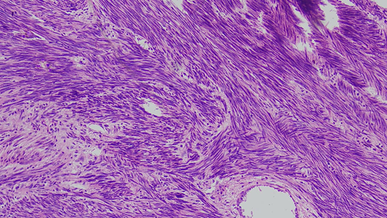
Figure 1: Atypia and osteoclast type multinucleate giant cells
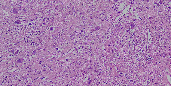
Figure 2: Chondrosarcomatous differentiation
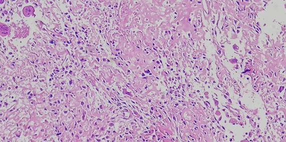
Figure 3: Osteosarcomatous differentiation; stromal osteoid deposits
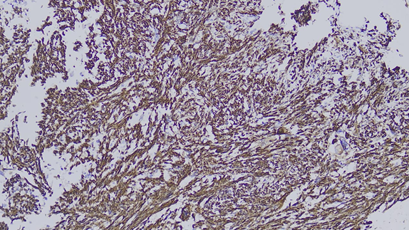
Figure 4: Tumor cells showing diffuse cytoplasmic positivity for SMA
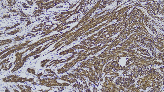
Figure 5: Tumor cells showing diffuse cytoplasmic positivity for H- caldesmon
DISCUSSION:
The present cross-sectional study evaluated the incremental diagnostic role of immunohistochemistry (IHC) in 120 soft-tissue sarcomas and related mesenchymal tumours. The findings demonstrated that integrating a morphology-directed IHC panel with routine histopathology substantially improved diagnostic certainty, lineage identification and histological subclassification. These observations support the contemporary concept that soft-tissue tumour diagnosis should be based on the integration of clinical context, anatomical location, morphology, immunophenotype and, where necessary, molecular alterations.
Diagnostic contribution of immunohistochemistry
In the present study, a definitive diagnosis could be established by morphology alone in 55.8% of cases, which increased to 90.8% after IHC, representing an absolute improvement of 35.0% (P<0.001). Similarly, specific histological subtype assignment increased from 51.7% to 88.3%, correct lineage identification increased from 63.3% to 92.5%, and high diagnostic confidence increased from 49.2% to 85.8%. IHC confirmed the initial diagnosis in 59.2%, refined it in 25.8% and changed it in 15.0%; thus, 40.8% of cases underwent a clinically relevant diagnostic modification.
Doyle et al. (2015)[1] emphasized that newer IHC markers improve the recognition of soft-tissue tumour entities that cannot be reliably separated using morphology and conventional lineage markers alone. This observation is consistent with the present finding that more than two-fifths of cases were refined or reclassified after IHC. Han et al. (2015)[2] similarly showed that combining traditional markers with newer molecular-surrogate antibodies markedly improved the classification of diagnostically difficult spindle-cell tumours.
Schaefer et al. (2018)[9] described IHC as particularly valuable for confirming lineage and detecting surrogate evidence of recurrent molecular alterations. Sbaraglia and Dei Tos (2019)[10] further stated that the pathological diagnosis of sarcoma is an integrated process rather than a purely morphological exercise. These views support the substantial increase in diagnostic certainty observed in the present investigation. The findings are also in agreement with Wei et al. (2017)[7], who recommended an algorithmic approach in which an initial broad panel excludes carcinoma, melanoma and lymphoma, followed by subtype-directed markers selected according to morphology and anatomical location.
The proportion of cases requiring molecular confirmation declined from 30.8% to 14.2% after IHC. Anderson and Hornick (2019)[11] explained that antibodies directed against proteins affected by recurrent genetic alterations can serve as practical surrogates for molecular testing. Examples include nuclear STAT6 for the NAB2::STAT6 fusion in solitary fibrous tumour, loss of H3K27me3 in a subset of malignant peripheral nerve sheath tumours and MDM2/CDK4 overexpression in atypical lipomatous tumour or well-differentiated and dedifferentiated liposarcoma. Anderson and Jo (2021)[15] similarly observed that molecular-surrogate IHC can provide rapid and cost-effective diagnostic information, especially where fluorescence in situ hybridization or sequencing is not readily available.
Nevertheless, the 9.2% of cases that remained inconclusive after IHC illustrate its limitations. Rottmann et al. (2023)[17] noted that staining may be focal, absent or overlapping and that several contemporary sarcoma entities are defined by gene fusions or other molecular alterations. Choi and Ro (2023)[18] therefore recommended molecular confirmation for equivocal lesions and for diagnoses requiring demonstration of a defining genetic event. Thus, IHC should not be considered a replacement for molecular testing in every case, but rather an efficient method of resolving many differential diagnoses and selecting cases requiring further investigation.
Demographic and anatomical spectrum
The mean age of the patients was 44.73±18.62 years, with the largest proportion belonging to the 40-59-year age group. Patients with high-grade sarcomas were significantly older than those with low- or intermediate-grade tumours (48.31 versus 39.17 years; P=0.008). A modest male predominance was observed, with males accounting for 55.8% of cases, although sex was not significantly associated with tumour grade.
Sbaraglia and Dei Tos (2019)[10] reported that adult-type soft-tissue sarcomas are most frequently encountered in middle-aged and older adults, whereas certain round-cell and rhabdomyoblastic sarcomas predominate in children and younger individuals. The wide age distribution in the current study reflected the inclusion of both adult and paediatric histological subtypes. Sbaraglia et al. (2021)[14] also highlighted that age is an important element in formulating the differential diagnosis because identical morphological patterns may represent different tumour entities in different age groups.
The lower extremity was the most frequent anatomical site, representing 35.8% of cases, followed by the upper extremity, trunk or chest wall, retroperitoneum or abdomen, pelvis or perineum, and head and neck. This pattern agrees with Harati et al. (2017)[8], who observed that the extremities, particularly the lower extremity, constitute a major site of adult soft-tissue sarcomas. In the present study, anatomical site was not significantly associated with grade (P=0.388), indicating that high- and low/intermediate-grade sarcomas occurred across all anatomical regions.
High-grade sarcomas comprised 60.8% of the study sample. The mean tumour size was 8.37 cm, and 64.2% measured more than 5 cm. High-grade sarcomas were significantly larger than low/intermediate-grade tumours (9.41 versus 6.76 cm; P=0.001). Tumours larger than 5 cm had approximately threefold greater odds of being high-grade (OR=2.97; 95% CI: 1.38-6.39), while deep-seated tumours had 2.88-fold greater odds of high grade. Harati et al. (2017)[8] likewise found that a substantial proportion of soft-tissue sarcomas were large and high-grade at diagnosis, and that tumour size and grade were important indicators of adverse biological behaviour.
Spindle-cell morphology was the most frequent pattern in the present study, accounting for 39.2%, followed by round-cell, pleomorphic, myxoid, epithelioid and vascular or mixed patterns. This distribution is compatible with the broad morphological approach described by Wei et al. (2017)[7] and Black et al. (2022)[16], who classified diagnostically challenging soft-tissue tumours into spindle-cell, epithelioid, round-cell, pleomorphic and myxoid patterns before applying targeted IHC panels.
Round-cell tumours had 3.45-fold higher odds and pleomorphic tumours had 8.15-fold higher odds of being high-grade than spindle-cell tumours. Tumour necrosis was strongly associated with high grade (OR=5.03; P<0.001), while high-grade tumours showed a markedly greater median mitotic count than low/intermediate-grade tumours (18 versus 6 per 10 high-power fields; P<0.001). Lymphovascular invasion was also significantly associated with high-grade disease (OR=3.14; P=0.020). These findings are biologically plausible because differentiation, mitotic activity and necrosis form the principal components of conventional sarcoma grading. The present results therefore demonstrate consistency between the recorded histomorphological features and the final grade.
Immunohistochemical expression patterns
Subtype-directed IHC panels generated discriminatory findings in 88.3% of cases. Vimentin was positive in 94.2%, demonstrating high sensitivity for mesenchymal differentiation; however, diffuse vimentin expression in undifferentiated pleomorphic sarcoma was not significantly different from that in other sarcomas (P=0.196). Wei et al. (2017)[7] and Black et al. (2022)[16] cautioned that vimentin is broadly expressed in mesenchymal tumours and some non-mesenchymal malignancies and, therefore, has limited value for specific subclassification. The lack of significant subtype discrimination in the current study reinforces that vimentin should be regarded as a screening rather than a lineage-defining marker.
Leiomyosarcoma was the most frequent subtype, accounting for 23.3%. Combined SMA and desmin positivity was present in 92.9%, while h-caldesmon was positive in 85.7%. Both panels demonstrated highly significant associations with leiomyosarcoma compared with other tumours (P<0.001). Doyle et al. (2015)[1] and Wei et al. (2017)[7] described the combined expression of SMA, desmin and h-caldesmon as supportive of smooth-muscle differentiation. They also emphasized that SMA alone lacks specificity because it may be expressed in myofibroblastic and pericytic tumours. The high combined positivity observed in the present study therefore supports the use of multiple myogenic markers with appropriate cytoplasmic staining patterns.
Among synovial sarcomas, diffuse nuclear TLE1 expression was present in 94.1%, while EMA and/or cytokeratin was detected in 82.4%. El Beaino et al. (2020)[13], in a systematic review and meta-analysis, reported pooled TLE1 sensitivity of approximately 94% and specificity of approximately 81% for synovial sarcoma. The current TLE1 positivity rate of 94.1% closely matches the pooled sensitivity reported by these investigators. However, TLE1 is not entirely specific and can be expressed in malignant peripheral nerve sheath tumour, solitary fibrous tumour and other spindle-cell sarcomas. Therefore, the combination of TLE1 with epithelial markers and molecular demonstration of an SS18::SSX fusion remains important in equivocal cases. Anderson and Jo (2021)[15] similarly advised that TLE1 should be interpreted as part of a panel rather than as an isolated confirmatory marker.
Myogenin and/or MyoD1 was positive in 92.9% of rhabdomyosarcomas and only 2.8% of comparator tumours. This strong association supports the value of nuclear myogenic regulatory proteins in confirming skeletal-muscle differentiation. Wei et al. (2017)[7] described myogenin and MyoD1 as more specific for rhabdomyoblastic differentiation than desmin, which can also be expressed in smooth-muscle and other mesenchymal tumours. The high expression rate in the current study is therefore consistent with their established diagnostic utility.
In liposarcoma, S100 and/or MDM2/CDK4 supported the diagnosis in 84.6% of cases. Clay et al. (2016)[6] found that MDM2 and CDK4 IHC can assist in diagnosing atypical lipomatous tumour or well-differentiated liposarcoma, although immunostaining is less sensitive and specific than detection of MDM2 amplification. Consequently, the present findings should be interpreted according to subtype: S100 may support adipocytic differentiation, while nuclear MDM2 and CDK4 are more useful for well-differentiated and dedifferentiated liposarcoma. In morphologically equivocal cases, MDM2 fluorescence in situ hybridization remains preferable.
S100 and/or SOX10 positivity was observed in 72.7% of malignant peripheral nerve sheath tumours (MPNSTs). This incomplete sensitivity is consistent with the known loss or focal expression of Schwannian markers in high-grade MPNST. Schaefer et al. (2016)[4] demonstrated that loss of H3K27me3 was diagnostically useful in a substantial subset of MPNSTs. Cleven et al. (2016)[5] similarly found that H3K27me3 loss supported MPNST over neurofibroma, cellular schwannoma and most undifferentiated sarcomas, although loss could also occur in certain mimics. Asano et al. (2017)[8] confirmed that complete H3K27me3 loss was moderately sensitive but highly specific for MPNST in an appropriate differential diagnosis. Thus, the S100/SOX10 findings in the present study support using these markers together with H3K27me3 and clinical evidence of nerve association or neurofibromatosis.
All nine gastrointestinal stromal tumours expressed DOG1, while 88.9% expressed CD117/KIT. Anderson and Jo (2021)[15] noted that DOG1 is highly sensitive for gastrointestinal stromal tumour and may remain positive in some KIT-negative cases. The current observation of universal DOG1 positivity but slightly lower CD117 positivity supports the complementary use of both markers. Nevertheless, DOG1 and KIT expression must be correlated with gastrointestinal location and morphology because occasional non-GIST tumours may express either marker.
All solitary fibrous tumours showed nuclear STAT6 expression, compared with 1.8% of comparator tumours. Han et al. (2015)[2] reported that nuclear STAT6 was a highly sensitive and specific marker for solitary fibrous tumour. Tai et al. (2015)[3] further demonstrated that STAT6 nuclear expression reflects the underlying NAB2::STAT6 fusion and is associated with the molecular pathogenesis of this tumour. The 100% positivity observed in the current study therefore agrees with previous evidence. The result also explains why solitary fibrous tumours showed relatively low exact morphology-only concordance: STAT6 allowed several non-specific spindle-cell diagnoses to be refined.
Concordance between morphology and final diagnosis
Exact agreement between the initial morphology-based diagnosis and final integrated diagnosis was 59.2%, with a Cohen’s kappa value of 0.52, indicating moderate agreement. After accepting refinements within the same lineage, overall agreement increased to 85.0% and kappa increased to 0.79, indicating substantial agreement. IHC produced a 35-percentage-point increase in definitive diagnosis, and the overall association between the initial and final classifications was statistically significant (P<0.001).
The moderate exact concordance does not imply poor morphological assessment. Rather, it reflects the considerable overlap among sarcoma subtypes and the increasing requirement for immunophenotypic or molecular definition. Sbaraglia et al. (2021)[14] emphasized that the contemporary WHO classification integrates morphology, IHC and molecular genetics and recognizes several entities that cannot be diagnosed reliably by morphology alone. Anderson et al. (2021)[12] similarly described the 2020 WHO classification as increasingly dependent on ancillary tests for distinguishing morphologically overlapping tumours.
Exact concordance was highest for leiomyosarcoma at 75.0%, probably because fascicular architecture, cigar-shaped nuclei and eosinophilic cytoplasm frequently provide recognizable smooth-muscle differentiation. Concordance was lower for undifferentiated pleomorphic sarcoma and solitary fibrous tumour. Undifferentiated pleomorphic sarcoma is a diagnosis of exclusion, requiring the elimination of epithelial, melanocytic, neural, myogenic and other specific lines of differentiation. Solitary fibrous tumour may overlap morphologically with numerous patternless or haemangiopericytoma-like spindle-cell neoplasms but becomes more readily identifiable after nuclear STAT6 staining.
Diagnostic refinement or change was required in 57.9% of pleomorphic tumours. Compared with spindle-cell tumours, pleomorphic tumours had more than threefold greater odds of diagnostic modification (OR=3.24; P=0.033). This is consistent with Schaefer et al. (2018)[9], who highlighted the central role of ancillary tests in excluding dedifferentiated liposarcoma, pleomorphic leiomyosarcoma, MPNST, sarcomatoid carcinoma and melanoma before diagnosing an undifferentiated pleomorphic sarcoma. Choi and Ro (2023)[18] further observed that molecular assays are particularly valuable when pleomorphic or undifferentiated morphology lacks a specific immunophenotype.
CONCLUSION:
Immunohistochemistry played a substantial diagnostic role in differentiating and classifying soft-tissue sarcomas. Its addition to routine histomorphology increased the proportion of cases with a definitive diagnosis from 55.8% to 90.8%, improved correct lineage identification from 63.3% to 92.5%, and enabled assignment of a specific histological subtype in 88.3% of cases. IHC refined the initial diagnosis in 25.8% and changed it in 15.0%, resulting in a clinically important diagnostic modification in 40.8% of cases. Subtype-directed panels particularly SMA, desmin and h-caldesmon for leiomyosarcoma; TLE1 and epithelial markers for synovial sarcoma; myogenin and MyoD1 for rhabdomyosarcoma; DOG1 and CD117 for gastrointestinal stromal tumour; and nuclear STAT6 for solitary fibrous tumour showed strong diagnostic associations. Agreement between the initial morphology-based and final diagnoses improved from moderate exact-category agreement (κ=0.52) to substantial agreement when lineage-consistent refinements were considered (κ=0.79). IHC was especially valuable in pleomorphic and morphologically ambiguous tumours. Therefore, an appropriately selected, morphology-directed IHC panel should be routinely integrated with clinical, radiological and histopathological findings. Molecular testing should remain available for equivocal cases and tumours defined by specific genetic alterations.
LIMITATIONS OF STUDY
This study had several limitations. First, its cross-sectional, single-centre design restricted the generalizability of the findings to institutions with different patient profiles, referral patterns and laboratory resources. Second, the relatively small number of cases within individual histological subtypes resulted in wide confidence intervals and limited detailed subtype-specific analyses. Third, antibody panels were selected according to the initial morphological differential diagnosis and were therefore not identical in all cases, which limited direct comparisons of marker performance across the entire study population. Fourth, molecular confirmation using fluorescence in situ hybridization, polymerase chain reaction or next-generation sequencing was not available for every diagnostically difficult or genetically defined tumour. The final integrated diagnosis might consequently have incorporated IHC itself, creating partial incorporation bias when the diagnostic contribution of IHC was assessed. Fifth, tissue fixation, antigen retrieval, tumour heterogeneity, focal marker expression and small biopsy size could have influenced staining results. Sixth, interobserver variability in the interpretation of morphology and IHC intensity was not assessed systematically. Seventh, the study did not evaluate the cost-effectiveness, turnaround time or incremental benefit of individual antibodies. Finally, the absence of clinical follow-up prevented correlation of IHC-based classification with treatment response, recurrence, metastasis and survival.
REFERENCES:
1. Doyle LA. Soft tissue tumor pathology: new diagnostic immunohistochemical markers. Semin Diagn Pathol. 2015;32(5): 329-340.
2. Han Y, Zhang Q, Yu X, Han X, Wang H, Xu Y, et al. Immunohistochemical detection of STAT6, CD34, CD99 and BCL-2 for diagnosing solitary fibrous tumors/hemangiopericytomas. Int J Clin Exp Pathol. 2015;8(10):13166-13175.
3. Tai HC, Chuang IC, Chen TC, Li CF, Huang SC, Kao YC, et al. NAB2-STAT6 fusion types account for clinicopathological variations in solitary fibrous tumors. Mod Pathol. 2015;28(10):1324-1335.
4. Schaefer IM, Fletcher CDM, Hornick JL. Loss of H3K27 trimethylation distinguishes malignant peripheral nerve sheath tumors from histologic mimics. Mod Pathol. 2016;29(1):4-13.
5. Cleven AHG, Sannaa GAA, Briaire-de Bruijn I, Ingram DR, van de Rijn M, Rubin BP, et al. Loss of H3K27 tri-methylation is a diagnostic marker for malignant peripheral nerve sheath tumors and an indicator for an inferior survival. Mod Pathol. 2016;29(6):582-590.
6. Clay MR, Martinez AP, Weiss SW, Edgar MA. MDM2 and CDK4 immunohistochemistry: should it be used in problematic differentiated lipomatous tumors? A new perspective. Am J Surg Pathol. 2016;40(12):1647-1652.
7. Wei S, Henderson-Jackson E, Qian X, Bui MM. Soft tissue tumor immunohistochemistry update: illustrative examples of diagnostic pearls to avoid pitfalls. Arch Pathol Lab Med. 2017;141(8):1072-1091.
8. Asano N, Yoshida A, Ichikawa H, Mori T, Nakamura M, Kawai A, et al. Immunohistochemistry for trimethylated H3K27 in the diagnosis of malignant peripheral nerve sheath tumours. Histopathology. 2017;70(3):385-393.
9. Schaefer IM, Hornick JL. Diagnostic immunohistochemistry for soft tissue and bone tumors: an update. Adv Anat Pathol. 2018;25(6):400-412.
10. Sbaraglia M, Dei Tos AP. The pathology of soft tissue sarcomas. Radiol Med. 2019;124(4):266-281.
11. Anderson WJ, Hornick JL. Immunohistochemical correlates of recurrent genetic alterations in sarcomas. Genes Chromosomes Cancer. 2019;58(2):111-123.
12. Anderson WJ, Doyle LA. Updates from the 2020 World Health Organization classification of soft tissue and bone tumours. Histopathology. 2021;78(5):644-657.
13. El Beaino M, Araujo DM, Lazar AJ, Lin PP. Diagnostic value of TLE1 in synovial sarcoma: a systematic review and meta-analysis. Sarcoma. 2020;2020:7192347.
14. Sbaraglia M, Bellan E, Dei Tos AP. The 2020 WHO classification of soft tissue tumours: news and perspectives. Pathologica. 2021;113(2):70-84.
15. Anderson WJ, Jo VY. Diagnostic immunohistochemistry of soft tissue and bone tumors: an update on biomarkers that correlate with molecular alterations. Diagnostics (Basel). 2021;11(4):690.
16. Black MA, Coffin CM, Hameed M, Thway K. Diagnosis of soft tissue tumors using immunohistochemistry as a surrogate for recurrent fusion oncoproteins. Semin Diagn Pathol. 2022;39(1):38-47.
17. Rottmann D, Abdulfatah E, Pantanowitz L. Molecular testing of soft tissue tumors. Diagn Cytopathol. 2023;51(1):12-25.
18. Choi JH, Ro JY. The recent advances in molecular diagnosis of soft tissue tumors. Int J Mol Sci. 2023;24(6):5934.