Surgical Correlation Of Multidetector Computed Tomography Findings In Non-Traumatic Acute Abdominal Emergencies.
- Manish Bhagat , Professor & Head of Department, Department of Radio-diagnosis, Sri Aurobindo Medical College & P.G. Institute, Indore.
- Radhika Sharda , PG Resident, Department of Radio-diagnosis, Sri Aurobindo Medical College & P.G. Institute, Indore.
- Jatin Walia , PG Resident, Department of Radio-diagnosis, Sri Aurobindo Medical College & P.G. Institute, Indore.
- Yash Yadav , PG Resident, Department of Radio-diagnosis, Sri Aurobindo Medical College & P.G. Institute, Indore.
- Faraz Ansari , PG Resident, Department of Radio-diagnosis, Sri Aurobindo Medical College & P.G. Institute, Indore.
- Rakesh Sangwan , PG Resident, Department of General Medicine, Sri Aurobindo Medical College & P.G. Institute, Indore.
Article Information:
Abstract:
Background: Non-traumatic acute abdomen encompasses a broad spectrum of gastrointestinal, hepatobiliary, pancreatic, inflammatory, infective, obstructive, and perforative emergencies. Accurate and timely diagnosis is essential for appropriate surgical management. Multidetector computed tomography (MDCT) provides rapid, high-resolution, multiplanar assessment of abdominal pathology. Aim: To evaluate the surgical correlation of MDCT findings in non-traumatic acute abdominal emergencies. Materials and Methods: This hospital-based prospective cross-sectional analytical observational study included 50 patients presenting with clinically suspected non-traumatic acute abdomen who underwent MDCT followed by laparotomy or laparoscopy. CT examinations were performed using a 128-slice MDCT scanner. Radiological findings were documented and correlated with intraoperative and available histopathological findings. Diagnostic performance was assessed using sensitivity, specificity, positive predictive value, negative predictive value, and overall accuracy. Results: Males constituted 66% of the study population, while 54% were below 50 years of age. Acute appendicitis was the commonest diagnosis (20%), followed by bowel obstruction and hollow-viscus perforation (14% each), acute cholecystitis (12%), and acute pancreatitis (10%). MDCT demonstrated important complications including appendicular perforation, abscess, bowel obstruction, pneumoperitoneum, inflammatory collections, and pancreatic necrosis. Overall, MDCT findings were concordant with the final diagnosis in 45 of 50 patients, yielding an overall diagnostic accuracy of 90%. The association between CT diagnosis and final diagnosis was statistically significant (p<0.001). MDCT demonstrated particularly high diagnostic performance in bowel obstruction, acute cholecystitis, and hollow-viscus perforation. Conclusion: MDCT demonstrated strong correlation with surgical and final diagnoses and accurately characterized diverse non-traumatic acute abdominal emergencies. Its rapid acquisition, excellent anatomical coverage, and ability to demonstrate complications make it an important imaging modality for timely diagnosis and surgical decision-making.
Keywords:
Article :
INTRODUCTION:
Non-traumatic acute abdomen is a common surgical emergency encompassing a wide spectrum of gastrointestinal, hepatobiliary, pancreatic, vascular, urological, gynaecological, and other intra-abdominal conditions. Patients may present with acute pain, vomiting, abdominal distension, fever, obstruction, haemorrhage, or peritonitis, while overlapping clinical manifestations may make a definitive diagnosis difficult on clinical assessment alone.[1]
Imaging plays a central role in the evaluation of these patients. Although radiography and ultrasonography remain useful in selected conditions, their diagnostic utility may be limited by operator dependence, bowel gas, patient habitus, and restricted anatomical coverage. Multidetector computed tomography (MDCT), with rapid acquisition, high spatial resolution, and multiplanar reconstruction, allows comprehensive assessment of the abdomen and pelvis and is particularly valuable in emergency settings. [2,3]
MDCT provides simultaneous evaluation of the bowel, solid organs, mesentery, retroperitoneum, peritoneal cavity, and vascular structures. Contrast-enhanced CT can demonstrate inflammatory changes, abnormal organ perfusion, ischaemia, necrosis, collections, and vascular abnormalities. Findings such as bowel or organ wall thickening, fat stranding, free fluid, abscess formation, pneumoperitoneum, and abnormal enhancement patterns help establish the diagnosis and determine disease severity. [4,5] In bowel emergencies, MDCT can identify the site and cause of obstruction, volvulus, strangulation, and intestinal ischaemia, while CT angiography is valuable in suspected mesenteric ischaemia.[5,6]
MDCT is also highly useful in detecting hollow-viscus perforation by demonstrating extraluminal air, free fluid, and the probable site of perforation. CT angiographic protocols further facilitate assessment of vascular emergencies, including aneurysmal rupture, arterial dissection, active bleeding, and associated organ ischaemia. [6,7] Accurate demonstration of the extent and complications of disease also assists surgeons in selecting appropriate operative or conservative management. [7,8]
Despite these advantages, CT utilization requires consideration of radiation exposure and potential adverse effects of iodinated contrast, particularly in patients with renal impairment. [8,9] Correlation of MDCT findings with operative findings is therefore important for determining its diagnostic reliability in acute surgical emergencies. The present study was undertaken to evaluate the surgical correlation of multidetector computed tomography findings in non-traumatic acute abdominal emergencies, with emphasis on the correlation between preoperative CT findings and intraoperative findings.
MATERIALS AND METHODS:
This hospital-based prospective cross-sectional analytical observational study was conducted in the Department of Radiodiagnosis, Sri Aurobindo Medical College and Postgraduate Institute (SAMC & PGI), Indore, Madhya Pradesh, India, over a period of 18 months from June 2024 to November 2025, following approval from the Institutional Ethics Committee.
Written informed consent was obtained from all participants or their legally authorized representatives before enrollment. Patient confidentiality was maintained using unique study identification numbers, and participation was voluntary without affecting routine clinical management. The potential risks associated with radiation exposure and intravenous contrast were considered against the clinical benefits of timely diagnosis, and CT examinations were performed as part of routine clinical care.
Inclusion and Exclusion Criteria
A total of 50 patients presenting with clinical features suggestive of non-traumatic acute abdomen were enrolled using consecutive purposive sampling. Patients of all age groups and either sex were eligible if MDCT was clinically indicated and they subsequently underwent surgical intervention by laparotomy or laparoscopy. The intraoperative findings were considered the reference standard for correlation with the MDCT findings.
Pregnant females, patients with known hypersensitivity to iodinated intravenous contrast, patients with impaired renal function, those presenting with traumatic abdominal injury, haemodynamically unstable patients who could not safely undergo CT despite resuscitation, and patients or their legal guardians who declined written informed consent were excluded.
Methodology
Participants and Sampling
Following written informed consent, demographic characteristics, presenting complaints, clinical examination findings, relevant laboratory investigations, MDCT findings, intraoperative findings, and final diagnosis were recorded using a predesigned structured proforma. Consecutive purposive sampling was used, and all eligible patients presenting during the study period were considered for enrollment until the predetermined sample size was achieved.
MDCT Examination and Image Analysis
All CT examinations were performed using a PHILIPS 128-slice multidetector CT scanner according to standardized departmental abdominal CT protocols. Pre-contrast images were obtained from the dome of the diaphragm to the pubic symphysis, followed by intravenous administration of non-ionic iodinated contrast media. Contrast-enhanced imaging was performed using arterial, portal venous, and delayed phases according to the clinical indication. Axial images were reconstructed with appropriate slice thickness, and multiplanar reformation (MPR), maximum intensity projection (MIP), and volume-rendered technique (VRT) images were generated whenever required.
The CT images were independently reviewed by two experienced radiologists who were blinded to the final surgical diagnosis. Appropriate abdominal, bone, and lung window settings were used for image interpretation. CT findings were systematically assessed according to the underlying pathology, including bowel wall thickening, fat stranding, free fluid, pneumoperitoneum, bowel obstruction, inflammatory changes, abscess formation, vascular abnormalities, organ involvement, and other relevant pathological findings. Any discrepancy between the radiologists was resolved by consensus.
Surgical Correlation
Patients were taken for surgical intervention based on the combined clinical and radiological assessment. Intraoperative findings were documented by the operating surgeon and subsequently compared with the preoperative MDCT findings. Where surgical specimens were obtained, histopathological examination was performed. The final diagnosis was established by integrating the intraoperative and available histopathological findings and was used as the reference diagnosis for evaluating the diagnostic performance of MDCT.
Outcome Measures
The primary outcomes were the spectrum of MDCT findings in non-traumatic acute abdominal emergencies, diagnostic characterization of the underlying pathology, and correlation of MDCT findings with intraoperative findings. Diagnostic performance was assessed using sensitivity, specificity, positive predictive value, negative predictive value, and overall diagnostic accuracy, where applicable. Secondary outcomes included the demographic profile of patients, presenting clinical features, etiological spectrum of non-traumatic acute abdomen, and the degree of agreement between MDCT diagnosis and the final surgical diagnosis.
Statistical Analysis
Data were entered into a structured master chart and analysed using R statistical software. Continuous variables were expressed as mean ± standard deviation for normally distributed data or median with interquartile range for non-normally distributed data. Categorical variables were presented as frequencies and percentages. Diagnostic performance of MDCT was assessed using two-by-two contingency tables by comparing the CT diagnosis with the surgical reference standard. Sensitivity, specificity, positive predictive value, negative predictive value, and overall diagnostic accuracy were calculated with 95% confidence intervals. The Chi-square test or Fisher's exact test, as appropriate, was used to assess associations between categorical variables and CT findings with surgical outcomes. A p-value <0.05 was considered statistically significant. Results were presented using tables, cross-tabulations, graphs, and descriptive summaries.
RESULTS:
A total of 50 patients with non-traumatic acute abdomen were included. The study population showed a male predominance, with 33 (66.0%) males and 17 (34.0%) females. Patients below 50 years constituted 27 (54.0%) cases. Diffuse abdominal pain was the commonest pain pattern, reported in 17 (34.0%) patients, followed by right iliac fossa pain in 9 (18.0%). Vomiting was the commonest associated symptom, occurring in 37 (74.0%) patients. Guarding or rigidity was absent in 40 (80.0%) patients, while 6 (12.0%) had guarding and 4 (8.0%) had board-like rigidity. [Table 1]
Table 1. Demographic and Clinical Profile of the Study Population
|
Variable |
Category |
n (%) |
|
Age |
<50 years |
27 (54.0) |
|
|
≥50 years |
23 (46.0) |
|
Sex |
Male |
33 (66.0) |
|
|
Female |
17 (34.0) |
|
Site of abdominal pain |
RUQ |
7 (14.0) |
|
|
Epigastric |
8 (16.0) |
|
|
RIF |
9 (18.0) |
|
|
LIF |
5 (10.0) |
|
|
Diffuse |
17 (34.0) |
|
|
Flank |
6 (12.0) |
|
Vomiting |
Present |
37 (74.0) |
|
|
Absent |
13 (26.0) |
|
Abdominal distension |
Present |
11 (22.0) |
|
|
Absent |
39 (78.0) |
|
Constipation/obstipation |
Present |
15 (30.0) |
|
|
Absent |
35 (70.0) |
|
Guarding/rigidity |
Absent |
40 (80.0) |
|
|
Present |
6 (12.0) |
|
|
Board-like |
4 (8.0) |
Acute appendicitis was the commonest clinical diagnosis, accounting for 10 (20.0%) cases. In these patients, periappendiceal fluid or collection was the most frequent associated CT finding, present in 5 (50.0%) cases. Among bowel obstruction cases, adhesive disease was the commonest cause, and small bowel obstruction accounted for 4 (57.1%) cases. In hollow-viscus perforation, pneumoperitoneum and free intraperitoneal fluid were present in all 7 (100.0%) cases. [Table 2]
Table 2. Clinical Diagnosis and CT Findings in Major Acute Abdominal Conditions
|
Diagnosis / CT parameter |
Category |
n (%) |
|
Clinical diagnosis |
Acute appendicitis |
10 (20.0) |
|
Bowel obstruction |
7 (14.0) |
|
|
Hollow-viscus perforation |
7 (14.0) |
|
|
Acute cholecystitis |
6 (12.0) |
|
|
Acute pancreatitis |
5 (10.0) |
|
|
Abdominal tuberculosis |
4 (8.0) |
|
|
Liver abscess |
3 (6.0) |
|
|
Ureteric calculi |
3 (6.0) |
|
|
Inflammatory bowel disease |
3 (6.0) |
|
|
Pyelonephritis |
2 (4.0) |
|
|
Acute appendicitis |
Perforation |
3 (30.0) |
|
Appendicolith |
4 (40.0) |
|
|
Periappendiceal fluid/collection |
5 (50.0) |
|
|
Abscess formation |
2 (20.0) |
|
|
Appendicular mass/phlegmon |
2 (20.0) |
|
|
Lymphadenopathy |
4 (40.0) |
|
|
Bowel obstruction |
Adhesive |
2 (28.6) |
|
Hernia-related |
1 (14.3) |
|
|
Malignant |
1 (14.3) |
|
|
Volvulus |
1 (14.3) |
|
|
Inflammatory/Crohn’s |
1 (14.3) |
|
|
Tubercular |
1 (14.3) |
|
|
Jejunum |
1 (14.3) |
|
|
Ileum/ileocecal region |
2 (28.6) |
|
|
Sigmoid colon |
2 (28.6) |
|
|
Caecum/large bowel |
1 (14.3) |
|
|
Small bowel, general |
1 (14.3) |
|
|
Small bowel obstruction |
4 (57.1) |
|
|
Large bowel obstruction |
3 (42.9) |
|
|
Perforation |
0 (0.0) |
|
|
Hollow-viscus perforation |
Duodenum |
2 (28.6) |
|
Stomach |
1 (14.3) |
|
|
Ileum |
2 (28.6) |
|
|
Sigmoid colon |
2 (28.6) |
|
|
Pneumoperitoneum |
7 (100.0) |
|
|
Free intraperitoneal fluid |
7 (100.0) |
|
|
Bowel wall defect visualized |
3 (42.9) |
|
|
Indirect signs only |
4 (57.1) |
|
|
Associated abscess |
2 (28.6) |
In acute cholecystitis, gallbladder distension was present in all 6 (100.0%) patients, while pericholecystic fluid or fat stranding was present in 5 (83.3%). In acute pancreatitis, interstitial oedematous pancreatitis was the predominant pattern, observed in 4 (80.0%) cases, and peripancreatic fluid collections were seen in 3 (60.0%). Among liver abscess cases, 2 (66.7%) were solitary and 1 (33.3%) was multiple; no rupture or portal/hepatic venous thrombosis was identified. [Table 3]
Table 3. CT Findings in Hepatobiliary and Pancreatic Emergencies
|
Condition / Parameter |
Category |
n (%) |
|
Acute cholecystitis |
Gallstones present |
3/6 (50.0) |
|
Gallstones absent |
3/6 (50.0) |
|
|
Gallbladder distension present |
6/6 (100.0) |
|
|
Pericholecystic fluid/fat stranding present |
5/6 (83.3) |
|
|
Mild inflammation |
3/6 (50.0) |
|
|
Moderate inflammation |
2/6 (33.3) |
|
|
Severe inflammation |
1/6 (16.7) |
|
|
Acute pancreatitis |
Homogeneous interstitial oedematous enhancement |
4/5 (80.0) |
|
Heterogeneous necrotizing enhancement |
1/5 (20.0) |
|
|
Peripancreatic fluid collection |
3/5 (60.0) |
|
|
Mild fat stranding |
2/5 (40.0) |
|
|
Moderate fat stranding |
2/5 (40.0) |
|
|
Severe fat stranding |
1/5 (20.0) |
|
|
Pleural effusion |
1/5 (20.0) |
|
|
Ascites |
1/5 (20.0) |
|
|
Liver abscess |
Single abscess |
2/3 (66.7) |
|
Multiple abscesses |
1/3 (33.3) |
|
|
Abscess rupture |
0/3 (0.0) |
|
|
Portal/hepatic vein thrombosis |
0/3 (0.0) |
|
|
THAD |
1/3 (33.3) |
All 4 (100.0%) abdominal tuberculosis cases demonstrated bowel wall thickening, necrotic mesenteric lymphadenopathy, and omental/peritoneal thickening. Bowel wall thickening was also present in all 3 (100.0%) inflammatory bowel disease cases, with terminal ileal involvement and asymmetrical disease in 2 (66.7%) each. Hydroureteronephrosis was present in all 3 (100.0%) ureteric calculus cases. Both pyelonephritis cases showed focal renal involvement, with moderate and severe perinephric fat stranding in 1 (50.0%) case each. [Table 4]
Table 4. CT Findings in Infective, Inflammatory and Urological Conditions
|
Condition / Parameter |
Category |
n (%) |
|
Abdominal tuberculosis |
Bowel wall thickening |
4/4 (100.0) |
|
Necrotic mesenteric lymphadenopathy |
4/4 (100.0) |
|
|
Ascites |
2/4 (50.0) |
|
|
Omental/peritoneal thickening |
4/4 (100.0) |
|
|
Bowel obstruction features |
1/4 (25.0) |
|
|
Inflammatory bowel disease |
Bowel wall thickening |
3/3 (100.0) |
|
Stricture formation |
2/3 (66.7) |
|
|
Fistulae |
1/3 (33.3) |
|
|
Mesenteric abscess |
1/3 (33.3) |
|
|
Terminal ileum involvement |
2/3 (66.7) |
|
|
Other site |
1/3 (33.3) |
|
|
Asymmetrical involvement |
2/3 (66.7) |
|
|
Symmetrical involvement |
1/3 (33.3) |
|
|
Bowel obstruction |
1/3 (33.3) |
|
|
Ureteric calculi |
Proximal ureter |
1/3 (33.3) |
|
Mid ureter |
1/3 (33.3) |
|
|
Distal ureter |
1/3 (33.3) |
|
|
Hydroureteronephrosis |
3/3 (100.0) |
|
|
Partial obstruction |
2/3 (66.7) |
|
|
Complete obstruction |
1/3 (33.3) |
|
|
Perinephric/periureteric fat stranding |
2/3 (66.7) |
|
|
Acute pyelonephritis |
Focal renal involvement |
2/2 (100.0) |
|
Diffuse renal involvement |
0/2 (0.0) |
|
|
Mild perinephric fat stranding |
0/2 (0.0) |
|
|
Moderate perinephric fat stranding |
1/2 (50.0) |
|
|
Severe perinephric fat stranding |
1/2 (50.0) |
|
|
Calculi |
0/2 (0.0) |
|
|
Emphysematous changes |
0/2 (0.0) |
|
|
Renal/perinephric abscess |
1/2 (50.0) |
|
|
Hydronephrosis |
0/2 (0.0) |
Bowel wall thickening was the most frequent overall CT finding, identified in 17 (34.0%) patients. Free intraperitoneal fluid was present in 10 (20.0%), mesenteric lymphadenopathy in 8 (16.0%), pneumoperitoneum and bowel dilatation in 7 (14.0%) each, and peritoneal thickening/enhancement in 4 (8.0%). The appendix was visualized in 49 (98.0%) patients. [Table 5]
Table 5. Overall CT Findings in Non-Traumatic Acute Abdomen
|
CT finding |
Present n (%) |
Absent n (%) |
|
Pneumoperitoneum |
7 (14.0) |
43 (86.0) |
|
Free intraperitoneal fluid |
10 (20.0) |
40 (80.0) |
|
Peritoneal thickening/enhancement |
4 (8.0) |
46 (92.0) |
|
Mesenteric lymphadenopathy |
8 (16.0) |
42 (84.0) |
|
Bowel dilatation |
7 (14.0) |
43 (86.0) |
|
Bowel wall thickening |
17 (34.0) |
33 (66.0) |
|
Appendix visualized |
49 (98.0) |
1 (2.0) |
CT findings correlated with the final diagnosis in 45 (90.0%) of the 50 patients. Complete agreement was observed for bowel obstruction, acute cholecystitis, acute pancreatitis, abdominal tuberculosis, ureteric calculi, and pyelonephritis. Concordance was 8/10 (80.0%) for acute appendicitis, 6/7 (85.7%) for hollow-viscus perforation, and 2/3 (66.7%) each for liver abscess and inflammatory bowel disease.
Table 6. Correlation Between CT Diagnosis and Final Diagnosis
|
Diagnosis |
CT diagnosis n |
Intraoperative/laboratory findings n |
Final diagnosis n |
Concordance |
|
Acute appendicitis |
10 |
8 |
8 |
8/10 (80.0) |
|
Bowel obstruction |
7 |
7 |
7 |
7/7 (100.0) |
|
Hollow-viscus perforation |
7 |
6 |
6 |
6/7 (85.7) |
|
Acute cholecystitis |
6 |
6 |
6 |
6/6 (100.0) |
|
Acute pancreatitis |
5 |
5 |
5 |
5/5 (100.0) |
|
Abdominal tuberculosis |
4 |
4 |
4 |
4/4 (100.0) |
|
Liver abscess |
3 |
2 |
2 |
2/3 (66.7) |
|
Ureteric calculi |
3 |
3 |
3 |
3/3 (100.0) |
|
Inflammatory bowel disease |
3 |
2 |
2 |
2/3 (66.7) |
|
Pyelonephritis |
2 |
2 |
2 |
2/2 (100.0) |
|
Total |
50 |
45 |
45 |
45/50 (90.0) |
Overall, 45 (90.0%) patients were correctly diagnosed by MDCT and 5 (10.0%) were categorized as incorrect or unconfirmed. The association between CT diagnosis and final diagnosis was statistically significant (Pearson chi-square = 45.0, df = 9, p <0.001; likelihood ratio = 42.731, df = 9, p <0.001). For overall non-traumatic acute abdomen, the reported sensitivity was 100.0%, PPV 90.0%, and diagnostic accuracy 90.0%. MDCT showed particularly high diagnostic performance for bowel obstruction and acute cholecystitis, with 100.0% sensitivity, specificity, PPV, NPV, and accuracy in both conditions. For hollow-viscus perforation, sensitivity was 100.0%, specificity 97.7%, PPV 85.7%, NPV 100.0%, and accuracy 98.0%. For liver abscess, sensitivity was 100.0%, specificity 97.9%, PPV 66.7%, NPV 100.0%, and accuracy 98.0%. [Table 7]
Table 7. Diagnostic Performance and Statistical Association of MDCT
|
Condition / Parameter |
TP |
FP |
FN |
TN |
Sensitivity (%) |
Specificity (%) |
PPV (%) |
NPV (%) |
Accuracy (%) |
|
Overall non-traumatic acute abdomen |
45 |
5 |
0 |
Not reported |
100.0 |
Not reported |
90.0 |
Not reported |
90.0 |
|
Acute appendicitis |
8 |
2 |
0 |
40 |
100.0 |
95.2 |
80.0 |
100.0 |
96.0 |
|
Bowel obstruction |
7 |
0 |
0 |
43 |
100.0 |
100.0 |
100.0 |
100.0 |
100.0 |
|
Hollow-viscus perforation |
6 |
1 |
0 |
43 |
100.0 |
97.7 |
85.7 |
100.0 |
98.0 |
|
Acute cholecystitis |
6 |
0 |
0 |
44 |
100.0 |
100.0 |
100.0 |
100.0 |
100.0 |
|
Liver abscess |
2 |
1 |
0 |
47 |
100.0 |
97.9 |
66.7 |
100.0 |
98.0 |
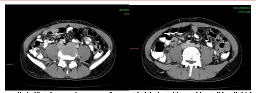
Figure 1. Appendix is dilated measuring approx. 8 mm, at 6 o’clock position and has mild wall thickening along with surrounding minimal fat stranding and few sub-cm lymph nodes, concerning for Acute Appendicitis.
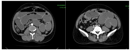
Figure 2. Moderately dilated jejunal and ileal loops with multiple air fluid levels. The transition point is in mid ileum with small bowel feces sign- Small Bowel Obstruction.
 .
.
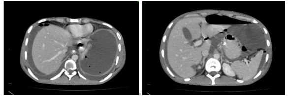
Figure 3. Multiple foci of free air seen in bilateral upper abdomen and anterior abdomen suggestive of Pneumo Peritoneum.
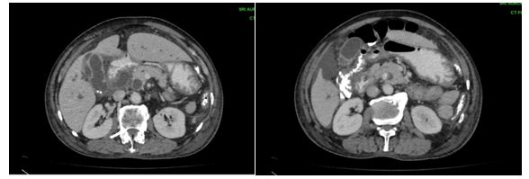
Figure 4. Gall bladder is grossly distended with 2-3 hyperdense calculi in its lumen with mild to moderate pericholecystic fluid. Also seen is hyperdense calculus in terminal CBD with resultant bilateral IHBR dilatation and fusiform dilatation of CBD. Findings suggestive of Acute Calculus Cholecystitis with Choledocholithiasis
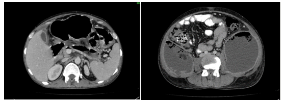
Figure 5. Body and tail of pancreas is replaced by well-defined thick wall collection communicating with the MPD. These findings are suggestive of Necrotizing Pancreatitis with multiple walled off collections and their extension in peritoneal cavity. Multiple foci of air are present within these collections which may be due to super-added infection. (Modified CTSI score of 10).
DISCUSSION:
The present study evaluated the role of multidetector computed tomography (MDCT) in patients presenting with non-traumatic acute abdomen, with particular emphasis on correlation of CT findings with intraoperative and final diagnoses. The study included 50 patients and demonstrated that MDCT provided a broad assessment of inflammatory, obstructive, perforative, infective, hepatobiliary, pancreatic, and urological causes of acute abdomen. Overall, CT findings correlated with the final diagnosis in 45 patients, giving an overall diagnostic accuracy of 90%. These findings support the important role of MDCT in the rapid evaluation and surgical decision-making of patients with acute abdominal emergencies.
In the present study, there was a male predominance, with males constituting 66% of the study population, while 54% of patients were below 50 years of age. This demographic pattern is comparable with previous studies describing acute abdominal emergencies as a frequent indication for urgent imaging in adult patients. Leschka et al. [9] emphasized the increasing importance of MDCT in the evaluation of acute abdominal pathology, while Paolantonio et al. [10] highlighted the need for rapid imaging in non-traumatic acute abdominal pain. The relatively broad age distribution in the present study may be explained by the heterogeneous disease spectrum, with appendicitis occurring more commonly in younger individuals and obstruction, perforation, and hepatobiliary disorders occurring across older age groups. Aschoff et al. [11] and Sultan et al. [12] similarly emphasized the usefulness of MDCT across different adult age groups, particularly for gastrointestinal obstruction and inflammatory emergencies.
Acute appendicitis was the most frequent diagnosis in the present study, accounting for 20% of cases. MDCT demonstrated perforation in 30%, appendicolith in 40%, periappendiceal fluid or collection in 50%, abscess formation in 20%, appendicular mass or phlegmon in 20%, and lymphadenopathy in 40% of patients. These findings demonstrate that MDCT was useful not only for diagnosing appendicitis but also for identifying complications that may influence surgical management. Leschka et al. [9] reported appendicitis as an important indication for emergency CT, while Kessner et al. [13] also identified appendicitis as a common cause of acute non-traumatic abdominal pain requiring CT. The identification of perforation, abscess, phlegmon, and periappendiceal collections is particularly relevant because these findings may influence the decision between immediate surgery and conservative treatment.
Bowel obstruction and hollow-viscus perforation each accounted for 14% of cases. Among obstruction cases, adhesive disease was the most common cause, while small bowel obstruction accounted for 57.1%. MDCT demonstrated the level, site, and underlying cause of obstruction, with no perforation identified. These findings are consistent with Idris et al. [14], who demonstrated the diagnostic value of MDCT in identifying the transition point and aetiology of small bowel obstruction. Sultan et al. [12] similarly reported the usefulness of MDCT in determining the cause and location of obstruction, while Zhou et al. [15] demonstrated good diagnostic performance of MDCT in small bowel obstruction. Kelkar et al. [16] also identified postoperative adhesions as an important cause of intestinal obstruction. The ability of MDCT to demonstrate the transition point, bowel dilatation, mesenteric changes, and complications such as ischaemia or perforation makes it particularly valuable in surgical planning.
Hollow-viscus perforation was characterized by pneumoperitoneum and free intraperitoneal fluid in all patients. The bowel wall defect was directly visualized in 42.9% of cases, while the remaining cases were diagnosed using indirect CT findings. Associated abscess formation was present in 28.6%. These observations emphasize the sensitivity of MDCT for detecting small quantities of extraluminal air and for localizing the probable site of perforation. Aschoff et al. [11] and Paolantonio et al. [10] emphasized the importance of MDCT in gastrointestinal perforation and other life-threatening abdominal emergencies. Leschka et al. [9] further highlighted the importance of thin-section imaging and multiplanar reconstruction in improving diagnostic confidence. Meeson et al. [17] also described the role of CT in identifying intra-abdominal collections and complications that may influence surgical or interventional management.
Hepatobiliary and pancreatic emergencies constituted an important component of the study. Acute cholecystitis accounted for 12% of cases, with gallbladder distension present in all patients and pericholecystic fluid or fat stranding in 83.3%. Gallstones were identified in 50% of cases. These findings indicate that inflammatory changes surrounding the gallbladder may be more informative than the presence of calculi alone. Leschka et al. [9] and Hammerstingl RM et al. [18] emphasized the value of optimized contrast-enhanced MDCT in demonstrating inflammatory changes and complications of hepatobiliary disease. Acute pancreatitis accounted for 10% of cases, with interstitial oedematous pancreatitis predominating and peripancreatic fluid collections present in 60%. These findings are consistent with the observations of Aschoff et al. [11] and Meeson et al. [17], who highlighted CT for assessment of pancreatic inflammation, necrosis, collections, and disease severity.
MDCT also demonstrated characteristic findings in abdominal tuberculosis and inflammatory bowel disease. All patients with abdominal tuberculosis showed bowel wall thickening, necrotic mesenteric lymphadenopathy, and omental or peritoneal thickening, while ascites occurred in 50%. In inflammatory bowel disease, bowel wall thickening was universal, with stricture formation in 66.7% and terminal ileal involvement in 66.7%. These findings support the observations of Aschoff et al. [11] regarding the usefulness of MDCT in demonstrating bowel wall abnormalities, strictures, mesenteric disease, and extraintestinal complications. Meeson et al. [17] further emphasized the role of CT in identifying inflammatory collections and complications.
Despite these findings, the present study had certain limitations, including the relatively small sample size and single-centre design, which may limit generalizability. Only surgically managed patients were included, and histopathological confirmation was not available in every case. Subgroup analysis was also limited by the small number of patients in individual diagnostic categories, and long-term outcomes, radiation dose, and cost-effectiveness were not assessed.
Overall, the present study demonstrated strong concordance between MDCT and final diagnosis, with an overall diagnostic accuracy of 90%. MDCT showed high performance in major emergencies, particularly obstruction, cholecystitis, perforation, and appendicitis. Its rapid acquisition, excellent anatomical coverage, and multiplanar imaging facilitated accurate diagnosis, complication detection, and timely surgical planning in non-traumatic acute abdominal emergencies.
CONCLUSION:
The present study demonstrates that MDCT is a valuable imaging modality in the evaluation of non-traumatic acute abdominal emergencies. It accurately identified a broad spectrum of inflammatory, obstructive, perforative, infective, hepatobiliary, pancreatic, and urological conditions. Its ability to define disease extent, complications, and anatomical localization showed strong correlation with final surgical diagnoses. MDCT therefore facilitates rapid diagnosis, appropriate surgical planning, and timely management, supporting its important role in contemporary emergency abdominal imaging.
REFERENCES:
1. Stoker J, van Randen A, Laméris W, Boermeester MA. Imaging patients with acute abdominal pain. Radiology. 2009 Oct;253(1):31-46.
2. Cartwright SL, Knudson MP. Evaluation of acute abdominal pain in adults. Am Fam Physician. 2008 Apr 1;77(7):971-8. PMID: 18441863.
3. van Randen A, Laméris W, van Es HW, van Heesewijk HP, van Ramshorst B, Ten Hove W, Bouma WH, van Leeuwen MS, van Keulen EM, Bossuyt PM, Stoker J, Boermeester MA; OPTIMA Study Group. A comparison of the accuracy of ultrasound and computed tomography in common diagnoses causing acute abdominal pain. Eur Radiol. 2011 Jul;21(7):1535-45.
4. Ahn SH, Mayo-Smith WW, Murphy BL, Reinert SE, Cronan JJ. Acute nontraumatic abdominal pain in adult patients: abdominal radiography compared with CT evaluation. Radiology. 2002 Oct;225(1):159-64.
5. Laméris W, van Randen A, van Es HW, van Heesewijk JP, van Ramshorst B, Bouma WH, ten Hove W, van Leeuwen MS, van Keulen EM, Dijkgraaf MG, Bossuyt PM, Boermeester MA, Stoker J; OPTIMA study group. Imaging strategies for detection of urgent conditions in patients with acute abdominal pain: diagnostic accuracy study. BMJ. 2009 Jun 26;338:b2431.
6. Sala E, Watson CJ, Beadsmoore C, Groot-Wassink T, Fanshawe TR, Smith JC, Bradley A, Palmer CR, Shaw A, Dixon AK. A randomized, controlled trial of routine early abdominal computed tomography in patients presenting with non-specific acute abdominal pain. Clin Radiol. 2007 Oct;62(10):961-9
7. Menke J. Diagnostic accuracy of multidetector CT in acute mesenteric ischemia: systematic review and meta-analysis. Radiology. 2010 Jul;256(1):93-101.
8. Hines J, Rosenblat J, Duncan DR, Friedman B, Katz DS. Perforation of the mesenteric small bowel: etiologies and CT findings. Emerg Radiol. 2013 Apr;20(2):155-61.
9. Leschka S, Alkadhi H, Wildermuth S, Marincek B. Multi-detector computed tomography of acute abdomen. Eur Radiol. 2005 Dec;15(12):2435-47
10. Paolantonio P, Rengo M, Ferrari R, Laghi A. Multidetector CT in emergency radiology: acute and generalized non-traumatic abdominal pain. Br J Radiol. 2016;89(1061):20150859
11. Aschoff AJ. MDCT of the abdomen. Eur Radiol. 2006 Nov;16 Suppl 7:M54-7
12. Sultan A, Hassan M, Ali M. Role of Multidetector Computed Tomography with Multiplanar and Curved Multiplanar Reformations in the Detection of Cause of Intestinal Obstruction: A Tertiary Care Experience. Cureus. 2020 Mar 29;12(3):e7464.
13. Kessner R, Barnes S, Halpern P, Makrin V, Blachar A. CT for Acute Nontraumatic Abdominal Pain-Is Oral Contrast Really Required? Acad Radiol. 2017 Jul;24(7):840-845
14. Idris M, Kashif N, Idris S, Memon WA, Tanveer UH, Haider Z. Accuracy of 64-slice multidetector computed tomography scan in detection of the point of transition of small bowel obstruction. Jpn J Radiol. 2012 Apr;30(3):235-41.
15. Zhou J, Cong R, Shi J, Chen F, Zhu J, Xiao J, Sheng M, Yang J, He B. Diagnostic significance of multidetector computed tomography (MDCT) in patients with small bowel obstruction: a meta-analysis. Jpn J Radiol. 2020 May;38(5):458-462
16. Kelkar A, Thakur VBS, Jeeson J. Evaluation of Multidetector Computed Tomography in the Diagnosis of Intestinal Obstruction. Cureus. 2024 Aug 5;16(8):e66244
17. Meeson S, Alvey CM, Golding SJ. Justifying multidetector CT in abdominal sepsis: time for review? Br J Radiol. 2009 Mar;82(975):190-7
18. Hammerstingl RM, Vogl TJ. Abdominal MDCT: protocols and contrast considerations. Eur Radiol. 2005 Dec;15 Suppl 5:E78-90.