Correlation Of Computed Tomography Findings With Histopathological Diagnosis In Gallbladder Lesions.
- Manish Bhagat , Professor & Head of Department, Department of Radio-diagnosis., Sri Aurobindo Medical College & P.G. Institute, Indore.
- Jatin Walia , PG Resident, Department of Radio-diagnosis, Sri Aurobindo Medical College & P.G. Institute, Indore.
- Radhika Sharda , PG Resident, Department of Radio-diagnosis, Sri Aurobindo Medical College & P.G. Institute, Indore.
- Yash Yadav , PG Resident, Department of Radio-diagnosis, Sri Aurobindo Medical College & P.G. Institute, Indore.
- Faraz Ansari , PG Resident, Department of Radio-diagnosis, Sri Aurobindo Medical College & P.G. Institute, Indore.
- Hiral Bishnoi , PG Resident, Department of Obstetrics and Gynecology, Sri Aurobindo Medical College & P.G. Institute, Indore.
Article Information:
Abstract:
Background: Gallbladder lesions encompass a broad spectrum of benign inflammatory conditions and malignant pathologies, with considerable overlap in their clinical and imaging manifestations. Computed tomography (CT) plays an important role in lesion characterization, assessment of local invasion, and differentiation between benign and malignant disease. Methods: This hospital-based cross-sectional observational study was conducted in the Department of Radiodiagnosis, Sri Aurobindo Medical College and Postgraduate Institute, Indore, over 18 months from June 2024 to November 2025. Patients with suspected gallbladder lesions who underwent CT and had histopathological correlation were evaluated. CT findings were assessed for lesion morphology, wall thickening, enhancement pattern, lesion location, mass formation, lymphadenopathy, infiltration, biliary dilatation, and hepatic involvement. Histopathology was considered the reference standard. Statistical associations were assessed using the Chi-square test, with p<0.05 considered statistically significant. Results: A total of 37 patients were evaluated, with female predominance (70.3%). The majority were aged 40–59 years (62.2%). Inflammatory lesions were the commonest clinical diagnosis (43.2%), and pain was the predominant symptom (59.5%). CT classified 73.0% of lesions as benign and 27.0% as malignant/suspicious. Diffuse wall thickening was the most frequent wall pattern (75.7%). Mass formation (p=0.001), lymphadenopathy (p<0.001), infiltration (p<0.001), and liver involvement (p=0.001) were significantly associated with malignancy. CT demonstrated 100% sensitivity, 96.4% specificity, 90% positive predictive value, 100% negative predictive value, and 97.3% overall diagnostic accuracy. Conclusion: CT is a valuable imaging modality for evaluating gallbladder lesions and differentiating benign from malignant pathology. Morphological features such as mass formation, lymphadenopathy, infiltration, and hepatic involvement are particularly useful indicators of malignancy. Histopathological correlation remains essential in indeterminate or suspicious lesions.
Keywords:
Article :
INTRODUCTION:
Gallbladder diseases constitute an important spectrum of hepatobiliary disorders, ranging from common benign conditions such as cholelithiasis and cholecystitis to potentially aggressive malignancies such as gallbladder carcinoma (GBC) [1,2]. These conditions may present with overlapping clinical manifestations, including right upper quadrant pain, fever, jaundice, dyspeptic symptoms, or may remain asymptomatic, making clinical differentiation difficult [1-3]. Chronic inflammatory conditions may further produce gallbladder wall thickening, fibrosis, calcification, and other morphological changes that can mimic malignant lesions. Similarly, gallbladder polyps and adenomyomatosis may demonstrate imaging appearances that overlap with early neoplastic disease [4].
Gallbladder carcinoma is an uncommon but highly aggressive malignancy, with a particularly high incidence in certain geographical regions, including northern India. It predominantly affects older individuals and shows a female predominance, with peak incidence during the sixth and seventh decades of life [5,6]. Its poor prognosis is largely related to late diagnosis, early invasion of the adjacent liver, and frequent lymphatic and hematogenous dissemination [7,8]. Early diagnosis is therefore essential for appropriate staging, assessment of resectability, and treatment planning [9].Ultrasonography (USG) remains the initial imaging modality for suspected gallbladder disease because it is widely available, inexpensive, non-invasive, free of ionizing radiation, and highly effective for detecting gallstones and inflammatory changes [10]. However, its diagnostic performance may be limited by operator dependence, obesity, overlying bowel gas, and difficulty in differentiating subtle malignant wall changes from benign inflammatory thickening [11]. Computed tomography (CT), particularly multidetector CT (MDCT), provides superior anatomical coverage, high spatial resolution, rapid acquisition, and multiplanar reconstruction, allowing comprehensive assessment of the gallbladder and surrounding structures [12]. CT can demonstrate gallbladder masses, focal or diffuse wall thickening, intraluminal lesions, hepatic invasion, lymphadenopathy, vascular involvement, peritoneal disease, and distant metastases, thereby contributing substantially to staging and assessment of surgical resectability [13,14]. It can also help distinguish benign from malignant lesions based on wall morphology, enhancement characteristics, preservation or loss of wall stratification, and local disease extension [15]. Furthermore, CT is valuable in detecting complications of acute cholecystitis, including gangrenous or emphysematous changes, perforation, abscess formation, and biliary obstruction [16].
Thus, systematic evaluation of gallbladder lesions on CT and correlation with histopathological findings are essential for improving diagnostic accuracy and facilitating early identification of malignancy. The present study was undertaken to evaluate the utility of computed tomography in gallbladder lesions, assess the spectrum of CT findings, and correlate these findings with histopathological diagnosis.
MATERIALS AND METHODS:
This hospital-based cross-sectional observational study was conducted in the Department of Radiodiagnosis, Sri Aurobindo Medical College and Postgraduate Institute (SAIMS), Indore, Madhya Pradesh, India, over 18 months from June 2024 to November 2025, following approval from the Institutional Ethics Committee.
Participant confidentiality was maintained using unique study identification numbers, and participation was voluntary without interference with routine clinical care. Written informed consent was obtained from all participants before enrollment.
Inclusion and Exclusion Criteria
A total of 37 patients with clinically suspected gallbladder lesions who underwent computed tomography (CT) examination and had histopathological evaluation available for correlation were enrolled using convenience sampling. Patients of any age and sex were eligible. Patients unwilling to provide informed consent, those on life support systems, and those with a history of abdominal trauma likely to confound CT findings were excluded. Histopathological examination of surgical or biopsy specimens was considered the reference standard for final diagnosis.
Methodology
Participants and Sampling
Following written informed consent, demographic characteristics, presenting complaints, clinical diagnosis, CT findings, and histopathological diagnosis were recorded using a predesigned structured proforma. Convenience sampling was used, and all eligible patients presenting during the study period were considered for enrollment until the predetermined sample size was achieved.
CT Examination and Image Analysis
All CT examinations were performed using a Philips Healthcare Incisive 128-slice multidetector CT scanner according to standardized departmental abdominal CT protocols. Axial images were acquired and reconstructed in coronal and sagittal planes. Multiplanar reformation (MPR), maximum intensity projection (MIP), and volume-rendered technique (VRT) images were generated whenever required. CT images were reviewed by experienced radiologists using appropriate window settings. The gallbladder was assessed for wall thickness and pattern of thickening, intraluminal or polypoidal lesions, enhancement characteristics, lesion margins, hepatic invasion, regional lymphadenopathy, biliary tract involvement, and distant metastases. CT-based diagnoses were subsequently correlated with histopathological findings, and lesions were categorized as benign or malignant according to the final histopathological diagnosis.
Histopathological Correlation
Histopathological examination of surgical or biopsy specimens was considered the reference standard for final diagnosis. The CT-based diagnosis was correlated with the corresponding histopathological diagnosis to determine the diagnostic performance of CT in characterization of gallbladder lesions.
Outcome Measures
The primary outcomes were the spectrum of CT findings in gallbladder lesions, characterization of benign and malignant lesions, assessment of local and adjacent organ involvement, and correlation of CT findings with histopathological diagnosis.
Statistical Analysis
Data were entered into Microsoft Excel and analyzed using appropriate statistical software. Continuous variables were expressed as mean ± standard deviation, while categorical variables were presented as frequencies and percentages. The Chi-square test or Fisher's exact test, as appropriate, was used to assess associations between CT-based and histopathological diagnoses. Diagnostic performance of CT was assessed using sensitivity, specificity, positive predictive value, negative predictive value, and overall diagnostic accuracy, where applicable. A p-value of <0.05 was considered statistically significant.
RESULTS:
Demographic and Clinical Characteristics: A total of 37 patients with gallbladder lesions were evaluated. There was a female predominance, with 26 (70.3%) females and 11 (29.7%) males. Most patients belonged to the 40–59-year age group (23, 62.2%), followed by those aged ≥60 years (11, 29.7%), while three (8.1%) were younger than 40 years. Inflammatory conditions constituted the most frequent clinical diagnosis (16, 43.2%), followed by suspected malignancy (13, 35.1%) and polyp/adenomyomatosis (8, 21.6%). Pain was the predominant presenting symptom (22, 59.5%), while jaundice was reported in two (5.4%) patients and other nonspecific symptoms in 13 (35.1%). [Table 1]
Table 1. Demographic and Clinical Characteristics of the Study Population
|
Characteristic |
Category |
Frequency |
Percentage (%) |
|
Sex |
Female |
26 |
70.3 |
|
Male |
11 |
29.7 |
|
|
Total |
37 |
100 |
|
|
Age group |
<40 years |
3 |
8.1 |
|
40–59 years |
23 |
62.2 |
|
|
≥60 years |
11 |
29.7 |
|
|
Total |
37 |
100 |
|
|
Clinical diagnosis |
Inflammatory |
16 |
43.2 |
|
Polyp/Adenomyomatosis |
8 |
21.6 |
|
|
Suspected malignancy |
13 |
35.1 |
|
|
Total |
37 |
100 |
|
|
Presenting symptoms |
Pain dominant |
22 |
59.5 |
|
Jaundice |
2 |
5.4 |
|
|
Others |
13 |
35.1 |
|
|
Total |
37 |
100 |
CT Diagnosis and Lesion Characteristics: On CT evaluation, 27 (73.0%) lesions were categorized as benign and 10 (27.0%) as malignant/suspicious. The reported distribution of benign lesion types included cholecystitis in 20 (71.4%) cases, adenomyomatosis in four (14.3%), and polyps in four (14.3%). The body was the most frequently involved location overall (20, 54.1%), followed by the neck (8, 21.6%) and fundus (6, 16.2%). Among malignant lesions, other locations constituted the largest proportion (4, 44.4%), followed by the fundus (3, 33.3%) and diffuse involvement (2, 22.2%); no malignant lesions involved the body or neck. [Table 2]
Table 2. CT Diagnosis, Benign Lesion Types, and Lesion Location
|
Parameter |
Category |
Frequency |
Percentage (%) |
|
CT diagnosis |
Benign |
27 |
73.0 |
|
Malignant/Suspected |
10 |
27.0 |
|
|
Total |
37 |
100 |
|
|
Type of benign lesion |
Cholecystitis |
20 |
71.4 |
|
Adenomyomatosis |
4 |
14.3 |
|
|
Polyp |
4 |
14.3 |
|
|
Overall CT location |
Body |
20 |
54.1 |
|
Fundus |
6 |
16.2 |
|
|
Neck |
8 |
21.6 |
|
|
Diffuse |
1 |
2.7 |
|
|
Others |
2 |
5.4 |
|
|
Total |
37 |
100 |
|
|
Benign lesion location |
Body |
1 |
3.6 |
|
Diffuse |
2 |
7.1 |
|
|
Fundus |
5 |
17.9 |
|
|
Neck |
2 |
7.1 |
|
|
Others |
18 |
64.3 |
|
|
Total |
28 |
100 |
|
|
Malignant lesion location |
Body |
0 |
0 |
|
Diffuse |
2 |
22.2 |
|
|
Fundus |
3 |
33.3 |
|
|
Neck |
0 |
0 |
|
|
Others |
4 |
44.4 |
|
|
Total |
9 |
100 |
CT Morphological Findings and Associated Features: Most lesions showed no significant enhancement (29, 78.4%), while six (16.2%) demonstrated hyperenhancement. Heterogeneous and layered enhancement were each observed in one (2.7%) case. No significant association was observed between enhancement pattern and malignancy (χ² = 6.37, p = 0.095). Diffuse wall thickening was the predominant pattern (28, 75.7%), followed by focal thickening (6, 16.2%). Gallstones were present in 19 patients and absent in 18, with no significant association between gallstones and malignancy (χ² = 1.55, p = 0.214). Biliary dilatation was also not significantly associated with malignancy (χ² = 0.78, p = 0.376). [Table 3]
Table 3. CT Morphological Findings and Association with Histopathology
|
CT feature |
Category |
Benign |
Malignant |
Total |
χ² |
p-value |
|
Enhancement |
None |
24 |
5 |
29 |
6.37 |
0.095 |
|
Hyperenhancement |
3 |
3 |
6 |
|
|
|
|
Others |
1 |
1 |
2 |
|
|
|
|
Total |
28 |
9 |
37 |
|
|
|
|
Wall thickening pattern |
Diffuse |
|
|
28 |
|
|
|
Focal |
|
|
6 |
|
|
|
|
Not specified |
|
|
3 |
|
|
|
|
Total |
|
|
37 |
|
|
|
|
Gallstones |
Absent |
12 |
6 |
18 |
1.55 |
0.214 |
|
Present |
16 |
3 |
19 |
|
|
|
|
Total |
28 |
9 |
37 |
|
|
|
|
Biliary dilatation |
Absent |
20 |
5 |
25 |
0.78 |
0.376 |
|
Present |
8 |
4 |
12 |
|
|
|
|
Total |
28 |
9 |
37 |
|
|
CT Features Associated with Malignancy: The presence of a mass lesion was significantly associated with malignancy (χ² = 11.16, p = 0.001). A mass was present in seven of nine malignant cases compared with five of 28 benign cases. Lymphadenopathy demonstrated a highly significant association with malignancy (χ² = 17.99, p < 0.001), being present in five malignant cases and absent in all benign cases. Infiltration was present in one benign lesion (3.6%) and six malignant lesions, demonstrating a strong association with malignancy (χ² = 17.68, p < 0.001). Liver involvement was also significantly associated with malignancy (χ² = 10.16, p = 0.001), occurring in three malignant cases and none of the benign cases. [Table 4]
Table 4. Association of Major CT Features With Histopathological Diagnosis
|
CT feature |
Category |
Benign |
Malignant |
Total |
χ² |
p-value |
|
Mass lesion |
Absent |
23 |
2 |
25 |
11.16 |
0.001 |
|
Present |
5 |
7 |
12 |
|||
|
Total |
28 |
9 |
37 |
|||
|
Lymphadenopathy |
Absent |
28 |
4 |
32 |
17.99 |
<0.001 |
|
Present |
0 |
5 |
5 |
|||
|
Total |
28 |
9 |
37 |
|||
|
Infiltration |
Absent |
27 |
3 |
30 |
17.68 |
<0.001 |
|
Present |
1 |
6 |
7 |
|||
|
Total |
28 |
9 |
37 |
|||
|
Liver involvement |
Absent |
28 |
6 |
34 |
10.16 |
0.001 |
|
Present |
0 |
3 |
3 |
|||
|
Total |
28 |
9 |
37 |
Histopathological Correlation and Diagnostic Performance of CT: Histopathological examination demonstrated 28 (75.7%) benign and nine (24.3%) malignant lesions. CT diagnosis showed a statistically significant association with histopathological diagnosis (χ² = 32.11, p < 0.001). CT classified all 28 histopathologically benign lesions as benign, while nine of nine malignant lesions were identified as malignant/suspicious. One lesion classified as malignant/suspicious on CT was benign on histopathology, and no malignant lesion was falsely classified as benign. CT therefore demonstrated a sensitivity of 100%, specificity of 96.4%, positive predictive value of 90%, negative predictive value of 100%, and overall diagnostic accuracy of 97.3%. [table 5]
Table 5. Histopathological Diagnosis, CT-Histopathological Correlation, and Diagnostic Performance
|
Parameter |
Category |
Frequency/Value |
Percentage (%) |
|
Histopathological diagnosis |
Benign |
28 |
75.7 |
|
Malignant |
9 |
24.3 |
|
|
Total |
37 |
100 |
|
|
CT diagnosis vs histopathology |
CT benign / Histopathology benign |
27 |
100.0 of CT-benign group |
|
CT benign / Histopathology malignant |
0 |
0 |
|
|
CT malignant/suspected / Histopathology benign |
1 |
10.0 of CT malignant/suspected group |
|
|
CT malignant/suspected / Histopathology malignant |
9 |
90.0 of CT malignant/suspected group |
|
|
Total |
37 |
100 |
|
|
Diagnostic performance |
Sensitivity |
100% |
— |
|
Specificity |
96.4% |
— |
|
|
Positive predictive value |
90% |
— |
|
|
Negative predictive value |
100% |
— |
|
|
Overall diagnostic accuracy |
97.3% |
— |
|
|
Association |
CT diagnosis vs histopathology |
χ² = 32.11 |
p < 0.001 |
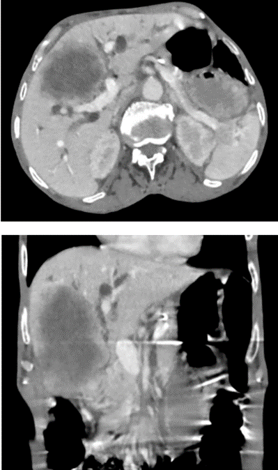
Figure 1. Contrast-enhanced CT demonstrating a large heterogeneously enhancing soft-tissue mass arising from the fundus and body of the gallbladder, with direct infiltration of adjacent hepatic segments IVa, IVb, V, and VIII. Moderate dilatation of the intrahepatic biliary radicles is also noted.
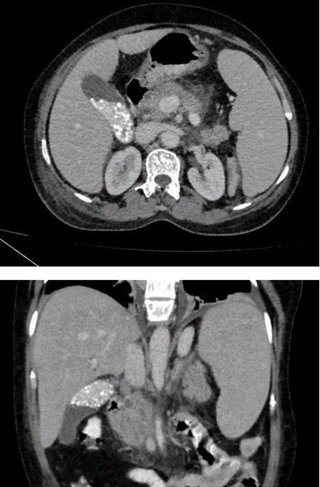
Figure 2. CT image demonstrating a distended gallbladder containing multiple calculi measuring approximately 2–4 mm in size, along with lithogenic sludge within the gallbladder lumen.
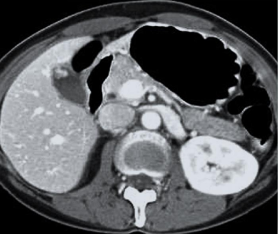
Figure 3.CT image demonstrating a well-defined, enhancing polypoidal mass arising from and adherent to the anterior wall of the gallbladder lumen, suggestive of a gallbladder polyp.
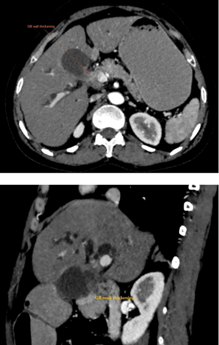
Figure 4. Contrast-enhanced CT demonstrating heterogeneously enhancing irregular wall thickening involving the neck of the gallbladder, extending into the proximal common bile duct (CBD) and common hepatic duct (CHD) through the cystic duct, with associated moderate intrahepatic biliary radicle dilatation.
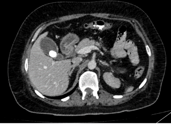
Figure 5.: CT image demonstrating a large solitary hyperdense calculus measuring approximately 1.8 cm within the gallbladder lumen, with an average gallbladder wall thickness of 3 mm.
DISCUSSION:
The present study evaluated the utility of computed tomography (CT) in the assessment of gallbladder lesions and correlated CT findings with histopathological diagnosis. The study population demonstrated a predominance of females (70.3%), with most patients aged 40–59 years (62.2%), followed by those aged ≥60 years (29.7%). This demographic pattern is consistent with the recognized epidemiology of gallbladder disease, particularly gallbladder carcinoma, which shows a female predominance and is more frequently encountered in the fifth to seventh decades of life [5,6,8]. The higher frequency among women may be related to hormonal and metabolic factors that predispose to gallstone formation and chronic gallbladder inflammation. Estrogen may increase biliary cholesterol saturation, whereas progesterone may reduce gallbladder motility, thereby promoting bile stasis and stone formation. Chronic inflammatory injury may subsequently contribute to dysplastic and neoplastic changes [17]. The relatively small proportion of patients younger than 40 years in the present study is also compatible with the reported age-related development of gallbladder pathology [18,19].
Inflammatory conditions were the most frequent clinical diagnosis in the present study (43.2%), followed by suspected malignancy (35.1%) and polyp/adenomyomatosis (21.6%). This predominance of inflammatory disease corresponds with the established clinical spectrum of gallbladder pathology, in which cholelithiasis and cholecystitis are common presentations [20,21]. Recurrent irritation by gallstones can result in mucosal inflammation, fibrosis, and gallbladder wall thickening, producing characteristic imaging abnormalities [20,22]. The relatively high proportion of patients with suspected malignancy also highlights the diagnostic overlap between inflammatory and neoplastic gallbladder disease. Gallbladder carcinoma may clinically and radiologically resemble chronic inflammatory disease, particularly in the presence of gallstones and chronic wall thickening [23-25].
Pain was the predominant presenting symptom, occurring in 59.5% of patients, whereas jaundice was reported in only 5.4%. This finding is consistent with previous literature identifying right upper quadrant abdominal pain as the commonest symptom of gallbladder disease [1,2]. Pain may result from gallbladder distension, cystic duct obstruction, biliary colic, or inflammation. The relatively low frequency of jaundice may reflect the fact that jaundice is generally associated with advanced disease, biliary obstruction, or hepatic and hilar involvement rather than being an early manifestation of gallbladder carcinoma [7,14]. A substantial proportion of patients (35.1%) had nonspecific symptoms, emphasizing the limitations of clinical assessment alone and the importance of imaging in the evaluation of suspected gallbladder pathology.
On CT, 73.0% of lesions were categorized as benign and 27.0% as malignant or suspicious. Among the benign lesions reported, cholecystitis was the predominant entity (71.4%), followed by adenomyomatosis and polyps (14.3% each). This distribution is consistent with the high prevalence of inflammatory gallbladder disease in routine clinical practice [21,26]. Chronic cholecystitis may demonstrate diffuse wall thickening and other inflammatory changes on CT [22]. Adenomyomatosis is a benign hyperplastic process that may occasionally mimic carcinoma, particularly when focal [27]. Gallbladder polyps are frequently benign cholesterol polyps, although larger or morphologically suspicious lesions may warrant evaluation for neoplasia [28]. CT assessment of lesion size, morphology, enhancement, wall irregularity, and local extension can therefore assist in characterization.
Gallstones were present in 19 patients and absent in 18, but their presence was not significantly associated with malignancy in the present study (p = 0.214). Gallstones are an established risk factor for chronic cholecystitis and gallbladder carcinoma, with persistent mucosal irritation contributing to the proposed inflammation-dysplasia-carcinoma sequence [5,20,29]. The lack of statistical significance in this study may be related to the relatively small sample size and the limited number of malignant cases. Moreover, gallstones are common in benign gallbladder disease and therefore have limited specificity as an isolated imaging marker of malignancy.
The body of the gallbladder was the most frequent overall site of lesion involvement (54.1%), followed by the neck (21.6%) and fundus (16.2%). Among malignant lesions, the fundus accounted for 33.3%, while no malignant lesion was separately localized to the body or neck. Lesion location is clinically relevant because the body and fundus are closely related to the adjacent hepatic parenchyma, particularly segments IV and V, and CT can demonstrate direct hepatic extension [19,30]. Localization also contributes to assessment of disease extent and surgical planning. The relatively greater representation of fundal involvement among malignant lesions is compatible with the reported predilection of gallbladder carcinoma for the fundus [23].
Most lesions showed no significant enhancement (78.4%), while hyperenhancement was observed in 16.2% and heterogeneous or layered enhancement in 2.7% each. Enhancement pattern was not significantly associated with malignancy (p = 0.095). This finding emphasizes the overlap between benign inflammatory and malignant processes. Inflammatory lesions may demonstrate enhancement because of hyperemia and vascular congestion, while malignant lesions may show heterogeneous enhancement related to tumor vascularity and necrosis [15,23,31]. Similar enhancement patterns may also occur in chronic cholecystitis, adenomyomatosis, and xanthogranulomatous cholecystitis, whereas early carcinoma may demonstrate only subtle enhancement [24,25]. Therefore, enhancement should not be interpreted in isolation, and assessment of wall morphology, local invasion, lymphadenopathy, and other associated findings remains essential.
Diffuse gallbladder wall thickening was the predominant pattern in the present study (75.7%), while focal thickening was observed in 16.2%. The predominance of diffuse thickening corresponds with the larger proportion of inflammatory lesions. Diffuse smooth wall thickening is well recognized in acute and chronic cholecystitis [16,22]. In contrast, focal or asymmetric irregular wall thickening is more suspicious for malignancy, particularly when associated with irregular enhancement, disruption of mural architecture, or hepatic infiltration [13,15]. Thus, the pattern and morphology of wall thickening are important components of CT-based characterization.
Several CT findings demonstrated a statistically significant association with malignancy. The presence of a mass lesion was significantly associated with malignant histopathology (χ² = 11.16, p = 0.001). Similarly, lymphadenopathy showed a strong association with malignancy (χ² = 17.99, p < 0.001). Infiltration was also significantly associated with malignancy (χ² = 17.68, p < 0.001), as was liver involvement (χ² = 10.16, p = 0.001). These findings are clinically important because a focal mass, adjacent organ invasion, and regional nodal disease are recognized imaging features of gallbladder carcinoma [13,14,23]. CT is particularly valuable because it can simultaneously demonstrate the primary lesion, hepatic invasion, lymphadenopathy, vascular involvement, and distant spread, thereby contributing to assessment of resectability and staging [12,14,19].
In contrast, biliary dilatation was not significantly associated with malignancy (p = 0.376). Similarly, the lack of a significant association for enhancement pattern and gallstones indicates that individual CT findings may have limited discriminatory value when considered in isolation. A systematic assessment combining morphology, wall characteristics, mass formation, local infiltration, lymphadenopathy, and hepatic involvement is therefore more appropriate.
Histopathology demonstrated 28 benign and nine malignant lesions. There was a statistically significant association between CT diagnosis and histopathological diagnosis (χ² = 32.11, p < 0.001). CT correctly categorized the histopathologically benign lesions in the CT-histopathology comparison and identified all nine malignant lesions as malignant/suspicious, with one false-positive case and no false-negative case. The resulting sensitivity was 100%, specificity was 96.4%, positive predictive value was 90%, negative predictive value was 100%, and overall diagnostic accuracy was 97.3%. These findings support the substantial diagnostic utility of CT in differentiating benign from malignant gallbladder lesions and are consistent with the established role of multidetector CT in evaluating gallbladder wall morphology, intraluminal lesions, hepatic invasion, regional nodes, and metastatic disease [12,14,32].
The high diagnostic performance observed in the present study should, however, be interpreted in the context of the relatively small study population and the limited number of malignant cases. CT findings may overlap between inflammatory and neoplastic conditions, particularly in chronic cholecystitis, adenomyomatosis, and xanthogranulomatous cholecystitis [24,25]. Consequently, histopathological examination remains essential for definitive diagnosis in suspicious or indeterminate lesions. Ultrasonography continues to serve as the initial imaging modality because of its availability and effectiveness in detecting gallstones and inflammatory changes, but its limitations include operator dependence, restricted field of view, bowel gas, and reduced assessment of extramural extension [11,33]. CT provides a broader anatomical assessment and is particularly valuable for defining local invasion, lymphadenopathy, biliary involvement, and distant disease [12,14].
Overall, the findings of this study demonstrate that CT is a useful modality for the comprehensive evaluation of gallbladder lesions. The predominance of benign inflammatory pathology was reflected by diffuse wall thickening and relatively limited enhancement, whereas mass formation, lymphadenopathy, infiltration, and hepatic involvement were significantly associated with malignancy. Correlation with histopathology demonstrated high diagnostic performance of CT, supporting its role not only in lesion characterization but also in determining disease extent and assisting preoperative planning. Nevertheless, because of the imaging overlap between certain benign and malignant lesions, CT findings should be interpreted in conjunction with clinical assessment and confirmed histopathologically when indicated.
CONCLUSION:
Computed tomography provides a valuable means of evaluating gallbladder lesions by demonstrating morphology, wall thickening, enhancement, lesion location, and anatomical extension. Benign inflammatory lesions, particularly cholecystitis, predominated, whereas mass formation, lymphadenopathy, infiltration, and hepatic involvement were significantly associated with malignancy. CT demonstrated excellent diagnostic performance in differentiating benign from malignant lesions. Although imaging overlap may occur between benign and malignant conditions, histopathological correlation remains essential for definitive diagnosis. CT therefore serves as an effective tool for lesion characterization, assessment of local invasion, staging, and preoperative planning, particularly in clinically or ultrasonographically indeterminate cases. Its systematic interpretation alongside clinical and histopathological findings can improve diagnostic confidence and guide appropriate patient management.
REFERENCES:
1. Gore RM, Yaghmai V, Newmark GM, Berlin JW, Miller FH. Imaging benign and malignant disease of the gallbladder. Radiol Clin North Am. 2002;40(6):1307-1323.
2. Chatterjee A, Lopes Vendrami C, Nikolaidis P, Mittal PK, Bandy AJ, Menias CO, et al. Uncommon intraluminal tumors of the gallbladder and biliary tract: spectrum of imaging appearances. Radiographics. 2019;39(2):388-412.
3. Khafaji MA, Bagasi JT, Albahiti SK, Alsayegh LA, Alsayyad SA, Algarni SS, et al. Accuracy of ultrasound and computed tomography in diagnosing acute cholecystitis patients in a tertiary care center in Saudi Arabia. Cureus. 2023;15(9):e44934.
4. Yu MH, Kim YJ, Park HS, Jung SI. Benign gallbladder diseases: imaging techniques and tips for differentiating with malignant gallbladder diseases. World J Gastroenterol. 2020;26(22):2967-2986.
5. Siegel RL, Miller KD, Jemal A. Cancer statistics, 2017. CA Cancer J Clin. 2017;67(1):7-30.
6. Gul P, Gul P, Altaf O, Kaimkhani TY, Asghar H. Spectrum of computed tomography findings in gall bladder carcinoma patients: a retrospective observational study from tertiary care oncology setup. Int Surg J. 2020;7(7):2091-2096.
7. George RA, Godara SC, Dhagat P, Som PP. Computed tomographic findings in 50 cases of gall bladder carcinoma. Med J Armed Forces India. 2007;63(3):215-219.
8. de Savornin Lohman E, de Bitter T, Verhoeven R, van der Geest L, Hagendoorn J, Haj Mohammad N, et al. Trends in treatment and survival of gallbladder cancer in the Netherlands: identifying gaps and opportunities from a nation-wide cohort. Cancers (Basel). 2020;12(4):918.
9. Rana P, Kalage D, Soundararajan R, Gupta P. Update on the role of imaging in the diagnosis, staging, and prognostication of gallbladder cancer. Indian J Radiol Imaging. 2024;35(2):218-233.
10. Zhang HP, Bai M, Gu JY, He YQ, Qiao XH, Du LF. Value of contrast-enhanced ultrasound in the differential diagnosis of gallbladder lesion. World J Gastroenterol. 2018;24(6):744-751.
11. Gerstenmaier JF, Hoang KN, Gibson RN. Contrast-enhanced ultrasound in gallbladder disease: a pictorial review. Abdom Radiol (NY). 2016;41(8):1640-1652. doi:10.1007/s00261-016-0729-4.
12. Grand D, Horton KM, Fishman EK. CT of the gallbladder: spectrum of disease. AJR Am J Roentgenol. 2004;183(1):163-170.
13. Mukthadira, Yasmin S, Farzana M, Islam M, Sharma NK. Role of computed tomography in differentiating between benign and malignant gallbladder masses with histopathological correlation. Community Based Med J. 2024;13(1):22-27.
14. Sahani DV, Kalva SP. Imaging the liver. Oncologist. 2004;9(4):385-397.
15. He WW, Zhu JG, Pylypenko D, Liu F, Wang M, Wu YF, et al. Differentiating benign from malignant gallbladder wall thickening in non-contrast MRI imaging: preliminary study of a combined diagnostic indicator. Medicine (Baltimore). 2022;101(40):e30861.
16. Singh T, Gupta P. Role of dual-energy computed tomography in gallbladder disease: a review. Journal of Gastrointestinal and Abdominal Radiology. 2022 Jul;5(02):107-13
17. Hundal R, Shaffer EA. Gallbladder cancer: epidemiology and outcome. Clin Epidemiol. 2014 Mar 7;6:99-109.
18. Bal MM, Ramadwar M, Deodhar K, Shrikhande S. Pathology of gallbladder carcinoma: current understanding and new perspectives. Pathol Oncol Res. 2015 Jul;21(3):509-25.
19. Ohtani T, Shirai Y, Tsukada K, Muto T, Hatakeyama K. Spread of gallbladder carcinoma: CT evaluation with pathologic correlation. Abdom Imaging. 1996 May-Jun;21(3):195-201.
20. Stinton LM, Shaffer EA. Epidemiology of gallbladder disease: cholelithiasis and cancer. Gut Liver. 2012 Apr;6(2):172-87.
21. Yadav S, Jategaonkar P, Bijlani M. Gallbladder polyps: an ambiguous cause of biliary colic. Ann Med Health Sci Res. 2014 Sep;4(Suppl 3):S332-3.
22. Bennett GL, Rusinek H, Lisi V, Israel GM, Krinsky GA, Slywotzky CM, Megibow A. CT findings in acute gangrenous cholecystitis. AJR Am J Roentgenol. 2002 Feb;178(2):275- 81.
23. Choi WS, Kim SH, Lee ES, Lee KB, Yoon WJ, Shin CI, Han JK. CT findings of gallbladder metastases: emphasis on differences according to primary tumors. Korean J Radiol. 2014 May-Jun;15(3):334-45.
24. John S, Moyana T, Shabana W, Walsh C, McInnes MDF. Gallbladder Cancer: Imaging Appearance and Pitfalls in Diagnosis. Can Assoc Radiol J. 2020 Nov;71(4):448-458.
25. Goshima S, Chang S, Wang JH, Kanematsu M, Bae KT, Federle MP. Xanthogranulomatous cholecystitis: diagnostic performance of CT to differentiate from gallbladder cancer. European journal of radiology. 2010 Jun 1;74(3):e79-83
26. Pilgrim CH, Groeschl RT, Pappas SG, Gamblin CT. An often overlooked diagnosis: imaging features of gallbladder cancer. Journal of the American College of Surgeons. 2013 Feb 1;216(2):333-9.
27. Gupta P, Marodia Y, Bansal A, Kalra N, Kumar-M P, Sharma V, Dutta U, Sandhu MS. Imaging-based algorithmic approach to gallbladder wall thickening. World J Gastroenterol. 2020 Oct 28;26(40):6163-6181
28. Ralls PW, Colletti PM, Lapin SA, Chandrasoma P, Boswell WD Jr, Ngo C, Radin DR, Halls JM. Real-time sonography in suspected acute cholecystitis. Prospective evaluation of primary and secondary signs. Radiology. 1985 Jun;155(3):767-71.
29. Hundal R, Shaffer EA. Gallbladder cancer: epidemiology and outcome. Clin Epidemiol. 2014 Mar 7;6:99-109.
30. Furlan A, Ferris JV, Hosseinzadeh K, Borhani AA. Gallbladder carcinoma update: multimodality imaging evaluation, staging, and treatment options. AJR Am J Roentgenol. 2008 Nov;191(5):1440-7.
31. Mathur M, Singh J, Singh DP, Kaur N, Gupta S, Haq S. Imaging Evaluation of Enhancement Patterns of Flat Gall Bladder Wall Thickening and Its Correlation with Clinical and Histopathological Findings. J Clin Diagn Res. 2017 Apr;11(4):TC07-TC11.
32. Seyal AR, Parekh K, Gonzalez-Guindalini FD, Nikolaidis P, Miller FH, Yaghmai V. Cross-sectional imaging of perforated gallbladder. Abdominal imaging. 2014 Aug;39(4):853-74
33. Gore RM, Thakrar KH, Newmark GM, Mehta UK, Berlin JW. Gallbladder imaging. Gastroenterology Clinics. 2010 Jun 1;39(2):265-87.