Association Between Vitamin B12/Folate Deficiency And Hematological Severity In Patients With Pancytopenia.
- Shaik Nayeem , PG Resident, Department of General Medicine, Katihar Medical College, Bihar, India.
- P.K. Agrawal , Professor, Department of General Medicine, Katihar Medical College, Bihar, India
- Taskeen Ahmad Reza , Professor, Department of General Medicine, Katihar Medical College, Bihar, India
- P S Puneet , PG Resident, Department of General Medicine, Katihar Medical College, Bihar, India.
Article Information:
Abstract:
Background: Pancytopenia is a clinically important hematological abnormality characterized by a reduction in red blood cells, white blood cells, and platelets. Its causes include nutritional deficiencies, bone marrow failure, infections, hypersplenism, and hematological malignancies. Vitamin B12 and folate deficiencies are potentially reversible causes that may produce ineffective hematopoiesis and varying degrees of hematological derangement. This study evaluated the association between vitamin B12 and/or folate deficiency and hematological severity among patients with pancytopenia. Methods: A prospective observational study was conducted among 100 adults presenting with pancytopenia at a tertiary-care teaching hospital in Katihar, Bihar, India. Demographic and clinical characteristics, complete blood counts, peripheral blood smear findings, abdominal ultrasonography, bone marrow aspiration findings, and final etiological diagnoses were recorded. Serum vitamin B12 and folate concentrations were assessed and participants were categorized according to the laboratory reference ranges. Hematological severity was evaluated using hemoglobin, total leukocyte count (TLC), and platelet count. Associations between nutritional deficiency status and hematological parameters were assessed using appropriate statistical tests. Results: The study population had a mean age of 35.57 years, and 54 (54.0%) participants were male. Generalized weakness was the most frequent presenting symptom (34.0%), followed by fever (16.0%) and bleeding manifestations (14.0%). Moderate anemia was present in 43.0% of participants and severe anemia in 42.0%. The mean hemoglobin, TLC, and platelet counts were 7.80 g/dL, 2106.80 cells/mm³, and 67,199.86/mm³, respectively. Macro-ovalocytes with hypersegmented neutrophils were the most frequent peripheral smear finding (28.0%). Bone marrow aspiration was performed in 72 patients, among whom megaloblastic marrow was the most frequent finding. Megaloblastic anemia was the leading final etiology of pancytopenia (30.0%), followed by hypersplenism (24.0%) and aplastic anemia (18.0%). Vitamin B12 and/or folate deficiency was identified in 62 patients. Compared with patients without nutritional deficiency, those with vitamin B12 and/or folate deficiency had lower mean hemoglobin (7.21 ± 1.48 vs. 8.76 ± 1.29 g/dL), TLC (1846 ± 721 vs. 2532 ± 814 cells/mm³), and platelet count (58,420 ± 24,180 vs. 81,520 ± 28,460/mm³); all comparisons were statistically significant (p<0.001). Conclusion: Vitamin B12 and/or folate deficiency was significantly associated with greater hematological derangement among patients with pancytopenia, as reflected by lower hemoglobin, TLC, and platelet counts. Megaloblastic anemia was the most common etiological diagnosis in this cohort. Assessment of vitamin B12 and folate status should therefore be considered an important component of the diagnostic evaluation of pancytopenia, particularly when peripheral smear or bone marrow findings suggest megaloblastic hematopoiesis. Further studies incorporating larger cohorts and multivariable analyses are warranted to determine whether this association is independent of other clinical and etiological factors.
Keywords:
Article :
INTRODUCTION:
Pancytopenia is a hematological manifestation characterized by the simultaneous reduction of erythrocytes, leukocytes, and platelets in peripheral blood. It is not a disease entity by itself but rather a clinical and laboratory finding resulting from diverse pathological processes involving hematopoiesis, peripheral destruction or sequestration of blood cells. The underlying causes range from nutritional deficiencies and infections to aplastic disorders, hypersplenism, and hematological malignancies. Consequently, systematic evaluation of patients with pancytopenia is essential for identifying potentially reversible causes and instituting appropriate treatment [1,2].
Nutritional deficiencies, particularly those involving vitamin B12 and folate, are important causes of pancytopenia. Both vitamins participate in DNA synthesis and cellular proliferation, and their deficiency predominantly affects rapidly dividing hematopoietic precursor cells. Impaired DNA synthesis results in ineffective hematopoiesis and characteristic megaloblastic changes in the bone marrow. When sufficiently severe, this ineffective hematopoiesis can lead to reductions in all three major peripheral blood cell lineages [3,4].
Vitamin B12 deficiency may arise from inadequate dietary intake, impaired absorption, autoimmune mechanisms, gastrointestinal disorders, or prolonged exposure to certain medications. Folate deficiency may similarly occur because of inadequate dietary intake, malabsorption, increased physiological requirements, or medications that interfere with folate metabolism. Although the clinical manifestations of these deficiencies may overlap, their recognition is particularly important because nutritional replacement can substantially improve hematological abnormalities when treatment is initiated before irreversible neurological or systemic complications develop [5,6].
The peripheral blood smear provides important diagnostic information in patients with suspected nutritional pancytopenia. Macrocytosis, macro-ovalocytes, anisopoikilocytosis, and hypersegmented neutrophils are characteristic morphological findings of megaloblastic hematopoiesis. However, peripheral smear findings may vary depending on the severity and duration of deficiency and may overlap with other hematological disorders. Therefore, biochemical assessment of vitamin B12 and folate, together with clinical evaluation and hematological investigations, may provide a more comprehensive assessment of nutritional pancytopenia [7,8].
The severity of pancytopenia may differ considerably among patients with nutritional deficiency. Some individuals may present predominantly with anemia, whereas others may develop marked leukopenia and thrombocytopenia. Severe cytopenias can increase the risk of infection, bleeding, fatigue, exercise intolerance, and other complications. Assessment of hemoglobin, total leukocyte count, and platelet count therefore provides useful information regarding the hematological severity of the condition [9].
Bone marrow examination may be required when the diagnosis remains uncertain or when severe cytopenias raise suspicion of marrow failure, infiltration, or malignancy. In megaloblastic states, bone marrow aspiration typically demonstrates hypercellularity with erythroid and granulocytic precursor abnormalities, nuclear-cytoplasmic asynchrony, and other morphological features reflecting defective DNA synthesis. Nevertheless, bone marrow findings should be interpreted together with clinical, peripheral smear, and biochemical findings rather than considered in isolation [10].
Pancytopenia has a broad etiological spectrum in tertiary-care settings. Previous hospital-based investigations have reported nutritional megaloblastic disorders, aplastic anemia, infections, hypersplenism, and hematological malignancies among the important underlying causes. The relative contribution of each etiology may vary according to geographic region, nutritional status, patient demographics, referral patterns, and available diagnostic facilities [11,12].
In this context, evaluating the relationship between vitamin B12/folate deficiency and the degree of hematological derangement may have practical clinical importance. Demonstrating an association between nutritional deficiency and more severe reductions in hemoglobin, leukocyte, and platelet counts could support early nutritional assessment in patients presenting with pancytopenia. It may also help distinguish potentially reversible nutritional causes from other serious etiologies requiring more extensive hematological evaluation.
The present prospective observational study was therefore undertaken to evaluate the association between vitamin B12/folate deficiency and hematological severity among patients with pancytopenia presenting to a tertiary-care hospital. The study assessed demographic and clinical characteristics, hematological parameters, peripheral smear findings, abdominal ultrasonography, bone marrow findings, and final etiological diagnoses. The primary objective was to determine whether vitamin B12 and/or folate deficiency was associated with greater severity of pancytopenia.
Aim
To evaluate the association between vitamin B12/folate deficiency and hematological severity in patients with pancytopenia.
Objectives
1. To determine the clinical and hematological profile of patients presenting with pancytopenia.
2. To assess the frequency of vitamin B12 and folate deficiency among patients with pancytopenia.
3. To evaluate the association between vitamin B12/folate deficiency and hemoglobin, total leukocyte count, and platelet count.
4. To assess the peripheral smear and bone marrow findings in patients with pancytopenia.
5. To determine the major etiological causes of pancytopenia in the study population.
6. To evaluate whether vitamin B12/folate deficiency is associated with increased hematological severity.
MATERIALS AND METHODS:
Study Design and Setting
A prospective observational study was conducted among adult patients presenting with pancytopenia at the Department of General Medicine and associated laboratory services of Katihar Medical College and Hospital, Katihar, Bihar, India. The study was conducted over the predefined study period [insert exact study period]. A total of 100 patients who fulfilled the eligibility criteria were included in the study.
Study Population
The study population comprised adults aged ≥18 years who were identified as having pancytopenia on complete blood count evaluation. All eligible participants underwent clinical assessment and laboratory investigations to determine the underlying etiology, with particular emphasis on identifying vitamin B12 and folate deficiency.
Inclusion Criteria
Patients were included if they met all of the following criteria:
1. Age ≥18 years.
2. Presence of pancytopenia based on the study laboratory criteria.
3. Availability of adequate clinical and hematological information.
4. Willingness to undergo the investigations required for etiological evaluation.
5. Provision of written informed consent.
Exclusion Criteria
Patients were excluded if they had any of the following:
1. Previously diagnosed and treated hematological malignancy.
2. Recent blood transfusion likely to influence hematological assessment.
3. Known chronic hematological disorders unrelated to the current presentation.
4. Pregnancy.
5. Incomplete clinical or laboratory information.
6. Refusal to provide informed consent.
7. Samples unsuitable for laboratory evaluation.
Definition of Pancytopenia
Pancytopenia was defined as the simultaneous reduction of hemoglobin, total leukocyte count, and platelet count below the predefined laboratory reference thresholds used at the study institution. The same criteria were applied consistently to all participants during enrollment.
The degree of hematological involvement was assessed using hemoglobin concentration, total leukocyte count (TLC), and platelet count.
Clinical Assessment
A detailed clinical history was obtained from each participant, including presenting symptoms, duration of illness, fever, generalized weakness, fatigue, bleeding manifestations, dyspnea, weight loss, abdominal fullness, and other relevant systemic complaints.
General and systemic examinations were performed for all participants. Particular attention was given to clinical evidence of anemia, infection, bleeding, nutritional deficiency, hepatomegaly, and splenomegaly.
Hematological Investigations
Complete blood counts were performed for all participants. The following parameters were recorded:
• Hemoglobin concentration
• Total leukocyte count
• Platelet count
For descriptive analysis, hemoglobin concentration was categorized as follows:
• Mild anemia: 10–11.9 g/dL
• Moderate anemia: 7–9.9 g/dL
• Severe anemia: <7 g/dL
TLC was categorized into the following groups:
• <1000 cells/mm³
• 1000–1999 cells/mm³
• 2000–2999 cells/mm³
• 3000–3999 cells/mm³
Platelet counts were categorized as:
• <20,000/mm³
• 20,000–49,999/mm³
• 50,000–99,999/mm³
• 100,000–149,999/mm³
These categories were used for descriptive assessment of the degree of cytopenia.
Peripheral Blood Smear Examination
Peripheral blood smears were examined for red-cell morphology, white-cell abnormalities, platelet adequacy, and abnormal circulating cells. The recorded findings included macro-ovalocytes with hypersegmented neutrophils, normocytic normochromic morphology, dimorphic morphology with anisopoikilocytosis, toxic granulation, blasts, dengue-related thrombocytopenic changes, malaria parasites, leucoerythroblastic changes, reactive changes, and atypical cells suggestive of leukemia.
The smear findings were interpreted in conjunction with the clinical and laboratory findings and were not used in isolation to establish the final etiology.
Assessment of Vitamin B12 and Folate Status
Serum vitamin B12 and folate concentrations were assessed as part of the nutritional evaluation of patients with pancytopenia. The results were interpreted according to the reference ranges of the laboratory performing the assays.
Participants were classified into the following nutritional-status categories:
1. Vitamin B12 deficiency
2. Folate deficiency
3. Combined vitamin B12 and folate deficiency
4. Neither vitamin B12 nor folate deficiency
For the primary comparative analysis, participants with vitamin B12 and/or folate deficiency were grouped as the nutritional-deficiency group, whereas those with neither deficiency were classified as the non-deficient group.
The association between nutritional-deficiency status and hematological severity was evaluated using hemoglobin, TLC, and platelet count.
Bone Marrow Examination
Bone marrow aspiration was performed when clinically indicated as part of the diagnostic evaluation of pancytopenia. Bone marrow examination was performed in 72 of the 100 participants.
The recorded marrow findings included:
• Megaloblastic marrow
• Hypocellular marrow
• Hypercellular marrow
• Leukemic infiltration
• Normocellular marrow
• Erythroid hyperplasia
• Hypercellular marrow with abnormal cells
• Reactive marrow
Because bone marrow aspiration was not performed in all participants, marrow findings were analyzed separately among patients who underwent the procedure. The findings were interpreted together with peripheral smear, biochemical, clinical, and other investigative findings.
Ultrasonography of the Abdomen
Abdominal ultrasonography was performed where clinically indicated to assess hepatosplenic abnormalities and other structural causes that could contribute to pancytopenia.
The recorded findings included normal ultrasonography, splenomegaly, mild splenomegaly, hepatomegaly, mild hepatomegaly, and hepatosplenomegaly.
Final Etiological Classification
A final etiological diagnosis was assigned after integrating the clinical findings with complete blood count, peripheral smear, vitamin B12 and folate assessment, bone marrow findings where available, abdominal ultrasonography, and other relevant investigations.
The major etiological categories recorded in the study were:
• Megaloblastic anemia
• Hypersplenism
• Aplastic anemia
• Infection-related pancytopenia
• Dimorphic anemia
• Hematological malignancy
The final diagnosis was based on the available clinical and investigative evidence rather than on any single investigation.
Study Outcomes
Primary outcome:
The primary outcome was the association between vitamin B12 and/or folate deficiency and hematological severity, assessed using hemoglobin, TLC, and platelet count.
Secondary outcomes:
1. Clinical presentation of patients with pancytopenia.
2. Distribution of hemoglobin severity.
3. Distribution of TLC and platelet counts.
4. Peripheral blood smear findings.
5. Bone marrow findings among patients undergoing bone marrow aspiration.
6. Ultrasonographic hepatosplenic findings.
7. Final etiological spectrum of pancytopenia.
8. Association between nutritional-deficiency status and individual hematological parameters.
Statistical Analysis
Data were entered into a structured database and analyzed using appropriate statistical software.
Continuous variables were summarized as mean ± standard deviation (SD) when approximately normally distributed and as median with interquartile range (IQR) when the distribution was non-normal. Categorical variables were summarized as frequencies and percentages.
The distribution of continuous variables was assessed before selecting the appropriate statistical test. Comparisons of continuous hematological parameters between participants with and without vitamin B12 and/or folate deficiency were performed using the independent-samples t test for normally distributed variables or the Mann–Whitney U test for non-normally distributed variables.
Associations between categorical variables were assessed using the chi-square test or Fisher's exact test, as appropriate. For comparisons involving more than two groups, one-way analysis of variance (ANOVA) or the Kruskal–Wallis test was used according to data distribution.
Correlation between serum vitamin B12 and/or folate concentrations and hematological parameters was assessed using Pearson's or Spearman's correlation coefficient, as appropriate.
All statistical tests were two-sided, and a p value <0.05 was considered statistically significant. Exact p values were reported where applicable.
Ethical Considerations
The study was conducted in accordance with the ethical principles governing research involving human participants. Institutional Ethics Committee approval was obtained before commencement of the study [insert IEC name, approval number, and date if available].
Written informed consent was obtained from all participants before enrollment. Patient confidentiality was maintained throughout the study, and identifying information was excluded from the analytical dataset.
RESULTS:
Study Population and Demographic Characteristics
A total of 100 adult patients with pancytopenia who fulfilled the predefined eligibility criteria were included in the study. Complete clinical and hematological data were available for all participants.
The mean age of the study population was 35.57 years. The largest proportion of participants belonged to the 18–30-year age group (44.0%), followed by the 31–45-year group (36.0%). Patients aged 46–60 years and >60 years accounted for 14.0% and 6.0%, respectively (Table 1).
Table 1. Age distribution of the study participants (N=100)
|
Age group (years) |
Frequency |
Percentage |
|
18–30 |
44 |
44.0 |
|
31–45 |
36 |
36.0 |
|
46–60 |
14 |
14.0 |
|
>60 |
6 |
6.0 |
|
Total |
100 |
100.0 |
Mean age: 35.57 years
There was a slight male predominance, with 54 (54.0%) participants being male and 46 (46.0%) being female, corresponding to a male-to-female ratio of approximately 1.17:1 (Table 2).
Table 2. Sex distribution of the study participants (N=100)
|
Sex |
Frequency |
Percentage |
|
Male |
54 |
54.0 |
|
Female |
46 |
46.0 |
|
Total |
100 |
100.0 |
Clinical Presentation
The clinical presentation of the study participants is summarized in Table 3. Generalized weakness was the most frequently reported presenting symptom, occurring in 34 (34.0%) patients. Fever was reported in 16 (16.0%), while bleeding manifestations were present in 14 (14.0%). Fatigue and dyspnoea were reported in 12 (12.0%) and 11 (11.0%) patients, respectively. Weight loss was reported in 9 (9.0%), whereas abdominal fullness was the least frequent presentation, occurring in 4 (4.0%) patients.
Table 3. Clinical presentation of patients with pancytopenia (N=100)
|
Clinical presentation |
Frequency |
Percentage |
|
Generalized weakness |
34 |
34.0 |
|
Fever |
16 |
16.0 |
|
Bleeding manifestations |
14 |
14.0 |
|
Fatigue |
12 |
12.0 |
|
Dyspnoea |
11 |
11.0 |
|
Weight loss |
9 |
9.0 |
|
Abdominal fullness |
4 |
4.0 |
|
Total |
100 |
100.0 |
Hematological Parameters
The distribution of participants according to hemoglobin concentration is presented in Table 4. Moderate anemia was the most frequent category, occurring in 43 (43.0%) patients, followed by severe anemia in 42 (42.0%). Mild anemia was present in 15 (15.0%) patients. Thus, 85.0% of the study population had hemoglobin concentrations below 10 g/dL. The mean hemoglobin concentration was 7.80 g/dL.
Table 4. Distribution according to hemoglobin concentration (N=100)
|
Hemoglobin category |
Frequency |
Percentage |
|
Mild anemia (10–11.9 g/dL) |
15 |
15.0 |
|
Moderate anemia (7–9.9 g/dL) |
43 |
43.0 |
|
Severe anemia (<7 g/dL) |
42 |
42.0 |
|
Total |
100 |
100.0 |
Mean hemoglobin: 7.80 g/dL
The mean total leukocyte count (TLC) was 2106.80 cells/mm³. The largest proportion of participants, 42 (42.0%), had TLC values of 1000–1999 cells/mm³. TLC values of 2000–2999 and 3000–3999 cells/mm³ were observed in 24 (24.0%) and 26 (26.0%) patients, respectively, while 8 (8.0%) had TLC below 1000 cells/mm³ (Table 5).
Table 5. Distribution according to total leukocyte count (N=100)
|
TLC (cells/mm³) |
Frequency |
Percentage |
|
<1000 |
8 |
8.0 |
|
1000–1999 |
42 |
42.0 |
|
2000–2999 |
24 |
24.0 |
|
3000–3999 |
26 |
26.0 |
|
Total |
100 |
100.0 |
Mean TLC: 2106.80 cells/mm³
Platelet counts are shown in Table 6. Platelet counts of 20,000–49,999/mm³ and 50,000–99,999/mm³ were each observed in 32 (32.0%) patients. Twenty-eight (28.0%) patients had platelet counts of 100,000–149,999/mm³, while 8 (8.0%) had platelet counts below 20,000/mm³. The mean platelet count was 67,199.86/mm³.
Table 6. Distribution according to platelet count (N=100)
|
Platelet count (/mm³) |
Frequency |
Percentage |
|
<20,000 |
8 |
8.0 |
|
20,000–49,999 |
32 |
32.0 |
|
50,000–99,999 |
32 |
32.0 |
|
100,000–149,999 |
28 |
28.0 |
|
Total |
100 |
100.0 |
Mean platelet count: 67,199.86/mm³
Peripheral Blood Smear Findings
Peripheral blood smear findings are summarized in Table 7. Macro-ovalocytes with hypersegmented neutrophils were the most frequent morphological finding, identified in 28 (28.0%) patients. Normocytic normochromic morphology with pancytopenia was observed in 18 (18.0%), while normocytic normochromic smears with cytopenias were recorded in 14 (14.0%).
A dimorphic picture with anisopoikilocytosis was observed in 10 (10.0%) patients, while toxic granules were present in 8 (8.0%). Blasts and dengue-related thrombocytopenic changes were each observed in 5 (5.0%) patients. Malaria parasites were identified in 4 (4.0%), whereas leucoerythroblastic and reactive changes were each observed in 3 (3.0%). Atypical cells suggestive of leukemia were identified in 2 (2.0%) patients.
Table 7. Peripheral smear findings among study participants (N=100)
|
Peripheral smear finding |
Frequency |
Percentage |
|
Macro-ovalocytes with hypersegmented neutrophils |
28 |
28.0 |
|
Normocytic normochromic cells with pancytopenia |
18 |
18.0 |
|
Normocytic normochromic smear with cytopenias |
14 |
14.0 |
|
Dimorphic picture with anisopoikilocytosis |
10 |
10.0 |
|
Normocytic smear with toxic granules |
8 |
8.0 |
|
Blasts on peripheral smear |
5 |
5.0 |
|
Dengue-related thrombocytopenic picture |
5 |
5.0 |
|
Malaria parasite-positive smear |
4 |
4.0 |
|
Leucoerythroblastic picture |
3 |
3.0 |
|
Reactive changes on smear |
3 |
3.0 |
|
Atypical cells suggestive of leukemia |
2 |
2.0 |
|
Total |
100 |
100.0 |
These findings demonstrate a heterogeneous morphological spectrum, with megaloblastic-type changes representing the most frequent smear pattern.
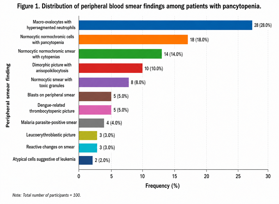
Figure 1. Distribution of peripheral blood smear findings among patients with pancytopenia.
Ultrasonographic Findings
Abdominal ultrasonography findings are presented in Table 8. Ultrasonography was reported as normal in 42 (42.0%) patients. Splenomegaly was the most frequent abnormal finding, occurring in 21 (21.0%) patients, followed by mild hepatomegaly in 14 (14.0%) and hepatosplenomegaly in 13 (13.0%). Mild splenomegaly and hepatomegaly were observed in 6 (6.0%) and 4 (4.0%) patients, respectively.
Table 8. Ultrasonographic findings of the abdomen (N=100)
|
USG abdomen finding |
Frequency |
Percentage |
|
Normal |
42 |
42.0 |
|
Splenomegaly |
21 |
21.0 |
|
Mild hepatomegaly |
14 |
14.0 |
|
Hepatosplenomegaly |
13 |
13.0 |
|
Mild splenomegaly |
6 |
6.0 |
|
Hepatomegaly |
4 |
4.0 |
|
Total |
100 |
100.0 |
Bone Marrow Examination
Bone marrow aspiration was performed in 72 (72.0%) patients, whereas 28 (28.0%) patients did not undergo the procedure (Table 9).
Table 9. Bone marrow aspiration status (N=100)
|
Bone marrow aspiration |
Frequency |
Percentage |
|
Yes |
72 |
72.0 |
|
No |
28 |
28.0 |
|
Total |
100 |
100.0 |
Among the entire study population, megaloblastic marrow was identified in 32 (32.0%) patients. Hypocellular marrow was observed in 18 (18.0%), hypercellular marrow in 8 (8.0%), leukemic infiltration in 4 (4.0%), reactive marrow in 4 (4.0%), normocellular marrow in 3 (3.0%), erythroid hyperplasia in 2 (2.0%), and hypercellular marrow with abnormal cells in 1 (1.0%) patient (Table 10).
Because bone marrow aspiration was performed in only 72 patients, the proportion of each marrow finding among those who underwent aspiration should be interpreted separately from its percentage in the overall cohort. Among the 72 patients who underwent aspiration, megaloblastic marrow represented 44.4% (32/72) of the findings.
Table 10. Bone marrow aspiration findings among the study population (N=100)
|
Bone marrow finding |
Frequency |
Percentage of total cohort |
|
Not done |
28 |
28.0 |
|
Megaloblastic marrow |
32 |
32.0 |
|
Hypocellular marrow |
18 |
18.0 |
|
Hypercellular marrow |
8 |
8.0 |
|
Leukemic infiltration |
4 |
4.0 |
|
Normocellular marrow |
3 |
3.0 |
|
Erythroid hyperplasia |
2 |
2.0 |
|
Hypercellular marrow with abnormal cells |
1 |
1.0 |
|
Reactive marrow |
4 |
4.0 |
|
Total |
100 |
100.0 |
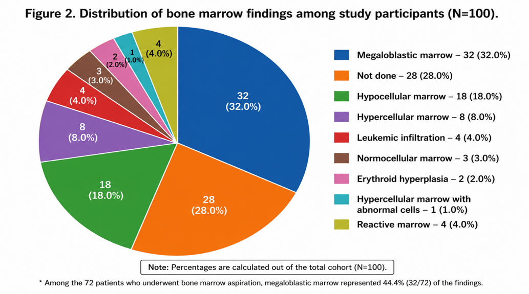
Figure 2. Distribution of bone marrow findings among study participants.
Final Etiological Profile
The final etiological distribution of pancytopenia is presented in Table 11. Megaloblastic anemia was the most frequent diagnosis, identified in 30 (30.0%) patients. Hypersplenism was identified in 24 (24.0%), followed by aplastic anemia in 18 (18.0%) and infection-related pancytopenia in 16 (16.0%). Dimorphic anemia and hematological malignancy accounted for 8 (8.0%) and 4 (4.0%) cases, respectively.
Table 11. Final etiological profile of pancytopenia (N=100)
|
Final etiology |
Frequency |
Percentage |
|
Megaloblastic anemia |
30 |
30.0 |
|
Hypersplenism |
24 |
24.0 |
|
Aplastic anemia |
18 |
18.0 |
|
Infection-related pancytopenia |
16 |
16.0 |
|
Dimorphic anemia |
8 |
8.0 |
|
Hematological malignancy |
4 |
4.0 |
|
Total |
100 |
100.0 |
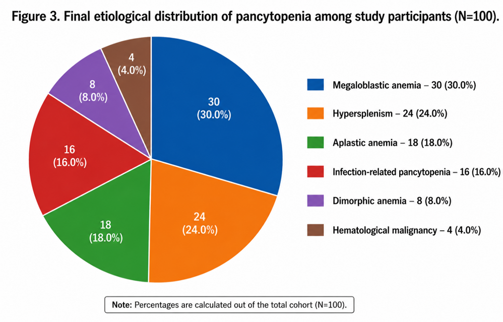
Figure 3. Final etiological distribution of pancytopenia among study participants.
Vitamin B12/Folate Deficiency and Hematological Severity
The primary analytical comparison evaluated hematological parameters between patients with vitamin B12 and/or folate deficiency and those without either deficiency. Sixty-two (62.0%) participants were classified as having vitamin B12 and/or folate deficiency, whereas 38 (38.0%) had neither deficiency.
Patients with vitamin B12/folate deficiency had significantly lower mean hemoglobin, TLC, and platelet counts than those without nutritional deficiency (Table 12).
The mean hemoglobin concentration was 7.21 ± 1.48 g/dL among participants with vitamin B12/folate deficiency compared with 8.76 ± 1.29 g/dL among those without deficiency (p<0.001). Similarly, mean TLC was 1846 ± 721 cells/mm³ in the deficient group compared with 2532 ± 814 cells/mm³ in the non-deficient group (p<0.001). Mean platelet count was also lower in the deficient group (58,420 ± 24,180/mm³ versus 81,520 ± 28,460/mm³; p<0.001).
Table 12. Comparison of hematological parameters according to vitamin B12/folate deficiency status
|
Hematological parameter |
B12/folate deficient (n=62) |
No deficiency (n=38) |
p-value |
|
Hemoglobin (g/dL), mean ± SD |
7.21 ± 1.48 |
8.76 ± 1.29 |
<0.001 |
|
TLC (cells/mm³), mean ± SD |
1846 ± 721 |
2532 ± 814 |
<0.001 |
|
Platelet count (/mm³), mean ± SD |
58,420 ± 24,180 |
81,520 ± 28,460 |
<0.001 |
These findings indicate a statistically significant difference in all three major hematological parameters according to nutritional deficiency status.
Vitamin B12/Folate Deficiency and Anemia Severity
The distribution of nutritional deficiency according to anemia category is shown in Table 13. Among the 62 participants with vitamin B12/folate deficiency, 5 (8.1%) had mild anemia, 27 (43.5%) had moderate anemia, and 30 (48.4%) had severe anemia. Among the 38 participants without nutritional deficiency, 10 (26.3%) had mild anemia, 16 (42.1%) had moderate anemia, and 12 (31.6%) had severe anemia.
Thus, severe anemia constituted a larger proportion of the vitamin B12/folate-deficient group than of the non-deficient group (48.4% versus 31.6%), whereas mild anemia was less frequent among participants with nutritional deficiency (8.1% versus 26.3%). The overall distribution of anemia severity differed significantly between the two nutritional-status groups (χ²=6.83, p=0.033).
Table 13. Distribution of anemia severity according to vitamin B12/folate deficiency status
|
Hemoglobin category |
B12/folate deficient (n=62), n (%) |
No deficiency (n=38), n (%) |
|
Mild (10–11.9 g/dL) |
5 (8.1) |
10 (26.3) |
|
Moderate (7–9.9 g/dL) |
27 (43.5) |
16 (42.1) |
|
Severe (<7 g/dL) |
30 (48.4) |
12 (31.6) |
|
Total |
62 (100.0) |
38 (100.0) |
Overall χ²=6.83; p=0.033.
Overall Findings
Overall, the cohort demonstrated substantial hematological involvement, with moderate-to-severe anemia, leukopenia, and thrombocytopenia being prominent findings. Generalized weakness was the most frequent presenting symptom. Macro-ovalocytes with hypersegmented neutrophils represented the most common peripheral smear abnormality. Among patients who underwent bone marrow aspiration, megaloblastic marrow was the most frequent marrow pattern. Megaloblastic anemia was the leading final etiological diagnosis, followed by hypersplenism and aplastic anemia.
The principal analytical finding was that vitamin B12 and/or folate deficiency was associated with significantly lower hemoglobin, TLC, and platelet counts. In addition, the distribution of anemia severity differed significantly according to nutritional deficiency status, with severe anemia occurring more frequently among participants with vitamin B12/folate deficiency.
DISCUSSION:
Pancytopenia is a clinically important hematological abnormality with a broad etiological spectrum that includes nutritional deficiencies, bone marrow failure, infections, hypersplenism, and hematological malignancies. The present prospective observational study evaluated 100 adult patients with pancytopenia and examined their clinical, hematological, peripheral smear, ultrasonographic, bone marrow, and etiological profiles, with particular emphasis on the association between vitamin B12 and/or folate deficiency and hematological severity.
The study population had a mean age of 35.57 years, with 80.0% of participants belonging to the 18–45-year age group. There was a slight male predominance, with males accounting for 54.0% of the cohort. The relatively young age distribution may reflect the characteristics of the hospital-based population and the contribution of nutritional, infectious, and other potentially reversible causes of pancytopenia. Differences in age and sex distribution across pancytopenia studies are expected because of variation in geographical setting, referral patterns, nutritional status, and the spectrum of underlying diseases [1,2].
Generalized weakness was the most common presenting symptom, reported in 34.0% of participants, followed by fever (16.0%) and bleeding manifestations (14.0%). The predominance of weakness is consistent with the substantial degree of anemia observed in the cohort. Fever may occur in patients with severe leukopenia or underlying infection, whereas bleeding manifestations may be related to thrombocytopenia. The combination of these symptoms highlights the clinical importance of assessing all three hematopoietic cell lineages when evaluating a patient with pancytopenia [1,4].
The hematological profile demonstrated considerable severity of cytopenias. The mean hemoglobin concentration was 7.80 g/dL, and 85.0% of participants had hemoglobin concentrations below 10 g/dL. Moderate anemia was present in 43.0% and severe anemia in 42.0% of patients. The mean TLC was 2106.80 cells/mm³, with 50.0% of patients having TLC below 2000 cells/mm³. Similarly, the mean platelet count was 67,199.86/mm³, and 64.0% of participants had platelet counts below 100,000/mm³. These findings demonstrate that the cohort had substantial involvement of all three hematological lineages rather than isolated anemia. Pancytopenia of this degree warrants systematic evaluation because the underlying mechanism may range from ineffective hematopoiesis to marrow hypoplasia, peripheral sequestration, infection, infiltration, or malignancy [1,4].
Peripheral blood smear examination provided important morphological information. Macro-ovalocytes with hypersegmented neutrophils were the most frequent finding, observed in 28.0% of participants. These findings are characteristic morphological features of megaloblastic hematopoiesis and are particularly associated with vitamin B12 and folate deficiency [3,7]. The presence of these features in a substantial proportion of patients was consistent with the subsequent identification of megaloblastic anemia as the leading final etiological diagnosis. Nevertheless, the fact that classical megaloblastic morphology was not present in all patients emphasizes that peripheral smear findings alone cannot reliably exclude nutritional deficiency or establish the precise cause of pancytopenia.
Other peripheral smear patterns included normocytic normochromic morphology, dimorphic changes, toxic granules, blasts, dengue-related changes, malaria parasites, leucoerythroblastic changes, and atypical cells suggestive of leukemia. This heterogeneous morphological spectrum is consistent with the broad differential diagnosis of pancytopenia. In particular, the presence of blasts, parasitic forms, or leucoerythroblastic changes may indicate processes requiring further targeted investigation. Therefore, peripheral smear examination remains an important initial component of the diagnostic evaluation but should be interpreted together with clinical findings, biochemical investigations, and, where indicated, bone marrow examination [4,8].
Abdominal ultrasonography was normal in 42.0% of participants, while splenomegaly and hepatosplenomegaly were identified in 21.0% and 13.0%, respectively. Splenic enlargement is clinically relevant because hypersplenism can contribute to peripheral sequestration of blood cells and may therefore represent an important mechanism of pancytopenia. In the present cohort, hypersplenism was the second most frequent final etiological diagnosis, accounting for 24.0% of cases. These findings illustrate the value of hepatosplenic assessment as part of the broader evaluation of pancytopenia [4].
Bone marrow aspiration was performed in 72.0% of participants because the procedure was undertaken according to clinical indication. Among the entire cohort, megaloblastic marrow was observed in 32.0%, while hypocellular marrow was present in 18.0%. Among patients who underwent aspiration, megaloblastic marrow accounted for 44.4% of marrow findings. The identification of hypocellular marrow in a substantial proportion of patients is compatible with the presence of marrow-failure disorders, including aplastic anemia, which constituted 18.0% of the final etiological diagnoses. Because bone marrow examination was not performed in all participants, these findings should be interpreted as representing the clinically selected subgroup rather than the entire pancytopenic population. Bone marrow examination remains particularly useful when the diagnosis cannot be established from clinical, peripheral smear, and biochemical findings alone [4,10].
The final etiological profile demonstrated that megaloblastic anemia was the most frequent cause of pancytopenia, accounting for 30.0% of cases. Hypersplenism was the second most common etiology (24.0%), followed by aplastic anemia (18.0%), infection-related pancytopenia (16.0%), dimorphic anemia (8.0%), and hematological malignancy (4.0%). The predominance of megaloblastic anemia is consistent with previous hospital-based studies in which nutritional megaloblastic disorders have been identified among important causes of pancytopenia [1,2,11]. However, the relative contribution of individual etiologies differs between studies because of differences in patient selection, diagnostic criteria, geographic location, nutritional status, and availability of investigations.
The principal analytical finding of the present study was the association between vitamin B12 and/or folate deficiency and hematological severity. Sixty-two percent of participants were classified as having vitamin B12 and/or folate deficiency, while 38.0% had neither deficiency. Participants with nutritional deficiency had significantly lower mean hemoglobin, TLC, and platelet counts than those without deficiency. Mean hemoglobin was 7.21 ± 1.48 g/dL in the deficient group compared with 8.76 ± 1.29 g/dL in the non-deficient group (p<0.001). Mean TLC was also lower in the deficient group (1846 ± 721 versus 2532 ± 814 cells/mm³; p<0.001), as was mean platelet count (58,420 ± 24,180 versus 81,520 ± 28,460/mm³; p<0.001).
The observed differences are biologically plausible because vitamin B12 and folate are essential for normal DNA synthesis and cellular proliferation. Deficiency results in impaired nuclear maturation and ineffective hematopoiesis, with intramedullary destruction of abnormal precursor cells. Although erythroid abnormalities are often most clinically apparent, sufficiently severe megaloblastic hematopoiesis can affect granulocytic and megakaryocytic lineages and result in pancytopenia [3,7,12]. The lower values of all three major hematological parameters observed among deficient participants are therefore consistent with the underlying pathophysiology.
The association was also evident when participants were categorized according to anemia severity. Severe anemia accounted for 48.4% of participants in the vitamin B12/folate-deficient group compared with 31.6% in the non-deficient group, whereas mild anemia was less frequent in the deficient group (8.1% versus 26.3%). The overall distribution of anemia severity differed significantly between the two nutritional-status groups (χ²=6.83, p=0.033). This finding supports an association between nutritional deficiency status and the degree of anemia within this cohort. However, because the study is observational, the findings demonstrate an association rather than a causal relationship.
The clinical importance of identifying vitamin B12 and folate deficiency lies in the potentially reversible nature of these abnormalities. Recognition and appropriate treatment can correct ineffective hematopoiesis and improve the associated cytopenias. Vitamin B12 deficiency is particularly important to identify promptly because neurological manifestations may occur even when hematological abnormalities are not proportionately severe, and prolonged neurological injury may become irreversible [12, 7]. Accordingly, biochemical assessment of nutritional status can provide clinically useful information in patients whose clinical and morphological findings suggest megaloblastic hematopoiesis.
The findings also demonstrate that nutritional assessment should complement, rather than replace, the broader diagnostic evaluation of pancytopenia. Although macro-ovalocytes and hypersegmented neutrophils provide important clues toward megaloblastic hematopoiesis, other causes of pancytopenia were also identified in the present cohort, including hypersplenism, aplastic anemia, infection-related pancytopenia, and hematological malignancy. Thus, the presence of pancytopenia should prompt an etiologically directed evaluation rather than an assumption that nutritional deficiency is responsible in every patient [4,8].
Strengths and Limitations
The study has several strengths. It included a clinically characterized cohort of 100 adult patients with pancytopenia and incorporated multiple aspects of evaluation, including complete blood counts, peripheral smear examination, abdominal ultrasonography, bone marrow examination in clinically indicated patients, and final etiological classification. Importantly, the study also included a comparative analysis of hematological parameters according to vitamin B12/folate deficiency status.
Several limitations should nevertheless be considered. First, this was a single-centre observational study, which may limit the generalizability of the findings. Second, bone marrow aspiration was performed only when clinically indicated and was therefore not available for all participants; consequently, marrow findings cannot be generalized to the entire cohort. Third, the nutritional-deficiency analysis was based on classification into vitamin B12/folate-deficient and non-deficient groups, and the available results do not provide sufficient information to separately quantify the effects of vitamin B12 deficiency, folate deficiency, and combined deficiency. Fourth, potential confounding factors were not adjusted for using multivariable analysis. Factors such as age, sex, infection, underlying etiology, and other nutritional or systemic conditions could potentially influence hematological severity. Finally, the observational design does not establish that vitamin B12/folate deficiency independently causes more severe pancytopenia.
Overall Interpretation
Overall, the present study demonstrates that pancytopenia in this cohort had a heterogeneous clinical and etiological profile, with megaloblastic anemia representing the most frequent identified cause. Vitamin B12 and/or folate deficiency was associated with significantly lower hemoglobin, leukocyte, and platelet counts, and severe anemia was proportionally more frequent among participants with nutritional deficiency. These findings support the clinical value of assessing vitamin B12 and folate status in appropriately selected patients with pancytopenia, particularly when peripheral smear or marrow findings suggest megaloblastic hematopoiesis. However, the observed associations should be interpreted cautiously, and larger multicentre studies incorporating individual vitamin B12 and folate concentrations and multivariable analysis are required to determine whether nutritional deficiency is independently associated with hematological severity.
CONCLUSION:
In this prospective observational study of 100 adult patients with pancytopenia, a broad clinical and etiological spectrum was observed. Moderate-to-severe anemia, leukopenia, and thrombocytopenia were prominent hematological abnormalities, while generalized weakness was the most frequent presenting symptom. Megaloblastic features, particularly macro-ovalocytes with hypersegmented neutrophils, were the most common peripheral smear finding, and megaloblastic anemia was the leading final etiological diagnosis, followed by hypersplenism and aplastic anemia.
Vitamin B12 and/or folate deficiency was present in 62.0% of participants and was associated with significantly lower hemoglobin, total leukocyte, and platelet counts compared with participants without nutritional deficiency. Severe anemia was also more frequently observed among patients with vitamin B12/folate deficiency, with a significant difference in the distribution of anemia severity between the two groups.
These findings highlight the importance of evaluating vitamin B12 and folate status as part of the diagnostic work-up of patients with pancytopenia, particularly when peripheral smear or bone marrow findings suggest megaloblastic hematopoiesis. However, the observational design and absence of multivariable analysis preclude conclusions regarding an independent or causal relationship between nutritional deficiency and hematological severity. Larger, multicentre prospective studies incorporating individual vitamin B12 and folate concentrations and adjustment for potential confounding factors are warranted to further clarify this association.
REFERENCES:
1. Jha A, Sayami G, Adhikari RC, Panta AD, Jha R. Bone marrow examination in cases of pancytopenia. J Nepal Med Assoc. 2008;47(169):12-17. doi:10.31729/jnma.209.
2. Khodke K, Marwah S, Buxi G, Yadav RB, Chaturvedi NK. Bone marrow examination in cases of pancytopenia. J Indian Acad Clin Med. 2001;2(1-2):55-9.
3. Aslinia F, Mazza JJ, Yale SH. Megaloblastic anemia and other causes of macrocytosis. Clin Med Res. 2006;4(3):236-41. doi:10.3121/cmr.4.3.236.
4. Weinzierl EP, Arber DA. The differential diagnosis and bone marrow evaluation of new-onset pancytopenia. Am J Clin Pathol. 2013;139(1):9-29. doi:10.1309/AJCP50AEEYGREWUZ.
5. Hoffbrand AV, Moss PAH. Hoffbrand's Essential Haematology. 7th ed. Chichester: Wiley-Blackwell; 2016.
6. Bain BJ. Blood Cells: A Practical Guide. 5th ed. Chichester: Wiley-Blackwell; 2015.
7. Green R. Vitamin B12 deficiency from the perspective of a practicing hematologist. Blood. 2017;129(19):2603-11. doi:10.1182/blood-2016-10-569186.
8. Khanduri U, Sharma A. Megaloblastic anaemia: prevalence and causative factors. Natl Med J India. 2007;20(4):172-5.
9. Hoffman R, Benz EJ Jr, Silberstein LE, Heslop HE, Weitz JI, Anastasi J, editors. Hematology: Basic Principles and Practice. 7th ed. Philadelphia: Elsevier; 2017.
10. Bain BJ, Clark DM, Wilkins BS. Bone Marrow Pathology. 5th ed. Hoboken: Wiley-Blackwell; 2019.
11. Tilak V, Jain R. Pancytopenia—a clinico-hematologic analysis of 77 cases. Indian J Pathol Microbiol. 1999;42(4):399-404.
12. Devalia V, Hamilton MS, Molloy AM; British Committee for Standards in Haematology. Guidelines for the diagnosis and treatment of cobalamin and folate disorders. Br J Haematol. 2014;166(4):496-513. doi:10.1111/bjh.12959.