Predictive Value of B-Scan Ultrasonography in Visual Prognosis of Patients with Mature Cataract.
- Meghana Patil , Associate Professor, Department of Ophthalmology, KLE Jagadguru Gangadhar Mahaswamigalu Moorusavirmath Medical College and Hospital, Hubli, KLE Academy of Higher Education and Research, Deemed to be University, Belagavi, Karnataka, India – 590010.
- Ambika Gayad , Associate Professor, Department of Radiology, KLE Jagadguru Gangadhar Mahaswamigalu Moorusavirmath Medical College and Hospital, Hubli, KLE Academy of Higher Education and Research, Deemed to be University, Belagavi, Karnataka, India – 590010.
- Mohan Bhavikatti , Professor, Department of Radiology, KLE Jagadguru Gangadhar Mahaswamigalu Moorusavirmath Medical College and Hospital, Hubli, KLE Academy of Higher Education and Research, Deemed to be University, Belagavi, Karnataka, India – 590010.
- Amrut Arvindrao Dambal , Vice Principal and Professor and HOD, Department of Biochemistry, KLE'S JGMM Medical College, Hubballi, India.
- Samata , Head of the Department, Department of Physiology, K H Patil Institute of Medical sciences, Gadag, India.
Article Information:
Abstract:
Background: Mature cataract obscures fundus visualization, making preoperative assessment of posterior segment pathology difficult. B-scan ultrasonography serves as a critical diagnostic tool in such cases and may help predict postoperative visual outcomes. Aim: To evaluate the predictive value of B-scan ultrasonography in determining visual prognosis in patients with mature cataract. Methods: A prospective observational study was conducted on 150 patients with mature cataract. All patients underwent preoperative B-scan ultrasonography. Findings were correlated with postoperative visual acuity and fundus examination at 6 weeks. Results: Out of 150 eyes, 112 (74.7%) showed normal B-scan findings, while 38 (25.3%) had posterior segment pathology. Patients with normal B-scan findings achieved significantly better postoperative visual acuity (≥6/18 in 82%) compared to those with abnormal findings (≤6/60 in 68%). Sensitivity, specificity, positive predictive value (PPV), and negative predictive value (NPV) of B-scan for predicting poor visual outcome were 61%, 96%, 90%, and 78%, respectively. Conclusion: B-scan ultrasonography is a reliable predictor of postoperative visual prognosis in mature cataract and should be routinely performed preoperatively.
Keywords:
Article :
INTRODUCTION:
Cataract remains the leading cause of reversible blindness worldwide. In advanced stages such as mature or hypermature cataract, the opacity of the lens precludes visualization of the posterior segment, limiting accurate preoperative evaluation.1 B-scan ultrasonography plays a crucial role in such cases by detecting posterior segment abnormalities like retinal detachment, vitreous hemorrhage, and posterior vitreous detachment. These findings significantly influence surgical planning and postoperative outcomes.2 Previous studies have demonstrated that B-scan has high specificity and good predictive value for posterior segment pathology in dense cataracts, thereby aiding prognosis assessment .3,4
B-scan ultrasonography has emerged as a reliable, non-invasive, and cost-effective imaging tool for evaluating the posterior segment in eyes with opaque media. Since its introduction by Karl Theodore Dussik and subsequent refinement in ophthalmology, B-scan has demonstrated high sensitivity and specificity in detecting posterior segment abnormalities.5 It provides real-time, two-dimensional cross-sectional images of intraocular structures, enabling clinicians to identify conditions that are otherwise obscured by dense cataracts. In resource-limited settings, where access to advanced imaging modalities such as Optical Coherence Tomography may be limited, B-scan remains indispensable.6
Several studies have emphasized the importance of preoperative B-scan evaluation in mature cataracts, reporting a significant incidence of unsuspected posterior segment pathology. These findings underscore its role not only in diagnosis but also in counseling patients regarding expected visual outcomes. For instance, detection of retinal detachment or extensive vitreoretinal pathology may indicate a guarded prognosis despite technically successful cataract surgery. Conversely, a normal B-scan examination is often associated with favorable postoperative visual recovery.7,8
Thus, B-scan ultrasonography plays a pivotal role in bridging the diagnostic gap created by media opacity. By enabling early detection of hidden ocular pathologies, it aids in surgical decision-making, risk stratification, and realistic patient counseling. Evaluating its predictive value in determining visual prognosis is therefore essential to optimize clinical outcomes and improve patient satisfaction in cases of mature cataract.
MATERIALS AND METHODS:
This prospective observational study was conducted at a tertiary care ophthalmology center and include 150 patients diagnosed with mature cataract. Patients aged above 40 years with clinically confirmed mature cataract and an obscured fundus view were enrolled in the study. Individuals with a history of ocular trauma, previous intraocular surgery, or known posterior segment pathology were excluded to avoid confounding factors affecting visual prognosis. All eligible patients underwent a detailed preoperative assessment, including measurement of visual acuity using the Snellen chart, slit-lamp biomicroscopy, and intraocular pressure evaluation. Since direct visualization of the posterior segment was not possible in mature cataracts, B-scan ultrasonography was performed in all cases to assess underlying ocular structures. Parameters evaluated on B-scan included the presence of vitreous opacities, posterior vitreous detachment (PVD), retinal detachment (RD), posterior staphyloma, and choroidal detachment.
Following preoperative evaluation, all patients underwent manual small incision cataract surgery (MSICS) with posterior chamber intraocular lens (PCIOL) implantation under standard aseptic conditions. Postoperative follow-up was conducted on day 1, at 1 week, and at 6 weeks after surgery. During each follow-up visit, patients were evaluated for best corrected visual acuity (BCVA) and fundus examination to identify any posterior segment abnormalities and correlate them with preoperative B-scan findings. The collected data was analyzed to determine the predictive value of B-scan ultrasonography in assessing visual outcomes in patients with mature cataract.
RESULTS:
A total of 150 patients with mature cataract were included in the study. As seen in table 1, the mean age of the participants was 61.5 ± 9.8 years, with a slight male predominance (82 males, 54.7%) compared to females (68, 45.3%).
As seen in table 2 , graph 1, preoperative B-scan ultrasonography revealed that the majority of patients (112, 74.7%) had normal posterior segment findings, while 38 patients (25.3%) showed abnormalities. Among the abnormal findings, vitreous opacity was the most common (18 cases, 12%), followed by posterior vitreous detachment (PVD) in 8 cases (5.3%), retinal detachment (RD) in 5 cases (3.3%), vitreous hemorrhage in 4 cases (2.7%), and posterior staphyloma in 3 cases (2%).
Postoperative visual outcomes demonstrated a strong correlation with preoperative B-scan findings. Among patients with normal B-scan results, a significant proportion (92, 82%) achieved good visual acuity (≥6/18), whereas only 6 patients (15.8%) with abnormal B-scan findings attained similar outcomes. Moderate visual acuity (6/24–6/60) was observed in 14 patients (12.5%) in the normal group and 6 patients (15.8%) in the abnormal group. Poor visual outcomes (<6/60) were markedly higher in the abnormal B-scan group (26 patients, 68.4%) compared to the normal group (6 patients, 5.3%) as seen in table 3, graph 2.
Further analysis of the predictive value of B-scan ultrasonography showed a sensitivity of 61% and a high specificity of 96%. The positive predictive value (PPV) was 90%, indicating a strong ability to correctly predict poor visual outcomes when abnormalities were present. The negative predictive value (NPV) was 78%, with an overall diagnostic accuracy of 84% as seen in graph 3. These findings suggest that B-scan ultrasonography is a reliable tool in predicting postoperative visual prognosis in patients with mature cataract.
Table 1: Demographic characteristics of the study participants
|
Parameter |
Values |
|
Total patients |
150 |
|
Mean age |
61.5 ± 9.8 years |
|
Male |
82 (54.7%) |
|
Female |
68 (45.3%) |
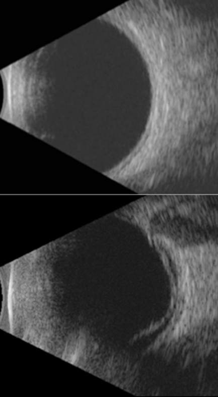
Figure 1 : Demonstrates the B-scan ultrasonography image of normal posterior segment ( left image) and posterior segment with retinal detachment (right image).
Table 2: Distribution of preoperative B-scan ultrasonography findings in eyes with mature cataract
|
Finding |
Number (n=150) |
Percentage |
|
Normal |
112 |
74.7% |
|
Vitreous opacity |
18 |
12% |
|
PVD |
8 |
5.3% |
|
RD |
5 |
3.3% |
|
Vitreous Haemorrhage |
4 |
2.7% |
|
Posterior Staphyloma |
3 |
2% |
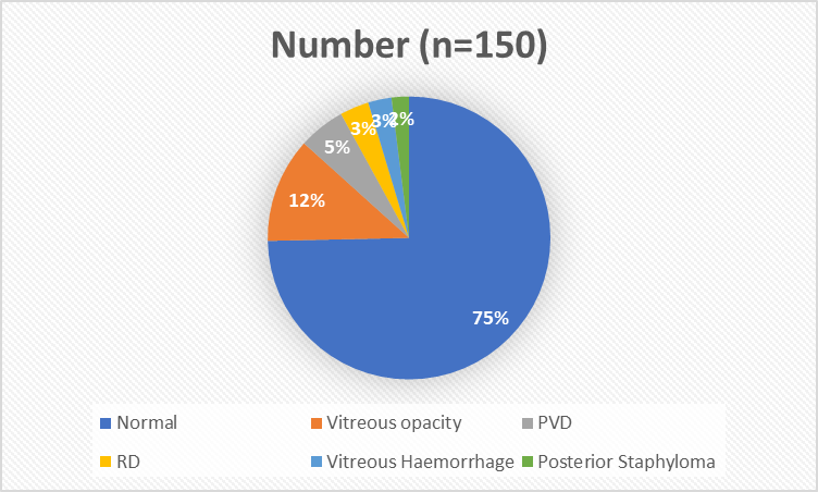
Graph 1: Pie chart depicting distribution of preoperative B-scan ultrasonography findings in eyes with mature cataract
Table 3: Association between preoperative B-scan ultrasonography findings and postoperative best-corrected visual acuity (BCVA).
|
Visual Activity |
Normal B-scan (n=112) |
Abnormal B-scan(n=38) |
|
≥6/18 |
92 (82%) |
6 (15.8%) |
|
6/24–6/60 |
14 (12.5%) |
6 (15.8%) |
|
<6/60 |
6 (5.3%) |
26 (68.4%) |
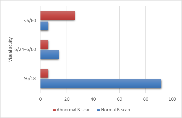
Graph 2: Bar diagram depicting association between preoperative B-scan ultrasonography findings and postoperative best-corrected visual acuity (BCVA).
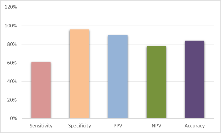
Graph 3: Predictive value of preoperative B-scan ultrasonography for postoperative visual prognosis
DISCUSSION:
The present study demonstrates that B-scan ultrasonography is an effective preoperative imaging modality for predicting postoperative visual outcomes in patients with mature cataract, particularly when direct visualization of the posterior segment is obscured. In our study, approximately 25% of eyes demonstrated posterior segment abnormalities, with vitreous opacities and posterior vitreous detachment being the most frequent findings. These abnormalities were associated with significantly poorer postoperative visual acuity, indicating the prognostic importance of preoperative ultrasonographic evaluation.
Our findings are comparable with those reported by Qureshi and Laghari, who evaluated dense cataracts and found posterior segment lesions in 23.8% of eyes. They concluded that B-scan ultrasonography was an indispensable investigation before cataract surgery because it identified retinal detachment, vitreous hemorrhage, and vitreous degeneration that altered surgical planning.9
Nzeh et al. reported posterior segment abnormalities in 19.6% of eyes with dense cataracts using ocular ultrasonography. The authors emphasized that routine preoperative B-scan examination significantly improves surgical decision-making by identifying occult posterior pathology that may otherwise remain undetected until after surgery.10
The observations of the present study are also consistent with those of Mohammadi et al., who demonstrated that approximately one-quarter of patients with dense cataracts had abnormal B-scan findings. They reported that patients with normal ultrasonographic findings achieved significantly better postoperative visual outcomes than those with retinal or vitreous pathology, confirming the prognostic role of B-scan ultrasonography.11
The diagnostic performance observed in our study, particularly the high specificity (96%) and positive predictive value (90%), agrees with the work of Ali and Rehman, who reported specificity approaching 95–96% and positive predictive values exceeding 90% for detecting clinically significant posterior segment lesions in mature cataracts. Their findings support the reliability of abnormal B-scan findings as strong predictors of poor postoperative vision.12
Comparable results were reported by Khanduri et al., who evaluated ultrasonographic findings in dense cataracts and concluded that B-scan demonstrated excellent diagnostic accuracy for retinal detachment, vitreous hemorrhage, and posterior vitreous detachment.13
Despite its excellent specificity, the sensitivity of B-scan ultrasonography in our study was only moderate. This finding is consistent with the observations of Coleman et al., who noted that although ophthalmic ultrasonography is highly reliable for detecting gross vitreoretinal pathology, subtle retinal breaks, localized macular disorders, and early epiretinal membrane formation may remain undetected.14
CONCLUSION:
B-scan ultrasonography is an essential preoperative investigation in mature cataract patients. It not only detects hidden posterior segment pathologies but also serves as a strong predictor of postoperative visual outcome.
REFERENCES:
1. Coleman DJ, Lizzi FL. Intraocular diagnosis and diagnostic ultrasound. Surv Ophthalmol. 1979;24(1):3-17. doi: 10.1016/0039-6257(79)90029-0
2. Qureshi MA, Laghari K. Role of B-scan ultrasonography in pre-operative evaluation of cataract patients. J Ayub Med Coll Abbottabad. 2010;22(4):31-33.
3. Anteby II, Blumenthal EZ, Zamir E, Waindim P. The role of preoperative ocular ultrasonography for patients with dense cataracts. Eur J Ophthalmol. 1998;8(4):222-224. doi: 10.1177/112067219800800406
4. Singh U, Shrestha GB. Role of B-scan ultrasonography in detecting posterior segment lesions in dense cataract. Nepal J Ophthalmol. 2011;3(2):146-149. doi: 10.3126/nepjoph.v3i2.5266
5. Mohamed A, Banerjee S, Ellis M. B-scan ultrasonography in ocular media opacities and posterior segment evaluation. Eye (Lond). 1997;11(Pt 5):676-680. doi: 10.1038/eye.1997.177
6. Byrne SF, Green RL. Ultrasound of the Eye and Orbit. 2nd ed. St Louis: Mosby; 2002.
7. American Academy of Ophthalmology. Basic and Clinical Science Course. Section 12: Retina and Vitreous. San Francisco (CA): American Academy of Ophthalmology; 2023.
8. Ossoinig KC. Standardized echography: basic principles, clinical applications, and results. Int Ophthalmol Clin. 1979;19(4):127-210. doi: 10.1097/00004397-197901940-00008
9. Qureshi MA, Laghari K. Role of B-scan ultrasonography in pre-operative cataract patients. Pak J Ophthalmol. 2010;26(2):83-86.
10. Nzeh DA, Owoeye JFA, Ademola-Popoola DS. Ocular ultrasonography in patients with dense cataract before cataract surgery. West Afr J Med. 2006;25(3):242-244. doi:10.4314/wajm.v25i3.28287.
11. Mohammadi A, Ghasemi-Rad M, Khodabandeh A. Diagnostic value of B-scan ultrasonography in dense cataract patients. Med Ultrason. 2011;13(4):267-270.
12. Ali Z, Rehman A. Diagnostic accuracy of B-scan ultrasonography in detecting posterior segment lesions in mature cataracts. J Coll Physicians Surg Pak. 2013;23(10):726-729.
13. Khanduri S, Agrawal S, Chhabra MK. Role of B-scan ultrasonography in preoperative evaluation of posterior segment in mature cataract. Delhi J Ophthalmol. 2015;26(2):101-105.
14. Coleman DJ. Reliability of ocular diagnostic ultrasonography. Ophthalmology. 1988;95(9):1325-1330. doi:10.1016/S0161-6420(88)33024-5.