Correlation Between Serum Ferritin Level And Degree Of Liver Fibrosis On Fibroscan In Patients With The Transfusion Dependent Thalassemia: A Cross-Sectional Study In Delhi.
- Rajiv Kumar yadav , DNB Resident, Department of Medicine, SDN Hospital
- Janet L. Thiek Nee Stephens , Senior Consultant & Professor Department of Medicine, SDN Hospital, Delhi
- Devmalya chakravartty , Senior Consultant & Professor Department of Medicine, SDN Hospital, Delhi
- Brij Gandhi , DNB Resident, Department of Medicine, SDN Hospital
Article Information:
Abstract:
Background: Transfusion-dependent thalassemia (TDT) is associated with progressive iron overload due to lifelong transfusion requirements, leading to hepatic iron deposition and fibrosis. Serum ferritin is widely used for monitoring iron burden, while FibroScan provides a non-invasive assessment of liver fibrosis. This study evaluated the correlation between serum ferritin levels and liver fibrosis assessed by FibroScan in patients with TDT. Methods: A hospital-based cross-sectional observational study was conducted at Swami Dayanand Hospital, Delhi, including 18 transfusion-dependent thalassemia patients. Demographic details, transfusion history, laboratory parameters, serial serum ferritin levels, liver stiffness measurement (LSM), CAP values, APRI, and FIB-4 scores were assessed. Correlation between serum ferritin index and LSM was evaluated using Spearman’s correlation analysis. Results: The mean age of participants was 19.44 ± 4.83 years, with male predominance (83.3%). The mean disease duration was 16.91 ± 3.03 years, and mean ferritin index was 2053.33 ± 1071.18 ng/mL. The mean liver stiffness measurement was 7.33 ± 0.94 kPa. Serum ferritin index demonstrated a significant positive correlation with LSM (ρ = 0.470, p = 0.049). Liver stiffness also showed a significant positive correlation with transfusion frequency (ρ = 0.518, p = 0.027). Conclusion: Serum ferritin levels correlate significantly with FibroScan-derived liver stiffness in TDT patients. Combined use of ferritin monitoring and FibroScan may aid in early detection and surveillance of hepatic fibrosis.
Keywords:
Article :
INTRODUCTION:
Thalassemia is a group of inherited hemoglobin disorders characterized by reduced or absent synthesis of one or more globin chains, resulting in ineffective erythropoiesis, chronic hemolysis, and varying degrees of anemia.[1] Among the different types, β-thalassemia, caused by pathogenic mutations affecting the β-globin gene (HBB), is one of the most prevalent forms worldwide, particularly in the Indian subcontinent. The clinical spectrum ranges from asymptomatic carrier states to severe forms such as transfusion-dependent thalassemia (TDT), requiring lifelong regular red blood cell transfusions for survival and prevention of anemia-related complications.[2]Transfusion-dependent thalassemia patients require repeated blood transfusions, usually beginning in early childhood, to maintain adequate hemoglobin levels and ensure normal growth and development. [3]However, long-term transfusion therapy results in progressive iron overload, as the human body lacks an effective physiological mechanism for iron excretion. In addition to transfusional iron accumulation, increased intestinal iron absorption secondary to ineffective erythropoiesis further contributes to excess iron deposition. Progressive iron accumulation leads to oxidative stress, cellular injury, and chronic inflammation, affecting multiple organs, particularly the liver, heart, and endocrine glands.[4]
The liver is the primary site of iron storage and is among the earliest organs affected by iron overload in patients with TDT. Excess hepatic iron deposition promotes hepatocellular injury, activation of hepatic stellate cells, and increased extracellular matrix deposition, resulting in progressive hepatic fibrosis. If untreated, hepatic fibrosis may advance to cirrhosis, portal hypertension, hepatic dysfunction, and increased risk of hepatocellular carcinoma. Therefore, early identification and monitoring of hepatic fibrosis are essential components in the long-term management of patients with transfusion-dependent thalassemia.[5]Serum ferritin is the most commonly used and easily available surrogate marker for assessing body iron burden in thalassemia patients. Serial monitoring of ferritin levels helps in evaluating iron overload and guiding iron chelation therapy.[6] However, ferritin is an acute-phase reactant and may be influenced by several factors, including infection, inflammation, and liver injury, which may limit its accuracy in predicting hepatic iron deposition and fibrosis severity.[7]Traditionally, assessment of hepatic fibrosis relied on liver biopsy, which remains a reference standard for histological evaluation. However, its invasive nature, risk of complications, sampling variability, and inability to be performed repeatedly limit its routine clinical application.[8] Transient elastography (FibroScan®) has emerged as a non-invasive, rapid, and reproducible method for evaluating liver stiffness, measured in kilopascals (kPa), which correlates with the degree of hepatic fibrosis. It provides a practical alternative for repeated assessment and monitoring of liver disease progression.[9]Previous studies have demonstrated variable correlations between serum ferritin levels and FibroScan-derived liver stiffness measurements in patients with thalassemia.[10] While some studies have reported significant associations between ferritin levels and hepatic fibrosis, others suggest that liver stiffness may also be influenced by inflammatory changes, liver enzymes, and other factors. Furthermore, data regarding the relationship between serum ferritin and FibroScan-based assessment of liver fibrosis among transfusion-dependent thalassemia patients, particularly from North India, remain limited.
Therefore, the present study was undertaken to evaluate the correlation between serum ferritin levels and the degree of liver fibrosis assessed by FibroScan in patients with transfusion-dependent thalassemia at Swami Dayanand Hospital, Delhi.
MATERIALS AND METHODS:
The present study was conducted as a hospital-based cross-sectional observational study at the Thalassemia Ward, Department of Medicine, Swami Dayanand Hospital, Delhi. The study was designed to evaluate the correlation between serum ferritin levels and the degree of liver fibrosis assessed by FibroScan in patients with transfusion-dependent thalassemia. The study used clinical, laboratory, and transient elastography data collected from eligible patients.The study was conducted over a period of four months, from August 2025 to November 2025.
Study Population
The study population consisted of patients diagnosed with transfusion-dependent thalassemia (TDT) who attended regular blood transfusion services at the thalassemia ward of Swami Dayanand Hospital, Delhi. All eligible patients aged more than 14 years were considered for inclusion in the study.
Study Objectives
The primary objective of the study was to evaluate the correlation between serum ferritin levels and FibroScan-derived liver stiffness measurements. Secondary objectives included assessment of the association between liver fibrosis and duration and frequency of blood transfusions and evaluation of the diagnostic utility of FibroScan for detecting clinically significant fibrosis in transfusion-dependent thalassemia patients.
Eligibility Criteria
Inclusion Criteria
Patients were included in the study if they fulfilled the following criteria:
1. Diagnosed cases of transfusion-dependent thalassemia receiving regular blood transfusions in the thalassemia ward.
2. Age more than 14 years.
Exclusion Criteria
Patients were excluded if they had:
1. Known chronic liver disease unrelated to transfusion-related iron overload, including autoimmune hepatitis, Wilson’s disease, alpha-1 antitrypsin deficiency, or hereditary hemochromatosis unrelated to transfusion.
2. Decompensated cirrhosis or gross ascites at the time of FibroScan examination.
3. Severe obesity (BMI >35 kg/m²) or anatomical limitations preventing reliable transient elastography measurements.
4. Acute hepatitis flare with ALT levels >5 times the upper limit of normal within six weeks of FibroScan assessment.
5. Pregnancy.
6. Incomplete clinical records or missing essential variables such as FibroScan values, serum ferritin levels, or chelation therapy details.
7. Acute infection or inflammatory conditions causing elevation of serum ferritin levels.
8. Patients unwilling to provide informed consent.
Data Collection
Data were collected using a structured proforma. Clinical history, demographic details, laboratory investigations, transfusion history, chelation therapy details, and FibroScan findings were recorded for each participant.Laboratory values closest to the FibroScan examination date and within a period of ±30 days were included for analysis. Previous three serum ferritin measurements performed at three-month intervals were also documented to calculate serial ferritin trends.
FibroScan Assessment
All participants underwent transient elastography using FibroScan®. Liver stiffness measurement (LSM) was recorded in kilopascals (kPa) as an indicator of hepatic fibrosis. Controlled attenuation parameter (CAP) values were also recorded to assess hepatic steatosis.
Calculation of Non-Invasive Fibrosis Scores
Non-invasive fibrosis indices were calculated using available clinical and laboratory parameters:
· Aspartate aminotransferase to Platelet Ratio Index (APRI)
· Fibrosis-4 (FIB-4) score
Statistical Analysis
Data collected from all participants were entered and analyzed using SPSS .21. Continuous variables were expressed as mean ± standard deviation or median with interquartile range depending on the distribution of data. Categorical variables were presented as frequency and percentage.The correlation between serum ferritin levels and FibroScan liver stiffness measurements was assessed using appropriate correlation tests based on data distribution. Associations between liver fibrosis parameters and clinical variables such as duration and frequency of transfusion were evaluated. Diagnostic performance of FibroScan parameters for detection of clinically significant fibrosis was assessed wherever applicable using sensitivity, specificity, and receiver operating characteristic (ROC) analysis.A p-value of <0.05 was considered statistically significant.
RESULTS:
A total of 18 patients with transfusion-dependent thalassemia were included in the study. The mean age of the study participants was 19.44 ± 4.83 years, with a median age of 19 years (IQR: 16–19.75) and an age range of 16–36 years. The majority of participants were aged 16 years (33.3%), followed by 19 years (27.8%). Male patients constituted the majority of the study population (83.3%; n=15), while females accounted for 16.7% (n=3). Anthropometric assessment showed a mean height of 154.61 ± 9.65 cm and mean weight of 44.14 ± 7.27 kg. Half of the participants were underweight (50.0%; n=9) and half had normal BMI (50.0%; n=9), with a mean BMI of 18.38 ± 1.76 kg/m² (Table 1).The mean age at diagnosis of thalassemia was 2.54 ± 3.61 years, with a median age at diagnosis of 1.10 years (IQR: 0.93–2.25). Most patients were diagnosed between 1–5 years of age (66.7%; n=12), followed by diagnosis before 1 year of age (27.8%; n=5). The mean duration of disease was 16.91 ± 3.03 years, with the majority of patients having disease duration between 16–20 years (72.2%; n=13). The mean number of blood transfusions received was 381.61 ± 185.69, with a median of 380.50 (IQR: 277–526.25) and a range of 55–685 transfusions (Table 2).The mean hemoglobin level among study participants was 8.36 ± 0.97 g/dL, with a median value of 8.50 g/dL (IQR: 8.13–9.00). The mean platelet count was 232.00 ± 125.54 ×10⁹/L. Biochemical evaluation demonstrated a mean ALT level of 57.72 ± 50.30 U/L and mean AST level of 54.50 ± 50.05 U/L. The mean total bilirubin level was 1.70 ± 0.97 mg/dL. The mean CRP level was 2.03 ± 0.89 mg/L, indicating absence of significant inflammatory elevation among the study participants (Table 3). The distribution of laboratory parameters is depicted graphically in Figure 1.Assessment of iron overload demonstrated elevated serial ferritin values among study participants. The mean ferritin values at three previous time points were 2464.28 ± 748.57 ng/mL (Ferritin-3), 3101.50 ± 1393.60 ng/mL (Ferritin-2), and 2500.28 ± 1359.71 ng/mL (Ferritin-1). The mean ferritin index calculated from serial ferritin measurements was 2053.33 ± 1071.18 ng/mL.FibroScan assessment showed a mean liver stiffness measurement (LSM) of 7.33 ± 0.94 kPa, with a median value of 7.30 kPa (IQR: 6.58–7.88). The mean CAP value was 208.28 ± 24.70 dB/m. The mean APRI score and FIB-4 score were 0.97 ± 1.28 and 1.20 ± 1.92, respectively (Table 4). The distribution of iron overload parameters and non-invasive liver assessment indices is illustrated in Figure 2.
To evaluate the association between iron overload and hepatic fibrosis, correlation analysis was performed between serum ferritin index and FibroScan-derived liver stiffness measurement. Serum ferritin index showed a positive correlation with liver stiffness measurement (LSM) with a Spearman correlation coefficient of ρ = 0.470, which was statistically significant (p = 0.049). This indicated that higher ferritin levels were associated with increased liver stiffness values, suggesting a relationship between iron burden and hepatic fibrosis (Table 5). The correlation pattern between ferritin index and LSM is presented in Figure 3.The relationship between liver stiffness measurement and transfusion-related factors was evaluated. LSM showed a positive correlation with duration of transfusion (ρ = 0.462; p = 0.053), although the association did not reach statistical significance. A statistically significant positive correlation was observed between LSM and frequency of transfusion (ρ = 0.518; p = 0.027), indicating that patients requiring more frequent transfusions had higher liver stiffness values (Table 6).
Table 1. Demographic and Anthropometric Characteristics of Study Participants (n = 18)
|
Variables |
Number (n) |
Percentage (%) |
|
Age (years) |
||
|
Mean ± SD |
19.44 ± 4.83 |
|
|
Median (IQR) |
19 (16–19.75) |
|
|
Range |
16–36 |
|
|
Sex |
||
|
Male |
15 |
83.3 |
|
Female |
3 |
16.7 |
|
Height (cm) |
||
|
Mean ± SD |
154.61 ± 9.65 |
|
|
Median (IQR) |
155 (150–158.75) |
|
|
Range |
130–175 |
|
|
Weight (kg) |
||
|
Mean ± SD |
44.14 ± 7.27 |
|
|
Median (IQR) |
44.25 (40–48.75) |
|
|
Range |
32–61 |
|
|
BMI (kg/m²) |
||
|
Underweight (<18.5) |
9 |
50.0 |
|
Normal weight (18.5–22.9) |
9 |
50.0 |
|
Mean ± SD |
18.38 ± 1.76 |
|
|
Median (IQR) |
18.3 (17.65–19.05) |
|
|
Range |
15–22.4 |
Table 2. Disease Profile and Transfusion Characteristics of Study Participants (n = 18)
|
Variables |
Number (n) |
Percentage (%) |
|
Age at Diagnosis (years) |
||
|
<1 year |
5 |
27.8 |
|
1–5 years |
12 |
66.7 |
|
6–10 years |
0 |
0.0 |
|
>10 years |
1 |
5.6 |
|
Mean ± SD |
2.54 ± 3.61 |
|
|
Median (IQR) |
1.10 (0.93–2.25) |
|
|
Duration of Disease (years) |
||
|
5–10 years |
1 |
5.6 |
|
11–15 years |
2 |
11.1 |
|
16–20 years |
13 |
72.2 |
|
>20 years |
2 |
11.1 |
|
Mean ± SD |
16.91 ± 3.03 |
|
|
Median (IQR) |
17.50 (15.56–18.38) |
|
|
Total Blood Transfusions |
||
|
Mean ± SD |
381.61 ± 185.69 |
|
|
Median (IQR) |
380.50 (277–526.25) |
|
|
Range |
55–685 |
Table 3. Laboratory Parameters of Study Participants (n = 18)
|
Laboratory Parameter |
Mean ± SD |
Median (IQR) |
Range |
|
Hemoglobin (g/dL) |
8.36 ± 0.97 |
8.50 (8.13–9.00) |
5.5–9.6 |
|
Platelet count (×10⁹/L) |
232.00 ± 125.54 |
223 (151.25–292.25) |
38–531 |
|
ALT (U/L) |
57.72 ± 50.30 |
45.50 (31.75–59.75) |
16–236 |
|
AST (U/L) |
54.50 ± 50.05 |
41 (38–50) |
20–241 |
|
Total bilirubin (mg/dL) |
1.70 ± 0.97 |
1.45 (1–2.10) |
0.5–3.8 |
|
CRP (mg/L) |
2.03 ± 0.89 |
1.95 (1.43–2.48) |
0.8–4.1 |
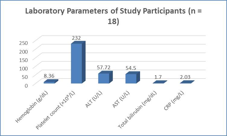
Figure 1 Laboratory Parameters of Study Participants (n = 18)
Table 4. Iron Overload Parameters and Non-Invasive Liver Assessment in Study Participants (n = 18)
|
Parameters |
Mean ± SD |
Median (IQR) |
Range |
|
Ferritin -3 (ng/mL) |
2464.28 ± 748.57 |
2414 (2057–3012) |
766–4000 |
|
Ferritin -2 (ng/mL) |
3101.50 ± 1393.60 |
2954.50 (2045–3579.75) |
1468–7120 |
|
Ferritin -1 (ng/mL) |
2500.28 ± 1359.71 |
2056.50 (1430.25–3579.50) |
711–5320 |
|
Ferritin Index (ng/mL) |
2053.33 ± 1071.18 |
1835.5 (1242.75–2837.25) |
503–4286 |
|
Liver Stiffness Measurement (LSM) (kPa) |
7.33 ± 0.94 |
7.30 (6.58–7.88) |
5.7–8.9 |
|
CAP (dB/m) |
208.28 ± 24.70 |
207.50 (191.25–218.75) |
173–265 |
|
APRI score |
0.97 ± 1.28 |
0.47 (0.33–0.54) |
0.17–4.78 |
|
FIB-4 score |
1.20 ± 1.92 |
0.47 (0.38–0.94) |
0.16–7.68 |
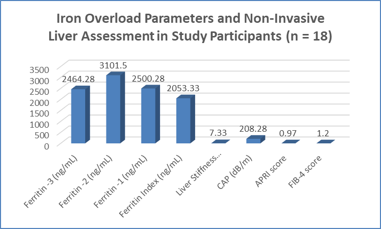
Figure 2 Iron Overload Parameters and Non-Invasive Liver Assessment in Study Participants (n = 18)
Table 5. Correlation Between Serum Ferritin Index and Liver Fibrosis Parameters (n = 18)
|
Variables Compared |
Spearman’s Correlation Coefficient (ρ) |
p-value |
|
Serum Ferritin Index vs Liver Stiffness Measurement (LSM, kPa) |
0.470 |
0.049 |
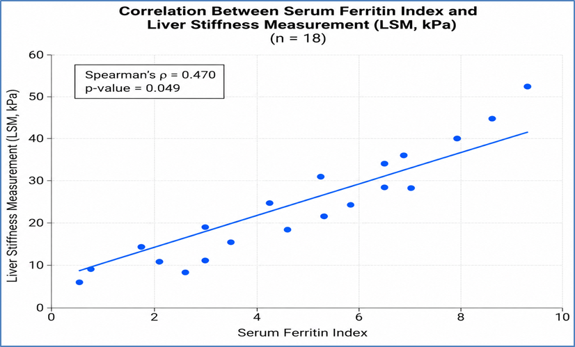
Figure 3 Correlation Between Serum Ferritin Index and Liver Fibrosis Parameters (n = 18)
Table 6. Correlation Between Liver Stiffness Measurement and Transfusion Characteristics (n = 18)
|
Variables Compared |
Spearman’s Correlation Coefficient (ρ) |
p-value |
|
LSM (kPa) vs Duration of transfusion (years) |
0.462 |
0.053 |
|
LSM (kPa) vs Frequency of transfusion |
0.518 |
0.027 |
DISCUSSION:
The present cross-sectional observational study evaluated the association between serum ferritin levels and liver fibrosis assessed by FibroScan in 18 patients with transfusion-dependent thalassemia (TDT). The study included assessment of demographic characteristics, transfusion history, biochemical parameters, serial ferritin levels, liver stiffness measurement (LSM), and non-invasive fibrosis indices. The major finding was a significant positive correlation between serum ferritin index and FibroScan-derived LSM (ρ = 0.470, p = 0.049), indicating that increasing iron burden was associated with increased hepatic stiffness and possible progression of fibrosis. Liver stiffness also showed a significant positive correlation with transfusion frequency (ρ = 0.518, p = 0.027), highlighting the role of cumulative transfusional iron exposure in hepatic injury.The mean age of participants was 19.44 ± 4.83 years, with male predominance (83.3%).
The mean age at diagnosis was 2.54 ± 3.61 years, and most patients (66.7%) were diagnosed between 1–5 years of age. The prolonged disease duration (16.91 ± 3.03 years) reflects the chronic nature of TDT and the long-term requirement for repeated transfusions. Chronic transfusion therapy, although essential for survival, results in progressive iron accumulation, which remains a major contributor to morbidity in these patients.The mean ferritin index in the present study was 2053.33 ± 1071.18 ng/mL, with persistently elevated serial ferritin values, indicating significant iron overload. Khan et al. (2023)[11] reported a median ferritin level of 1881 ng/mL among 91 TDT patients and demonstrated a significant correlation between serum ferritin and transient elastography-derived liver stiffness (r = 0.43, p = 0.001), supporting the association between systemic iron burden and hepatic fibrosis. Similarly, Atmakusuma et al.[12] observed a significant correlation between ferritin and liver stiffness (r = 0.651, p <0.001), suggesting that ferritin may reflect iron-mediated hepatic injury.In the present study, the significant correlation between serum ferritin index and LSM supports the hypothesis that chronic iron deposition contributes to hepatic fibrosis development. Ali et al. (2023)[13] reported a similar positive correlation between serum ferritin and FibroScan-assessed fibrosis (r = 0.287, p = 0.033). Khan et al.[11] also demonstrated a significant relationship between transient elastography values and ferritin levels (rₛ = 0.43, p = 0.001). However, Pipaliya et al.[14] found no significant correlation between ferritin and elastography (r = 0.19, p = 0.11), suggesting that ferritin alone may not accurately represent hepatic iron deposition due to the influence of inflammation, liver injury, and chelation status.
The mean LSM in the present study was 7.33 ± 0.94 kPa, suggesting mild hepatic stiffness, while the mean CAP value was 208.28 ± 24.70 dB/m, indicating minimal steatosis. FibroScan offers an important advantage in TDT patients because it is non-invasive, reproducible, and suitable for repeated monitoring. Fraquelli et al.[15] demonstrated a significant correlation between transient elastography and histological fibrosis (r = 0.73, p = 0.003), supporting its role in hepatic fibrosis assessment.The association between LSM and transfusion frequency observed in this study further emphasizes the contribution of cumulative transfusion exposure to hepatic fibrosis. Frequent transfusions promote iron deposition, oxidative stress, hepatocellular injury, and subsequent fibrosis. Therefore, combined monitoring of serial ferritin levels and FibroScan parameters may provide a practical strategy for early detection of hepatic complications and optimization of iron chelation therapy in TDT patients, particularly where MRI-based liver iron quantification is unavailable.
CONCLUSION:
The present study demonstrated a significant positive correlation between serum ferritin index and FibroScan-derived liver stiffness measurement in patients with transfusion-dependent thalassemia, suggesting that increasing iron burden is associated with progressive hepatic fibrosis. Liver stiffness also showed a significant association with transfusion frequency, highlighting the role of cumulative transfusional iron exposure in hepatic injury. FibroScan, along with serial serum ferritin monitoring, may serve as a useful non-invasive approach for early detection and monitoring of liver fibrosis in TDT patients. Larger prospective studies with longer follow-up and MRI-based iron assessment are required to validate these findings.
LIMITATIONS
The present study had a relatively small sample size (n = 18), which may limit the statistical power and generalizability of the findings. The cross-sectional design restricted assessment of temporal changes and causal relationships between iron overload and hepatic fibrosis progression. The absence of liver biopsy or MRI-based liver iron concentration assessment limited direct validation of FibroScan findings. Further large-scale prospective multicentric studies are required to confirm these observations.
REFERENCES:
1. Cao A, Galanello R. Beta-thalassemia. Genet Med. 2010;12(2):61-76.
2. Taher AT, Weatherall DJ, Cappellini MD. Thalassaemia. Lancet. 2018;391(10116):155-167.
3. De Sanctis V, Kattamis C, Canatan D, Soliman AT, Elsedfy H, Karimi M, et al. β-thalassemia distribution in the old world: an ancient disease seen from a historical standpoint. Mediterr J Hematol Infect Dis. 2017;9(1):e2017018.
4. Khaliq S. Thalassemia in Pakistan. Hemoglobin. 2022;46(1):12-14.
5. Taher AT, Saliba AN. Iron overload in thalassemia: different organs at different rates. Hematology Am Soc Hematol Educ Program. 2017;2017(1):265-271.
6. Bou-Fakhredin R, Bazarbachi AH, Chaya B, Sleiman J, Cappellini MD, Taher AT, et al. Iron overload and chelation therapy in non-transfusion dependent thalassemia. Int J Mol Sci. 2017;18(12):2778.
7. Musallam KM. Iron overload in non-transfusion-dependent thalassemia. Thalass Rep. 2013;3(1):e11.
8. Padeniya P, Ediriweera D, De Silva AP, Niriella M, Premawardhena A. The association between steatosis and liver damage in transfusion-dependent beta thalassaemia patients. Br J Haematol. 2023;200(5):517-523.
9. Majd Z, Haghpanah S, Ajami GH, Matin S, Namazi H, Bardestani M, et al. Serum ferritin levels correlation with heart and liver MRI and LIC in patients with transfusion-dependent thalassemia. Iran Red Crescent Med J. 2015;17(4):e24959.
10. Parakh N, Chandra J. Correlation of transient elastography with MRI T2* and serum ferritin levels in children with transfusion-dependent thalassemia. Indian Pediatr. 2022;59(12):929-932.
11. Khan H, Panjwani V, Al Rahbi S, Eltigani A, Qureshi RN, Unissa K, et al. Correlation of Transient Elastography with Liver Iron Concentration and Serum Ferritin Levels in Patients with Transfusion-Dependent Thalassemia Major from Oman. Mediterr J Hematol Infect Dis. 2023 Sep 1;15(1):e2023048.
12. Atmakusuma TD, Lubis AM. Correlation of Serum Ferritin and Liver Iron Concentration with Transient Liver Elastography in Adult Thalassemia Intermedia Patients with Blood Transfusion. J Blood Med. 2021 Apr 15;12:235-243.
13. Ali MS, Borhany M, Butt AJ, Munawar Ali R, Kashif S, Wahaj M, et al. Correlation Between Serum Ferritin and Degree of Hepatic Fibrosis on Fibroscan in Thalassemic Patients. Cureus. 2023 Jul 18;15(7):e42069.
14. Pipaliya N, Solanke D, Parikh P, Ingle M, Sharma R, Sharma S, et al. Comparison of Tissue Elastography With Magnetic Resonance Imaging T2* and Serum Ferritin Quantification in Detecting Liver Iron Overload in Patients With Thalassemia Major. Clin Gastroenterol Hepatol. 2017 Feb;15(2):292-298.e1.
15. Fraquelli M, Cassinerio E, Roghi A, Rigamonti C, Casazza G, Colombo M, et al. Transient elastography in the assessment of liver fibrosis in adult thalassemia patients. Am J Hematol. 2010 Aug;85(8):564-8.