Evaluation of Paediatric Pancytopenia Through Haematological Parameters and Bone Marrow Aspiration: An Observational Study.
- Angelee Keisham , PG 3rd year Resident, Department of Pathology, Gajra Raja Medical College, Gwalior (M.P.)
- Gajendra Pal Singh Uike , Professor and Head, Department of Pathology, Gajra Raja Medical College, Gwalior (M.P.)
- Ankit Kumar , PG 3rd year Resident, Department of Pathology, Gajra Raja Medical College, Gwalior (M.P.)
- Neetu Sharma , Professor, Department of Pediatrics, Gajra Raja Medical College, Gwalior (M.P.)
Article Information:
Abstract:
Background: Pancytopenia in children represents a significant haematological challenge with a heterogeneous etiological spectrum ranging from reversible nutritional deficiencies to life-threatening marrow failure syndromes and malignancies. Early identification of reversible causes such as nutritional anaemia can dramatically improve outcomes, while prompt diagnosis of aplastic anaemia or leukemia allows for early referral and initiation of definitive therapy. Aim: To find the aetiology of paediatric pancytopenia through a comprehensive evaluation of haematological parameters, vitamin profiles, and bone marrow aspiration. Methods: An observational study was conducted at a tertiary care center over 1.5 years, including 96 paediatric patients (aged ≤19 years) presenting with pancytopenia. Evaluation included clinical assessment, complete blood counts, reticulocyte counts, peripheral blood smears, serum vitamin B12 and folic acid levels, and bone marrow aspiration. Data were analyzed using appropriate statistical methods (p<0.05 considered significant). Results: The majority of patients were in the 11-15 years age group (47.92%). Pallor was the predominant clinical presentation (95.83%). Haematological profiling revealed severe cytopenias, with a mean haemoglobin of 6.08 ± 1.48 g/dL and a low mean reticulocyte count of 0.44 ± 0.21%. Macrocytic anaemia was the most common peripheral smear finding (42.71%). Vitamin B12 and folic acid deficiencies were highly prevalent (64.58% and 67.71%, respectively), with a significant co-occurrence (p<0.001). Bone marrow aspiration identified megaloblastic anaemia as the leading aetiology (36.46%), followed by aplastic anaemia (28.13%), and acute leukemia/ALL (12.50%). Age-wise stratification revealed acute leukemia and aplastic anaemia were predominant in younger children (1-10 years), whereas megaloblastic anaemia dominated in adolescents (11-19 years). Conclusion: Nutritional megaloblastic anaemia is the leading, treatable cause of paediatric pancytopenia in this setting, underscoring the critical need for routine vitamin B12 and folate screening. However, the substantial burden of aplastic anaemia and acute leukaemia necessitates prompt bone marrow evaluation to prevent diagnostic delay and guide definitive management.
Keywords:
Article :
INTRODUCTION:
Pancytopenia represents a significant haematological abnormality characterized by the simultaneous reduction of erythrocytes, leukocytes, and platelets below the lower limit of normal for age and sex.[1] Rather than constituting a disease entity in itself, pancytopenia is a clinicopathological manifestation of a wide array of underlying processes that primarily or secondarily involve the bone marrow.[2] In the paediatric population, the occurrence of pancytopenia is of particular clinical importance, as it reflects a spectrum of conditions ranging from reversible nutritional deficiencies and transient infection-induced marrow suppression to severe, life-threatening disorders such as acquired aplastic anaemia and haematological malignancies.[3,4] Children typically present with non-specific clinical features– including pallor, prolonged fever, asthenia, and bleeding manifestations– making a high index of suspicion and systematic evaluation essential for timely diagnosis and therapeutic intervention.[5,6]
The etiological profile of pancytopenia in children differs markedly from that observed in adults, necessitating age-specific diagnostic strategies.[7,8] In developing nations, paediatric pancytopenia is strongly influenced by nutritional and environmental determinants, with nutritional megaloblastic anaemia frequently emerging as a predominant aetiology.[9,10] Conversely, in tertiary referral settings, serious marrow pathologies such as acute lymphoblastic leukemia (ALL) and aplastic anaemia contribute substantially to the disease burden.[11,12] Unlike adult leukemias, which often present with marked leucocytosis, paediatric acute leukemias frequently manifest with pancytopenia due to rapid and extensive marrow infiltration, complicating early clinical differentiation.[13,14]
The initial evaluation of pancytopenia relies heavily on complete blood counts (CBC), reticulocyte response, and peripheral blood smear examination to characterize the severity and morphological patterns of the cytopenias.[15,16,17] However, definitive etiological diagnosis frequently necessitates bone marrow evaluation. Bone marrow aspiration provides direct morphological insight into marrow cellularity, architectural preservation, lineage maturation, and the presence of atypical or infiltrative elements, successfully distinguishing between ineffective haematopoiesis, marrow failure, and malignant replacement.[18,19,20]
Within the context of nutritional cytopenias, deficiencies in vitamin B12 and folic acid lead to impaired DNA synthesis and profound intramedullary precursor apoptosis, manifesting as ineffective haematopoiesis.[4,14] Despite being completely reversible with appropriate supplementation, megaloblastic pancytopenia can closely mimic malignant or aplastic states clinically and haematologically, highlighting a critical diagnostic gap that must be addressed rapidly to avert unnecessary invasive procedures and morbidity.[4,21] The present study was designed to evaluate the aetiology of pancytopenia in the paediatric age group through a detailed analysis of haematological parameters, peripheral smear morphology, serum vitamin B12/folate status, and bone marrow aspiration findings. The primary aim was to diagnose and establish the aetiology of pancytopenia, with specific objectives to evaluate bone marrow findings and determine the relationship between serum vitamin B12 and folic acid status in affected children.
Therefore, the present study aimed to evaluate the aetiological spectrum of paediatric pancytopenia through a structured assessment of haematological parameters, peripheral smear morphology, serum vitamin B12 and folate status, and bone marrow aspiration findings in a tertiary care setting.
Aim and objectives
The primary aim of this study was to determine the aetiological spectrum of paediatric pancytopenia through an integrated assessment of peripheral haematological parameters and bone marrow aspiration. The specific objectives were twofold: first, to characterise the morphological patterns and diagnostic yield of bone marrow aspiration in identifying underlying bone marrow pathologies; and second, to investigate the prevalence of, and correlation between, serum vitamin B12 and folate deficiencies within this clinical cohort.
MATERIALS AND METHODS:
Study Design and Study Setting: This prospective and retrospective observational study was conducted in the Department of Pathology in collaboration with the Department of Paediatrics at Gajra Raja Medical College and JA Group of Hospitals, Gwalior, India. These institutions serve as major tertiary care referral centers.
Study Period and Duration: The study spanned a period of one and a half years, from April 2024 to September 2025.
Study Population and Sampling: The target population comprised paediatric patients presenting with confirmed pancytopenia. A calculated sample size of 96 participants was established based on previously published prevalence data for aplastic anaemia (28.3%) in paediatric pancytopenia, utilizing a 5% level of significance and 9% absolute error. Sampling was performed by consecutively enrolling all eligible cases meeting the inclusion criteria during the study period.
Inclusion and Exclusion Criteria: The study included paediatric patients up to 19 years of age whose routine CBC demonstrated pancytopenia (defined as a simultaneous reduction in haemoglobin, total leukocyte count, and platelet count below age-appropriate reference ranges) and who subsequently underwent peripheral blood smear and bone marrow examination. Patients currently receiving chemotherapy, those with a history of recent blood transfusion, and individuals aged over 19 years were excluded to eliminate confounding variables and morphological artifacts.
Study Procedures: Following detailed clinical history and physical examination (assessing for pallor, fever, weakness, bleeding manifestations, hepatomegaly, splenomegaly, and lymphadenopathy), peripheral blood samples were collected in EDTA-anticoagulated vials. Haematological parameters were analyzed utilizing an automated 3-part haematology analyzer (Z3 series). Peripheral blood smears were manually prepared, stained with Leishman stain, and evaluated microscopically for red cell morphology and the presence of atypical cells. Reticulocyte counts were calculated manually. Serum vitamin B12 and folic acid levels were quantified using standard biochemical assays prior to marrow evaluation.
Bone marrow aspiration was performed from the posterior superior iliac crest under local anaesthesia with strict aseptic precautions. Aspirated marrow smears were stained with Leishman stain and independently assessed for cellularity, myeloid-to-erythroid ratio, lineage maturation, and the presence of abnormal cells or dysplasia.
Statistical Analysis Plan: Data were entered into Microsoft Excel and analyzed using SPSS software version 22.0. Continuous numerical variables were expressed as mean ± standard deviation (SD). Categorical variables were presented as frequencies (n) and percentages (%). Associations between categorical variables were assessed utilizing the Chi-square (χ²) test. A p-value of <0.05 was considered statistically significant.
Ethical Considerations: The study protocol obtained formal approval from the Institutional Ethics Committee (IEC) of Gajra Raja Medical College, Gwalior. Written informed consent/assent was secured from the parents or legal guardians of all participating children prior to inclusion.
RESULTS:
Table 1. Age distribution of study participants
|
Variable |
Number of Participants (n=96) |
Percentage (%) |
Chi-square, p- value |
|
|
Age range (in years) |
1-5 years |
10 |
10.42 |
χ² = 36.58, p < 0.001 |
|
6-10 years |
29 |
30.21 |
||
|
11-15 years |
46 |
47.92 |
||
|
16-19 years |
11 |
11.46 |
||
|
Gender |
Male |
43 |
44.79 |
χ² = 1.04, p = 0.31 |
|
Female |
53 |
55.21 |
||
|
TOTAL |
96 |
100 |
|
|
A total of 96 paediatric patients with pancytopenia were evaluated. The demographic distribution demonstrated a significant age-related variance, with the highest frequency of cases observed in the 11-15 years age group (47.92%), and the lowest in the 1-5 years age group (10.42%) (p<0.001). There was a slight female preponderance (55.21% vs. 44.79%); however, this gender distribution was not statistically significant (p=0.31).
Table 2. Haematological Parameters of Study Participants (Mean ± SD)
|
Parameter |
Mean |
SD |
|
Hb (g/dL) |
6.08 |
1.48 |
|
WBC (10³/µL) |
2.26 |
0.78 |
|
Platelets (10³/µL) |
35.23 |
18.20 |
|
MCV (fL) |
87.57 |
10.91 |
|
MCH (pg) |
29.22 |
3.27 |
|
RDW-CV (%) |
18.76 |
8.73 |
|
Retic count (%) |
0.44 |
0.21 |
The cohort exhibited severe cytopenias across all lineages. The mean haemoglobin was 6.08 ± 1.48 g/dL, indicating profound anaemia. Mean leukocyte and platelet counts were significantly depressed at 2.26 ± 0.78 × 10³/µL and 35.23 ± 18.20 × 10³/µL, respectively.
Table 3. Distribution of Types of Pancytopenia in Peripheral Smear Pattern among Study Participants
|
Types of pancytopenia |
Number (n=96) |
Percentage (%) |
Chi-square, p- value |
|
Normocytic Normochromic |
32 |
33.33 |
χ² = 54.30, p < 0.001 |
|
Macrocytic |
41 |
42.71 |
|
|
Dimorphic |
9 |
9.38 |
|
|
Microcytic Hypochromic |
8 |
8.33 |
|
|
Others (mixed nutritional, leukemia) |
6 |
6.25 |
|
|
TOTAL |
96 |
100 |
Morphologically, macrocytic anaemia was the most frequent peripheral smear pattern (42.71%), followed by normocytic normochromic anaemia (33.33%) (p<0.001).
Table 4. Distribution of Atypical Cells, Vitamin B12 and Folic Acid in Peripheral Smear among Study Participants
|
Variable |
Number (n=96) |
Percentage (%) |
Chi-square, p- value |
|
|
Atypical cells |
Present |
12 |
12.50 |
χ² = 54.00, p < 0.001 |
|
Absent |
84 |
87.50 |
||
|
Vitamin B12 |
Deficient |
62 |
64.58 |
χ² = 8.17, p = 0.004 |
|
Normal |
34 |
35.42 |
||
|
Folic acid |
Deficient |
65 |
67.71 |
χ² = 12.04, p < 0.001 |
|
Normal |
31 |
32.29 |
||
|
TOTAL |
96 |
100 |
|
|
Atypical cells were absent in the majority of peripheral smears (87.50%), though present in 12.50% of the cohort. Biochemical evaluation revealed a high prevalence of nutritional deficiencies. Vitamin B12 deficiency was confirmed in 64.58% of the participants, while folic acid deficiency was present in 67.71%.
Table 5. Association between Vitamin B12 Levels and Folic Acid Status
|
|
Normal folic acid |
Folic acid deficiency |
Chi-square, p- value |
|
Normal vitamin B12 |
27 |
7 |
χ² = 53.45, p < 0.001 |
|
Vitamin B12 deficiency |
4 |
58 |
A highly significant association was observed between the two; 58 out of 62 patients with Vitamin B12 deficiency exhibited concurrent folic acid deficiency (p<0.001).
Table 6. Bone Marrow Findings among Study Participants
|
Bone Marrow Findings |
Number of Participants (n=96) |
Percentage (%) |
Chi-square, p- value |
|
Megaloblastic |
35 |
36.46 |
χ² = 39.38, p < 0.001 |
|
Aplastic |
27 |
28.13 |
|
|
Mixed nutritional Anemias |
8 |
8.33 |
|
|
Acute Leukemia / ALL |
12 |
12.50 |
|
|
Infective Etiology / Reactive Plasmacytosis |
8 |
8.33 |
|
|
Other (IDA, Megakaryocytosis) |
6 |
6.25 |
|
|
TOTAL |
96 |
100 |
Bone marrow examination established definitive aetiologies for the pancytopenic presentations. Megaloblastic anaemia was the most frequent diagnosis (36.46%), followed closely by aplastic anaemia (28.13%) and acute leukemia/ALL (12.50%).
Table 7. Distribution of Clinical Features among Study Participants
|
Clinical features |
Number of Participants (n=96) |
Percentage (%) |
Chi-square, p- value |
|
Weakness |
57 |
59.38 |
χ² = 122.39, p < 0.001 |
|
Fever |
60 |
62.50 |
|
|
Pallor |
92 |
95.83 |
|
|
Lymphadenopathy |
18 |
18.75 |
|
|
Splenomegaly |
34 |
35.42 |
|
|
Hepatomegaly |
18 |
18.75 |
|
|
Petechiae |
13 |
13.54 |
|
|
TOTAL |
96 |
100 |
Clinically, pallor was nearly universal (95.83%), followed by fever (62.50%) and generalized weakness (59.38%). Organomegaly, including splenomegaly (35.42%) and hepatomegaly (18.75%), was also noted.
Table 8. Age-wise Most Common Type of Pancytopenia and Bone Marrow Findings
|
Age Range |
Number of patients (n=96) |
Common type of pancytopenia |
Common bone marrow aspirate finding |
|
1-5 years |
10 |
Normocytic Normochromic |
Acute Leukemia / ALL |
|
6-10 years |
29 |
Normocytic Normochromic |
Aplastic |
|
11-15 years |
46 |
Macrocytic |
Megaloblastic |
|
16-19 years |
11 |
Macrocytic |
Megaloblastic |
Age-stratified analysis revealed distinct etiological patterns: younger children (1-5 years) predominantly presented with normocytic normochromic smear patterns secondary to acute leukemia, whereas the 6-10 years group was largely affected by aplastic anaemia. Conversely, adolescents (11-19 years) primarily exhibited macrocytic pancytopenia resulting from megaloblastic marrow changes.
Bone Marrow Aspiration Findings
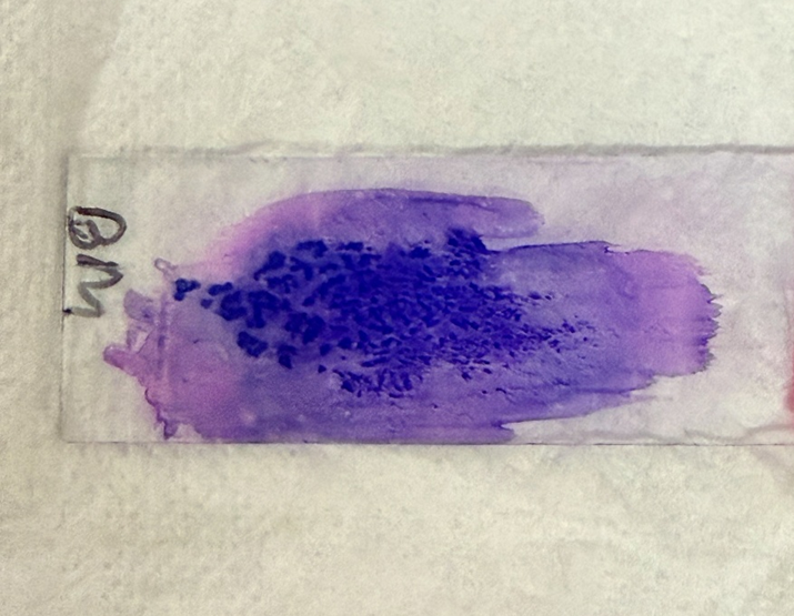
BONE MARROW ASPIRATION SLIDE SHOWING MARROW PARTICLES
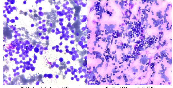
Subleukemic leukemia 40X Erythroid Hyperplasia 40X
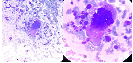
Megakaryocyte 40x Megakaryocyte oil emersion
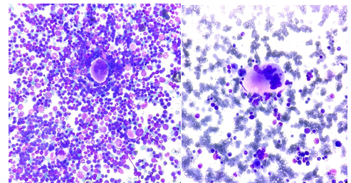
Hemophagocytosis, 10x Hemophagocytosis, 40x
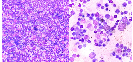
Hypercellular marrow with megaloblast 10X Megaloblast in Oil emersion
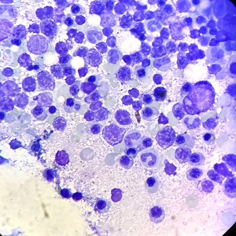
Plasmacytosis
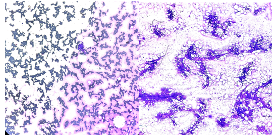
Hypocellular Marrow 10x Hypocellular Marrow 10x
Peripheral smear findings
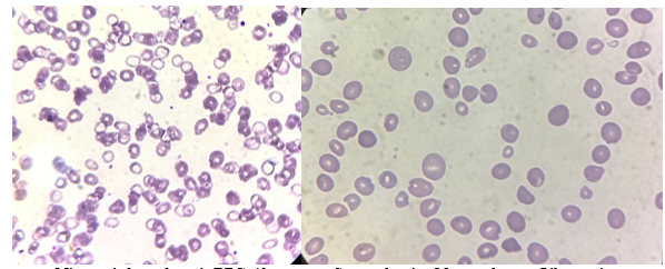
Microcytic hypochromic RBC 40x Smear showing Macrovalocytes Oil emersion
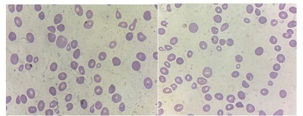
Dimorphic blood picture Peripheral smear showing anisocytosis
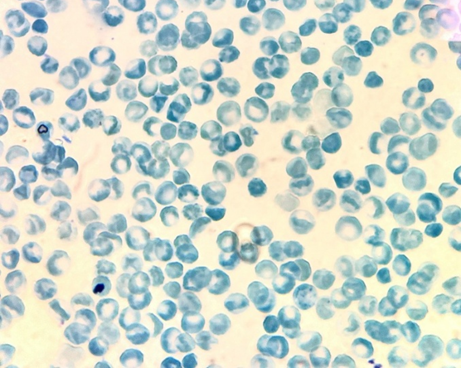
Rectic stain showing Low rectic count 40X
DISCUSSION:
The accurate evaluation of pancytopenia in the paediatric age group remains a major clinical challenge due to the condition's broad etiological spectrum and overlapping morphological manifestations. The primary objective of this study was to diagnose the aetiology of paediatric pancytopenia utilizing a synthesis of haematological parameters, vitamin profiling, and bone marrow aspiration. Our findings unequivocally establish nutritional deficiency– specifically megaloblastic anaemia secondary to combined B12 and folate depletion– as the predominant cause of pancytopenia in this demographic, followed closely by aplastic anaemia and acute leukemia.
Demographic analysis revealed a significant predisposition for pancytopenia in the 11-15 years age cohort (47.92%). This adolescent skew likely reflects periods of accelerated growth, heightened physiological demands, and often, marginal dietary intake leading to nutritional exhaustion. In contrast to our findings, Duong et al. (2020) in Vietnam observed peak incidences in much younger cohorts (6 months to 6 years), where hemophagocytic lymphohistiocytosis (58.3%) and acute lymphoblastic leukemia (26.0%) were the primary culprits.[22] This divergence underscores the profound influence of regional nutritional standards, underlying population genetics, and tertiary referral biases on the epidemiology of paediatric cytopenias.[5,9] Furthermore, our study identified a slight, albeit statistically insignificant, female preponderance (55.21%). While some previous Indian studies, such as those by Khan et al. (2018) in Kashmir and Chandel et al. (2025) in Uttar Pradesh, reported a mild male dominance,[23,19] the gender neutrality in our cohort suggests that biological sex is not a primary mechanistic determinant for pancytopenia in our study population.
Haematological evaluation in our cohort highlighted severe tri-lineage depression. The mean haemoglobin was critically low (6.08 ± 1.48 g/dL), correlating clinically with the near-universal presentation of pallor (95.83%). The presence of a depressed reticulocyte count (0.44 ± 0.21%) across the cohort clearly pointed to a primary bone marrow production failure– either through hypocellularity, ineffective haematopoiesis, or malignant infiltration– rather than peripheral consumptive processes.[15,16] Peripheral smear examination offered essential qualitative direction. Macrocytic morphology was the most frequently observed pattern (42.71%), strongly aligning with the marrow diagnosis of megaloblastic anaemia. This finding is consistent with Kumar et al. (2023) from Eastern India, who noted macrocytic predominance coupled with a 64% incidence of megaloblastic marrow.[24]
The biochemical assays in the current study elucidate the prominent role of micronutrient deficiencies in Indian paediatric cytopenias. Vitamin B12 deficiency was found in 64.58% of children, and folate deficiency in 67.71%. Crucially, we demonstrated a highly significant association (p<0.001) between the two parameters; 93.5% of children with B12 deficiency exhibited concurrent folate depletion. This combined nutritional deficit drives severe ineffective erythropoiesis and intramedullary haemolysis, manifesting as prominent anisocytosis (RDW-CV 18.76 ± 8.73%) and mixed morphological patterns.[25] Similar complex nutritional intersections were reported by Sahay and Ramesh (2018), where combined deficiency states surpassed isolated deficiencies as the principal aetiology of pancytopenia.[10]
Bone marrow aspiration proved indispensable, confirming megaloblastic anaemia as the single most common aetiology (36.46%). This finding is highly comparable to the data reported by Agrawal et al. (2021) in Jhansi (49.23% megaloblastic) and Shah et al. (2017) in Gujarat (35%).[26,27] However, our data sharply contrast with the study by Sana and Rashid (2022) in Karachi, Pakistan, where acquired aplastic anaemia represented an overwhelming 49% of paediatric pancytopenia cases.[28] In our cohort, aplastic anaemia was the second most frequent finding (28.13%), highlighting the persistent threat of primary marrow failure syndromes in the paediatric age group.
Importantly, our data highlight critical age-specific etiological transitions. Children between 1 and 5 years predominantly displayed normocytic normochromic smear patterns, with bone marrow evaluation confirming acute leukemia (ALL) as the dominant underlying pathology in this sub-cohort. This aligns with paediatric oncology paradigms where young children with ALL may present sub-acutely with marrow replacement and pancytopenia rather than classic leucocytosis.[13] As age advanced into the 6-10 years bracket, aplastic states became paramount. In adolescents (11-19 years), the physiological stress of puberty intersected with dietary inadequacy, shifting the paradigm heavily toward megaloblastic anaemia. This stratification provides a highly practical clinical heuristic for paediatricians initiating diagnostic work-ups.
The presence of atypical cells on peripheral smears (12.50%) correlated perfectly with the ultimate bone marrow diagnosis of acute leukemia (12.50%), reinforcing the smear's utility as an initial gatekeeper. Nevertheless, the absence of atypical cells in 87.50% of the pancytopenic cohort emphasizes that peripheral blood alone is insufficiently sensitive to exclude early marrow infiltration or failure, mandating bone marrow aspiration for definitive structural and functional assessment.[2,20]
Strengths and Limitations
The primary strength of this study lies in its comprehensive, multimodal diagnostic approach, integrating clinical findings, haematological indices, vitamin assays, and marrow morphology in a standardized cohort of 96 paediatric patients. The inclusion of the B12/folate correlation analysis adds robust pathophysiological insight into the mechanisms of macrocytic pancytopenia in adolescents. However, the study has notable limitations. The single-center, hospital-based design introduces inherent referral bias, potentially skewing the severity of the presenting aetiologies. Additionally, while bone marrow aspiration was diagnostic in most cases, adjunctive diagnostic modalities– such as trephine biopsy, flow cytometry, and cytogenetic profiling– were not uniformly employed. These tools are often critical for sub-classifying hypocellular myelodysplastic syndromes or complex constitutional marrow failure syndromes, representing an avenue for future investigation.
CONCLUSION:
Paediatric pancytopenia in our tertiary care setting is predominantly driven by reversible nutritional deficiencies, particularly combined Vitamin B12 and folate depletion, which manifests most frequently as megaloblastic anaemia in adolescents. However, a substantial proportion of young children present with pancytopenia secondary to life-threatening marrow failure (aplastic anaemia) or malignant infiltration (acute leukaemia). This duality highlights the clinical imperative for a structured diagnostic protocol. While complete blood counts and peripheral smears provide initial directional clues, prompt bone marrow aspiration remains the definitive diagnostic gold standard, ensuring that critical malignant or aplastic conditions are not overlooked and that easily reversible nutritional cytopenias are managed swiftly and appropriately.
REFERENCES:
1. Chiravuri S, De Jesus O. Pancytopenia. 2023 Aug 23. In: StatPearls [Internet]. Treasure Island (FL): StatPearls Publishing; 2026 Jan–. PMID: 33085293.
2. Weinzierl EP, Arber DA. Bone marrow evaluation in new-onset pancytopenia. Hum Pathol. 2013 Jun;44(6):1154-64.
3. Bhatnagar SK, Chandra J, Narayan S, Sharma S, Singh V, Dutta AK. Pancytopenia in children: etiological profile. J Trop Pediatr. 2005;51(4):236–239.
4. Erlacher M, Strahm B. Missing Cells: Pathophysiology, Diagnosis, and Management of (Pan)Cytopenia in Childhood. Front Pediatr. 2015 Jul 13;3:64. doi: 10.3389/fped.2015.00064. PMID: 26217651; PMCID: PMC4500095.
5. Nadig ND, Rajan P, Kolahalam VSK, Peddi S, Rathi T, Choudhary L, Agrawal M, Lohakare T, Mittal G. Investigating the Clinico-Etiological Profile of Pediatric Pancytopenia: A Prospective Study in a Tertiary Care Center. Cureus. 2024 Aug 26;16(8):e67875. doi: 10.7759/cureus.67875. PMID: 39328596; PMCID: PMC11426950.
6. Gupta N, Chalotra S, Parihar RK, Sharma P. Ramifications of pancytopenia in children in a tertiary care center in North Indian. J Appl Hematol. 2023;14(2):95-100.
7. Jan AZ, Zahid B, Ahmad S, Gul Z. Pancytopenia in children: A 6-year spectrum of patients admitted to Pediatric Department of Rehman Medical Institute, Peshawar. Pak J Med Sci. 2013;29(5):1153–1157.
8. Saleem U, Tariq H, Alam S, Zafar AF, Tanveer U, Khan MH. Etiological spectrum of pancytopenia in adults based on hematological parameters and bone marrow studies. Expert Rev Hematol. 2024;17(11):861-869.
9. Zubair AB, Razzaq MT, Hashmi AW, Ali SMY, Israr MM, Sadiq SM, Khan MF, Haider Z, Sabir M, Kaneez M. Clinical Characteristics and Etiological Spectrum of Pancytopenia in Pediatric Age Group: A Cross-Sectional Outlook From a Developing Country. Cureus. 2022 Aug 10;14(8):e27842. doi: 10.7759/cureus.27842. PMID: 36110464; PMCID: PMC9462587.
10. Sahay S, Ramesh ST. Study of hematological parameters and bone marrow in pancytopenia. J Med Sci. 2018;4(4):111-114.
11. Alim M, Verma N, Kumar A, Pooniya V, Rahman RA. Etio-hematological profile and clinical correlates of outcome of pancytopenia in children: experience from a tertiary care center in North India. Cureus. 2021;13(6):e15404.
12. Danewa A, Kalra M, Sachdeva A, Sachdeva P, Bansal D, Bhat S, Sachdeva D, Rani S, Yadav SP, Katewa S, Kumar A, Muniratnam D, Agarwal BR, Seth T, Mahajan A, Dua V, Kharya G, Misra R, Desai D, Gunasekaran V, Srivastava V. Diagnosis and Management of Acquired Aplastic Anemia: Consensus Statement of Indian Academy of Pediatrics. Indian Pediatr. 2022 Jun 15;59(6):467-475. Epub 2022 Feb 1. PMID: 35105820.
13. Kulkarni KP, Marwaha RK. Acute lymphoblastic leukemia with pancytopenia at presentation: clinical correlates, prognostic impact, and association with survival. J Pediatr Hematol Oncol. 2013;35(7):573–576.
14. Sharma R, Nalepa G. Evaluation and management of chronic pancytopenia. Pediatr Rev. 2016;37(3):101–113.
15. Das J, Khonglah Y, Tiewsoh I, Chowdhury Z, Barman H. Utility of reticulocyte indices in the diagnosis of pancytopenia. J Family Med Prim Care. 2022;11(4):1335-1340.
16. Avinash Priyadarshi, Ankur Priyadarshi, Sadaf Tanweer, Sonalika Roy, Ankita Kumari, Dharmendra Kumar, Asitava Deb Roy. Importance of absolute reticulocyte count in evaluation of pancytopenia: an experience of a tertiary care center in eastern India. Int J Acad Med Pharm. 2025;7(3):997-1001.
17. Chew S, Kamangar M. Approach to pancytopenia: From blood tests to the bedside. Clin Med. 2024;24(5):100235.
18. Gnanaraj J, Parnes A, Francis CW, Go RS, Takemoto CM, Hashmi SK. Approach to pancytopenia: Diagnostic algorithm for clinical hematologists. Blood Rev. 2018;32(5):361-367.
19. Chandel A, Jain M, Gupta K, Varshney A, Sharma PK. Diagnostic Utility of Bone Marrow Examination in Pancytopenia Cases in Tertiary Care Centre. Indian J Public Health Res Dev. 2025;16(3):112-118.
20. Tomasian A, Jennings JW. Bone marrow aspiration and biopsy: techniques and practice implications. Skeletal Radiol. 2022;51(1):81-88.
21. Leng YL, Zhao YQ, Li HJ, Gu Y, Han Y, Ding G, Zhang X, Li X, Feng HD, Lin Z, Zhao XW, Wang HM. Differentiating primary bone marrow failure syndromes in children: a retrospective analysis of early clinical and laboratory features. Front Pediatr. 2025 Jun 5;13:1557212. doi: 10.3389/fped.2025.1557212. PMID: 40538928; PMCID: PMC12176817.
22. Duong M, An DT, My LT. Pancytopenia in children at hematology department, children hospital 1: clinico-haematological profiles and bone marrow examination–a case series. J Health Sci. 2020;1(1):18-26.
23. Khan, S. P., Geelani, S., Khan, F. P., Ali, N., Akhter, S., Shah, S., Bashir, N., & Rasool, J. (2018). Evaluation of pancytopenia on bone marrow aspiration- study at a tertiary care center in Kashmir valley, India. International Journal of Advances in Medicine, 5(4), 946–949.
24. Kumar SS, Kumar NR, Majha M, Kumar PP, Manoranjan B, Sabita P. A prospective clinico-etiological study of 100 cases of pancytopenia in a tertiary care hospital in Eastern India. Panacea J Med Sci. 2023;11(2):236-240.
25. Sarbay H, Ay Y. Evaluation of children with macrocytosis: clinical study. Pan Afr Med J. 2018;31:54.
26. Agrawal P, Singh Y, Agrawal N. Haematological Evaluation in Patients of Pancytopenia by Bone Marrow Aspiration. J Evol Med Dent Sci. 2021;10(19):1398-1403.
27. Shah P, Patel RD, Gamit B, Gheewala S. Bone marrow examination in cases of pancytopenia. Int J Res Med Sci. 2017;5(4):1494-1498.
Sana N, Rashid A. Bone marrow biopsy, an effective diagnostic modality for pancytopenia among paediatric and adult population. JPMA J Pak Med Assoc. 2022;72(9):1815-1819.