Head and Neck lesions through lens of FNAC: A Diagnostic Perspective.
- Miral Dalsania , Senior resident, Department of Pathology, GMERS Medical college & Hospital, Junagadh, Gujarat, India
- Nikita Macchi , Assistant professor, Department of Pathology, GMERS Medical college & Hospital, Junagadh, Gujarat, India
- Hardik Makwana , Associate professor, Department of Pathology, GMERS Medical college & Hospital, Junagadh, Gujarat, India
- Nayana Lakum , Head of the department and Professor, Department of Pathology, GMERS Medical college & Hospital, Junagadh, Gujarat, India
Article Information:
Abstract:
Background: Fine-needle aspiration cytology (FNAC) is a simple, minimally invasive, rapid, and cost-effective diagnostic method for the initial evaluation of palpable superficial head-and-neck lesions. Assessment of demographic, anatomical, and cytological patterns can assist in establishing a timely diagnosis and guiding appropriate clinical management. Objective: To assess the pattern of FNAC diagnoses among patients with superficial head-and-neck lesions, with emphasis on age, gender, duration of swelling, anatomical distribution, diagnostic categories, and FNAC adequacy. Methods: This retrospective, hospital-based descriptive study was conducted in the Department of Pathology at a tertiary care centre. FNAC records of 464 patients with palpable superficial head-and-neck lesions evaluated during the preceding three-year period were reviewed. FNAC diagnoses were categorized as inflammatory, benign neoplasm, malignant, or inadequate/non-diagnostic. Data regarding age, gender, duration of swelling, anatomical site, diagnostic category, and adequacy were analyzed using frequencies and percentages. Results: The 21–30-year age group was predominant (28.0%), with a slight female predominance (51.5%). Cervical lymph nodes were the most common anatomical site (31.5%), followed by other superficial head-and-neck sites (28.9%) and thyroid lesions (22.0%). Inflammatory lesions constituted the largest diagnostic category (45.3%), followed by benign neoplasms (33.2%) and malignant lesions (14.7%). The overall FNAC adequacy rate was 93.1%. Site-specific variation was observed, with inflammatory lesions predominating in cervical lymph nodes (58.9%), while benign neoplasms predominated in thyroid (59.8%), parotid (45.9%), and submandibular (46.7%) lesions. Conclusions: FNAC showed high adequacy (93.1%) and predominantly inflammatory and benign lesions, supporting its usefulness as a practical, minimally invasive first-line diagnostic modality for superficial head-and-neck swellings.
Keywords:
Article :
INTRODUCTION:
Fine-needle aspiration cytology (FNAC) is a simple, minimally invasive, rapid, and cost-effective diagnostic technique widely used for the initial evaluation of palpable superficial lesions. It provides valuable cytomorphological information with minimal discomfort and can help differentiate inflammatory, benign, and malignant processes before more invasive diagnostic procedures are undertaken. Its accessibility, rapid turnaround time, and diagnostic utility have established FNAC as an important component of routine pathology practice. (1,2)
Head-and-neck lesions comprise a heterogeneous group of disorders involving cervical lymph nodes, thyroid, salivary glands, skin, and other superficial soft tissues. These lesions show considerable variation in their clinical presentation and cytomorphological spectrum according to anatomical site, age, duration of swelling, and underlying pathology. Since many head-and-neck lesions are readily accessible for aspiration, FNAC provides an effective initial diagnostic approach and can assist in guiding subsequent clinical management. Studies from tertiary-care centres have demonstrated the usefulness of FNAC in evaluating head-and-neck and neck masses, including correlation with histopathological findings. (1-5)
Cervical lymphadenopathy represents one of the common indications for FNAC in head-and-neck pathology. Reactive, inflammatory, granulomatous, metastatic, and lymphoid lesions may present with similar clinical features, and FNAC can assist in differentiating these conditions and identifying patients who require further investigation or tissue biopsy. (6,7) Thyroid and salivary-gland swellings also constitute important components of routine FNAC practice. Cytological evaluation of thyroid nodules can help identify lesions requiring further intervention and provides useful correlation with histopathology, while FNAC of salivary-gland lesions offers valuable preoperative diagnostic information. (9-11)
The cytological spectrum of head-and-neck lesions varies across different populations and healthcare settings. Previous studies have reported differences in the relative frequency of lymph-node, thyroid, salivary-gland, skin, and other soft-tissue lesions, as well as variation in their diagnostic categories. (4,12-14) Studies involving pediatric and adult populations have further demonstrated that age and anatomical distribution influence the pattern of lesions encountered in FNAC practice. (13,14) Retrospective evaluation of routinely maintained cytology records can therefore provide useful information regarding the demographic profile, duration of swelling, anatomical distribution, cytological diagnosis, and adequacy of FNAC over a defined period.
Although FNAC is an established diagnostic modality, histopathological examination remains important in selected cases, particularly when cytological findings are suspicious, malignant, inadequate, or discordant with clinical or radiological assessment. Previous studies comparing FNAC with histopathology have demonstrated good diagnostic agreement while also highlighting limitations related to sampling, lesion heterogeneity, and overlapping cytomorphological features. (1,2,5,15-17) Such cyto-histopathological correlation is particularly useful for assessing the diagnostic reliability of FNAC in routine practice.
Therefore, the present study was undertaken to retrospectively evaluate the pattern of FNAC diagnoses among patients with palpable superficial head-and-neck swellings over a three-year period, with emphasis on age, gender, duration of swelling, anatomical distribution, FNAC diagnostic categories, adequacy of aspiration, and available histopathological correlation. The study aims to describe the diagnostic spectrum of head-and-neck lesions encountered in routine pathology practice and to assess the usefulness of FNAC as an initial diagnostic investigation.
MATERIALS AND METHODS:
This retrospective, hospital-based descriptive study was conducted in the Department of Pathology at a tertiary care centre. FNAC records of 464 patients with clinically palpable superficial head-and-neck lesions evaluated during the preceding three-year period were retrospectively reviewed. Patients of all age groups and both genders were included. Breast and axillary lesions, deep-seated lesions requiring image-guided FNAC, and records with incomplete clinical information were excluded. FNAC had been performed under aseptic conditions using 22–23-gauge needles attached to 10-mL disposable syringes, with multiple passes from different areas of the lesion. At least four smears were prepared for each case and stained with May-Grünwald Giemsa and Papanicolaou stains. Cytological diagnoses were categorized as inflammatory, benign neoplasm, malignant, or inadequate/non-diagnostic. Relevant information including age, gender, duration of swelling, anatomical site, FNAC diagnostic category, and adequacy of aspiration was retrieved from pathology records. Duration of swelling was categorized as <1 month, 1–6 months, and >6 months. FNAC adequacy was assessed based on the availability of sufficient cellular material for cytological interpretation. Data were compiled and analysed using SPSS version 22.0. Descriptive statistics were expressed as frequencies and percentages.
RESULTS:
in the retrospective analysis. The age-wise and gender-wise distribution of the study population is presented in Figure 1. The 21–30 years age group had the highest representation, accounting for 28.0% of all cases, followed by the 31–40 years age group (22.0%). Overall, females constituted 51.5% of the study population, while males accounted for 48.5%, indicating a slight female predominance. The remaining cases were distributed across the other age groups, demonstrating that head-and-neck swellings were encountered across a broad age range.
The anatomical distribution of the swellings is presented in Figure 2. Cervical lymph nodes were the most frequently involved anatomical site, accounting for 31.5% of all cases. This was followed by other superficial head-and-neck sites (28.9%) and thyroid lesions (22.0%). Submandibular and parotid lesions accounted for 9.7% and 8.0%, respectively. Thus, cervical lymph-node, other superficial head-and-neck, and thyroid lesions together constituted the majority of the cases evaluated by FNAC.
Table 1: Duration of Swelling Before FNAC (N = 464)
|
Duration Category |
Number of Cases (n) |
Percentage (%) |
|
<1 month |
110 |
23.7 |
|
1–6 months |
244 |
52.6 |
|
>6 months |
110 |
23.7 |
|
Total |
464 |
100.0 |
Table 1 shows the distribution of cases according to the recorded duration of swelling before FNAC. The majority of cases, 244 (52.6%), had a swelling duration of 1–6 months. A total of 110 (23.7%) cases had swellings for less than one month, while an equal number, 110 (23.7%), had swellings persisting for more than six months. Overall, 354 (76.3%) cases had a swelling duration of six months or less, whereas nearly one-fourth of the cases had a duration exceeding six months.
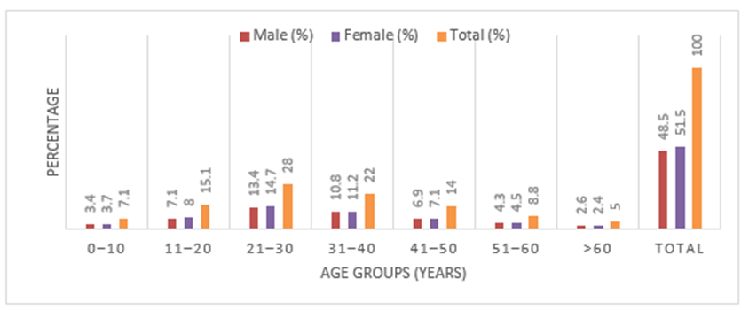
Figure 1: Age and Gender Distribution of Study Participants (N = 464)
Table 2: FNAC Adequacy Rate* and Diagnostic Categories (N = 464)
|
FNAC Diagnostic Category |
Number of Cases (n) |
Percentage (%) |
|
Inflammatory |
210 |
45.3 |
|
Benign Neoplasm |
154 |
33.2 |
|
Malignant |
68 |
14.7 |
|
Inadequate / Non-diagnostic |
32 |
6.9 |
|
Total |
464 |
100.0 |
* Overall FNAC adequacy rate: (210 + 154 + 68) / 464 × 100 = 93.1%
Table 2 demonstrates the distribution of FNAC diagnoses among the 464 cases. Inflammatory lesions constituted the largest diagnostic category, with 210 (45.3%) cases, followed by benign neoplasms in 154 (33.2%) cases. Malignant lesions were identified in 68 (14.7%) cases. In 32 (6.9%) cases, the aspirates were inadequate or non-diagnostic. Accordingly, adequate and diagnostically interpretable aspirates were obtained in 432 of 464 cases, giving an overall FNAC adequacy rate of 93.1%. The diagnostic categories demonstrate that inflammatory and benign lesions accounted for the majority of the FNAC diagnoses, while malignant lesions represented a smaller proportion of the overall study population.
Table 3: FNAC Diagnostic Categories by Anatomical Site (N = 464)
|
Anatomical Site |
Inflammatory n (%) |
Benign Neoplasm n (%) |
Malignant n (%) |
Inadequate n (%) |
Total n |
|
Cervical lymph node |
86 (58.9%) |
38 (26.0%) |
14 (9.6%) |
8 (5.5%) |
146 |
|
Thyroid |
22 (21.6%) |
61 (59.8%) |
13 (12.7%) |
6 (5.9%) |
102 |
|
Parotid region |
16 (43.2%) |
17 (45.9%) |
1 (2.7%) |
3 (8.1%) |
37 |
|
Submandibular region |
18 (40.0%) |
21 (46.7%) |
3 (6.7%) |
3 (6.7%) |
45 |
|
Other superficial head-and-neck sites |
68 (50.7%) |
17 (12.7%) |
37 (27.6%) |
12 (9.0%) |
134 |
|
Total |
210 (45.3%) |
154 (33.2%) |
68 (14.7%) |
32 (6.9%) |
464 (100%) |
Table 3 shows the distribution of FNAC diagnostic categories according to anatomical site. Cervical lymph-node lesions were predominantly inflammatory, with 86 (58.9%) cases, followed by benign neoplasms in 38 (26.0%) and malignant lesions in 14 (9.6%) cases. Among thyroid lesions, benign neoplasms predominated, accounting for 61 (59.8%) cases, followed by malignant lesions in 13 (12.7%) and inflammatory lesions in 22 (21.6%) cases. In the parotid region, benign neoplasms constituted the largest category with 17 (45.9%) cases, closely followed by inflammatory lesions with 16 (43.2%) cases. Similarly, submandibular lesions showed a predominance of benign neoplasms, accounting for 21 (46.7%) cases, followed by inflammatory lesions in 18 (40.0%) cases. Among other superficial head-and-neck sites, inflammatory lesions were predominant, comprising 68 (50.7%) cases, while malignant lesions accounted for 37 (27.6%) cases. Overall, the findings demonstrate marked variation in the cytological diagnostic pattern according to the anatomical site of the swelling.
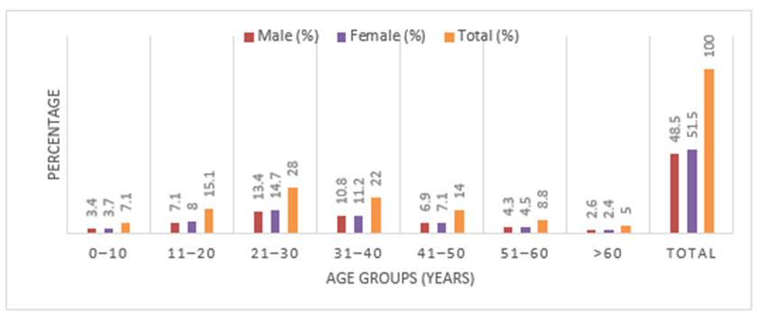
Figure 2: Anatomical Site Distribution of Swellings (N = 464)
DISCUSSION:
In the present retrospective study, the 21–30-year age group was predominant, accounting for 28.0% of the study population, followed by the 31–40-year group (22.0%). A slight female predominance was observed, with females comprising 51.5% and males 48.5% of cases. Cervical lymph nodes were the most frequent anatomical site (31.5%), followed by other superficial head-and-neck sites (28.9%) and thyroid lesions (22.0%). Shah and Gandhi reported a similar predominance of the 21–30-year age group (31%) and female predominance (59%), with lymph nodes being the commonest site (43%), followed by thyroid lesions (31%). (2) Nyamgoudar et al. also reported a substantial representation of young adults and lymph-node lesions in their FNAC series. (14) The predominance of young and middle-aged patients in the present study may reflect the common occurrence of benign, inflammatory, and lymph-node-related swellings in these age groups. The observed anatomical distribution further demonstrates the important contribution of cervical lymph nodes and thyroid lesions to routine head-and-neck FNAC practice.
The present study showed that 244 (52.6%) patients had swelling for 1–6 months, while 110 (23.7%) had a duration of less than one month and an equal number had swellings persisting for more than six months. Thus, 76.3% of the recorded cases had a swelling duration of six months or less at the time of FNAC. Sahni et al. emphasized the importance of duration of head-and-neck swelling in establishing the differential diagnosis and correlating clinical findings with cytological assessment. (6) The variation in duration observed in the present study may reflect differences in the nature of lesions, progression of disease, awareness, and timing of presentation to healthcare services. A prolonged duration does not necessarily indicate malignancy, as several benign and inflammatory lesions may persist for extended periods. Therefore, duration should be interpreted together with anatomical site and cytological findings during FNAC evaluation.
In the present study, inflammatory lesions constituted the largest FNAC diagnostic category, accounting for 210 (45.3%) cases, followed by benign neoplasms in 154 (33.2%) and malignant lesions in 68 (14.7%) cases. Only 32 (6.9%) aspirates were inadequate or non-diagnostic, giving an overall FNAC adequacy rate of 93.1%. Thus, inflammatory and benign lesions together constituted 78.5% of all recorded cases, demonstrating the predominance of non-malignant pathology among superficial head-and-neck swellings. Thapa et al. reported that benign and non-neoplastic lesions constituted 88.1% of head-and-neck FNAC cases, while malignant lesions accounted for 10.8%. (17) Sahni et al. similarly reported inflammatory lesions as an important component of head-and-neck masses. (6) Akinmoladun et al. also demonstrated the usefulness of FNAC in characterizing a broad spectrum of head-and-neck lesions. (12) The high adequacy rate observed in the present study supports the practical utility of FNAC as a minimally invasive first-line investigation for superficial head-and-neck swellings.
Considerable variation was observed in the FNAC diagnostic pattern according to anatomical site. Cervical lymph-node lesions were predominantly inflammatory, with 86 (58.9%) cases, followed by benign neoplasms (26.0%) and malignant lesions (9.6%). Qader et al. highlighted the diagnostic usefulness of FNAC in cervical lymphadenopathy, particularly in differentiating reactive and malignant lesions. (7) Ton Eryilmaz et al. also demonstrated the diagnostic value of lymph-node FNAC. (8) Thyroid lesions in the present study were predominantly benign neoplasms (59.8%), while malignant lesions accounted for 12.7%. Similar findings regarding the usefulness of FNAC in thyroid swellings have been reported by Bhise et al. and Abdullahi et al. (9,10) Parotid and submandibular lesions also showed a predominance of benign neoplasms, accounting for 45.9% and 46.7%, respectively. This pattern is consistent with the established role of FNAC in the evaluation of salivary-gland lesions, as reported by Kasinathan et al. and Rameeza and Hemalata. (3,11) Other superficial head-and-neck sites showed predominantly inflammatory lesions (50.7%), while malignant lesions accounted for 27.6%. The site-specific differences observed in the present study emphasize the importance of anatomical location in interpreting FNAC findings and recognizing the expected diagnostic spectrum of head-and-neck lesions.
CONCLUSION:
This retrospective study demonstrates that FNAC is a useful, minimally invasive, and practical first-line diagnostic tool for evaluating palpable head-and-neck swellings. Inflammatory lesions were the predominant diagnostic category (45.3%), followed by benign neoplasms (33.2%) and malignant lesions (14.7%), with a high overall FNAC adequacy rate of 93.1%. Cervical lymph nodes were the most frequently involved anatomical site, followed by other superficial head-and-neck sites and thyroid lesions. The diagnostic pattern varied considerably according to anatomical site, highlighting the importance of site-specific cytomorphological assessment. Overall, the findings support the continued use of FNAC as an accessible and effective initial investigation for the evaluation and diagnostic characterization of superficial head-and-neck lesions in routine pathology practice.
REFERENCES:
1. Bayrak BY, Paksoy N. Diagnostic accuracy of fine-needle aspiration cytology for extrathyroidal head-and-neck lesions performed by a cytopathologist with the assistance of radiologist: A single-center study. Cytojournal. 2025 Jun 2;22:57. doi: 10.25259/Cytojournal_247_2024. PMID: 40708833; PMCID: PMC12289109. https://pmc.ncbi.nlm.nih.gov/articles/PMC12289109/
2. Shah Y, Gandhi S. A Prospective Study of Comparison Between Fine Needle Aspiration Cytology (FNAC) with Histopathological Diagnosis (HPE) in Neck Masses. Indian J Otolaryngol Head Neck Surg. 2023 Sep;75(3):1447-1453. doi: 10.1007/s12070-022-03461-y. Epub 2023 Feb 25. PMID: 37636617; PMCID: PMC10447841. https://pmc.ncbi.nlm.nih.gov/articles/PMC10447841/
3. Kasinathan B, Manohar B, Ganapathy H. Diagnostic Accuracy of Fine Needle Aspiration Cytology (FNAC) in Salivary Gland Lesions with Histopathological Examination (HPE) Correlation in a Tertiary Care Centre in Southern India. Indian J Otolaryngol Head Neck Surg. 2023 Jun;75(2):871-879. doi: 10.1007/s12070-023-03550-6. Epub 2023 Feb 10. PMID: 37275112; PMCID: PMC10234911. https://pubmed.ncbi.nlm.nih.gov/37275112/
4. S JD, K BA, G VA. Study of fine needle aspiration cytology of palpable head and neck lesions in tertiary care centre [Internet]. Indian J Pathol Oncol. 2018 [cited 2026 Aug 15];5(3):375-381. Available from: https://ijpo.co.in/archive/volume/5/issue/3/article/17715
5. Chandra Prakash Kumawat, Yogesh Kumar Gupta, Archana Singh. Cytological Spectrum of Lesions in Neck Swelling with Histopathological Correlation in RUHS Medical College and Hospital. International Journal of Pharmaceutical and Clinical Research. 2022Nov30;14(11):282–7. doi:10.5281/zenodo.13267561 https://zenodo.org/records/13267561
6. Sahni D, Kaur A, Bansal A, Chawla S, Bedi N, Sahni P. A prospective study to assess clinicopathological correlation of head and neck masses. Int J Community Med Public Health [Internet]. 2024 Jul. 30 [cited 2026 Aug. 15];11(8):3107-12. Available from: https://www.ijcmph.com/index.php/ijcmph/article/view/12678
7. Bhise SV, Shaikh A, Hippargekar PM, Kothule S. A Prospective Study of Ultrasonographic and FNAC Correlation of Thyroid Swellings with Histopathology. Indian J Otolaryngol Head Neck Surg. 2022 Oct;74(Suppl 2):1942-1948. doi: 10.1007/s12070-020-01922-w. Epub 2020 Jun 24. PMID: 36452634; PMCID: PMC9702162. https://pubmed.ncbi.nlm.nih.gov/36452634/
8. Abdullahi IM, Yasin NA, Dirken ES, Mohamoud AM, Guler I, Adani AA. Comparative study of fine needle aspiration cytology and histopathology in thyroid nodules at a tertiary care hospital: First report from Somalia. Asian J Surg. 2023 Oct;46(10):4202-4207. doi: 10.1016/j.asjsur.2022.11.097. Epub 2022 Dec 8. PMID: 36504151. https://pubmed.ncbi.nlm.nih.gov/36504151/
9. Rameeza A, Hemalata M. Fine-needle aspiration cytology of salivary gland lesions. J Oral Maxillofac Pathol. 2022 Jan-Mar;26(1):52-56. doi: 10.4103/jomfp.jomfp_496_20. Epub 2022 Mar 31. PMID: 35571296; PMCID: PMC9106261. https://pmc.ncbi.nlm.nih.gov/articles/PMC9106261/
10. Qader K, Qader M, Mubarak A, Alaradi K. Diagnostic Accuracy of Fine-Needle Aspiration Cytology in Cervical Lymphadenopathies at a Tertiary Care Center in the Kingdom of Bahrain. Cureus. 2024 Jun 11;16(6):e62150. doi: 10.7759/cureus.62150. PMID: 38993430; PMCID: PMC11238613. https://pmc.ncbi.nlm.nih.gov/articles/PMC11238613/
11. Ton Eryilmaz O, Ucak R, Ozagari AA, Kabukcuoglu F. Diagnostic value of lymph node fine-needle aspiration cytology. Cytojournal. 2021 Apr 12;18:8. doi: 10.25259/Cytojournal_1_2020. PMID: 34221097; PMCID: PMC8248011. https://pubmed.ncbi.nlm.nih.gov/34221097/
12. Akinmoladun VI, Gbolahan OO, Aladelusi TO, Ogun GO, Ajani MA. Diagnostic Accuracy of Fine-Needle Aspiration Cytology in Head and Neck Lesions from a Tertiary Health Facility in Southwestern Nigeria. Niger Med J. 2020 Nov-Dec;61(6):303-306. doi: 10.4103/nmj.NMJ_65_20. Epub 2020 Dec 19. PMID: 33888925; PMCID: PMC8040946. https://pmc.ncbi.nlm.nih.gov/articles/PMC8040946/
13. Wadone MM, Masgal MM, Anita AM, Rajesh PS. A Study of Cytomorphological Spectrum of Head and Neck Lesions in Pediatric Age Group. J Cytol. 2023 Apr-Jun;40(2):75-80. doi: 10.4103/joc.joc_93_22. Epub 2023 May 22. PMID: 37388403; PMCID: PMC10305904. https://pmc.ncbi.nlm.nih.gov/articles/PMC10305904/
14. Nyamgoudar S, Mugni A, BH R. Spectrum of Head and Neck lesions diagnosed by Fine needle aspiration cytology. European Journal of Cardiovascular Medicine. 2024 Apr 1;14(2):386. https://openurl.ebsco.com/EPDB%3Agcd%3A2%3A25822274/detailv2?sid=ebsco%3Aplink%3Acrawlergcd&id=ebsco%3Agcd%3A177979223&crl=c&jrnl=20424884&link_origin=scholar.google.com
15. Meher Ullah M, Ahmed J, Mondal SR, Aman A, Qurashi SA, Alam ABMK. Comparison of fine needle aspiration cytology and histopathology in neck mass diagnosis. Int Surg J [Internet]. 2025 Jun. 25 [cited 2026 Aug. 15];12(7):1085-91. Available from: https://www.ijsurgery.com/index.php/isj/article/view/11203
16. Dr. karthikeyan, Dr. pratheep karthick, Dr. Jahid Husain S, Dr. Radhakrishnan nair, Prof. Dr. senguttuvan. “Analysis Of Correlation Of Fine Needle Aspiration Cytology With Histopathology Finding In Thyroid Lesion”. SEEJPH [Internet]. 2025 Jan. 25 [cited 2026 Aug. 15];:4421-7. Available from: https://seejph.com/index.php/seejph/article/view/6797
17. Thapa S, Ghosh A, Ghartmagar D, Regmi S, Talwar O. Utility of fine needle aspiration cytology in head and neck lesions and its correlation with histopathology- a study in tertiary care hospital. J Pathol Nep [Internet]. 2020 Sep. 30 [cited 2026 Aug. 15];10(2):1733-41. Available from: https://www.nepjol.info/index.php/JPN/article/view/30255