Peripheral Vertigo Through a Biochemical and Electrophysiological Lens: A Clinical Study.
- Aberna Govarthanaraj , Associate professor, Department of ENT, Madha Medical College and Research Institute, Kovur, Chennai - 600128. Tamil Nadu, India.
- Prathish Kumar , Associate professor, Department of General medicine, Madha Medical College and Research Institute, Kovur, Chennai - 600128. Tamil Nadu, India.
- Ranjana Kumari , Professor and Head, Department of ENT, Madha Medical College and Research Institute, Kovur, Chennai - 600128. Tamil Nadu, India.
Article Information:
Abstract:
Background: Peripheral vertigo is a common presentation in otorhinolaryngology practice. Calcium homeostasis governs otoconial integrity, and thyroid hormones influence inner-ear metabolism and neural conduction, yet these biochemical factors and objective otolith testing are seldom evaluated together. Objectives: To assess the association between serum calcium levels and vestibular evoked myogenic potential (VEMP) parameters, and between thyroid function parameters and VEMP parameters, in patients with peripheral vertigo. Methods: A hospital-based cross-sectional study was conducted on 160 patients aged 18–80 years with clinically diagnosed peripheral vertigo attending the ENT outpatient department. All underwent history, bedside vestibular examination, serum total calcium, thyroid profile (TSH, free T3, free T4), and cervical and ocular VEMP. Pearson correlation, chi-square test and multiple linear regression were applied; p < 0.05 was significant. Results: Mean age was 45.0 ± 11.7 years and 92 patients (57.5%) were women. Benign paroxysmal positional vertigo (BPPV) was the commonest diagnosis (92; 57.5%). Hypocalcaemia was present in 54 (33.8%), hypothyroidism in 35 (21.9%) and hyperthyroidism in 8 (5.0%). VEMP was abnormal in 71 (44.4%). Serum calcium correlated positively with cVEMP P13–N23 amplitude (r = +0.337) and negatively with P13 latency (r = -0.361) (both p < 0.001). TSH correlated negatively with cVEMP amplitude (r = -0.321) and positively with P13 latency (r = +0.395) (both p < 0.001). Abnormal VEMP occurred in 37/54 (68.5%) hypocalcaemic versus 34/106 (32.1%) normocalcaemic patients (χ² = 19.25, p < 0.001), and in 28/43 (65.1%) with thyroid dysfunction versus 43/117 (36.8%) euthyroid patients (χ² = 10.25, p = 0.001). On regression, calcium (β = 14.04) and TSH (β = -1.97) independently predicted cVEMP amplitude (adjusted R² = 0.198). Conclusion: Lower serum calcium and higher TSH were independently associated with reduced VEMP amplitude and prolonged latency. Serum calcium and thyroid profile, combined with VEMP, add objective value to the evaluation of peripheral vertigo and identify potentially correctable metabolic and endocrine contributors.
Keywords:
Article :
INTRODUCTION:
Vertigo is among the commonest symptoms encountered in otorhinolaryngology practice, and the majority of cases arise from disorders of the peripheral vestibular apparatus. In a population-based neurotological survey, benign paroxysmal positional vertigo (BPPV) alone accounted for 8% of individuals with moderate or severe dizziness, with a lifetime prevalence of 2.4% and a one-year incidence of 0.6%.¹ Peripheral vertigo encompasses BPPV, Ménière's disease, vestibular neuritis and labyrinthitis, and although the diagnosis is largely clinical, positional testing and bedside examination alone cannot quantify the degree of otolith dysfunction or identify a systemic cause.²
Calcium homeostasis is central to vestibular physiology. The otoconia of the utricle and saccule are calcium carbonate crystals embedded in a protein matrix, and their formation, maintenance and resorption depend on tightly regulated endolymphatic calcium concentration; disturbance of this equilibrium promotes otoconial degeneration and dislodgement, the accepted substrate of BPPV.³ Consistent with this, reduced bone mineral density and low vitamin D — the principal regulator of calcium metabolism — have repeatedly been demonstrated in patients with idiopathic BPPV.⁴ Thyroid hormones exert a parallel influence on the inner ear by modulating endolymphatic homeostasis, cochleovestibular microcirculation and neural conduction, and by interacting with parathyroid hormone and calcium metabolism; hypothyroidism has accordingly been linked with an increased risk of recurrent peripheral vertigo.⁵
Vestibular evoked myogenic potentials (VEMP) provide the only widely available objective measure of otolith function. Cervical VEMP (cVEMP) assesses the saccule and inferior vestibular nerve through the vestibulocollic reflex, while ocular VEMP (oVEMP) assesses the utricle and superior vestibular nerve; prolonged P13 and N23 latencies, reduced P13–N23 amplitude, interaural asymmetry or an absent response indicate otolith or vestibular nerve dysfunction, and may be demonstrable before the bedside examination becomes abnormal.⁶
Although serum calcium, thyroid dysfunction and VEMP abnormalities have each been studied in isolation, few studies have examined them together in the same cohort, and the extent to which biochemical derangement is reflected in measurable otolith dysfunction remains undefined. The present study was therefore undertaken to evaluate the correlation between serum calcium levels, thyroid function tests and VEMP parameters in patients presenting with peripheral vertigo, with the specific objectives of assessing the association between serum calcium and VEMP parameters (P13 and N23 latencies and P13–N23 amplitude), and of determining the association between thyroid function parameters (TSH, free T3 and free T4) and VEMP parameters, so that a combined biochemical and electrophysiological approach to the evaluation of peripheral vertigo can be appraised.
MATERIALS AND METHODS:
This hospital-based cross-sectional study was conducted in the Department of Otorhinolaryngology, Madha Medical College and Research Institute, Chennai, over 18 months, after Institutional Ethics Committee approval and in accordance with the Declaration of Helsinki and the ICMR National Ethical Guidelines (2017); written informed consent was obtained from every participant.
Consecutive patients aged 18–80 years attending the outpatient department with giddiness, in whom peripheral vertigo was diagnosed clinically, were enrolled. Patients with central vertigo, previous ear surgery, chronic otitis media, known metabolic bone disease, those on calcium supplementation, and those with severe systemic or neurological illness were excluded. BPPV, Ménière's disease and vestibular neuritis were diagnosed according to established criteria.⁷⁻⁹ The sample size of 160 was derived, patients were recruited by consecutive sampling.
Each participant underwent a structured history and general, otological and vestibular examination including spontaneous and gaze-induced nystagmus, head impulse test, Dix–Hallpike manoeuvre, supine head roll, Romberg and Unterberger tests.
Venous samples were analysed on standard automated analysers for total serum calcium (normal 8.6–10.2 mg/dL) and thyroid profile (TSH 0.4–4.0 mIU/L, free T3 2.3–4.2 pg/mL, free T4 0.8–1.8 ng/dL). Hypothyroidism was defined as a raised TSH with low or low-normal free T4, and hyperthyroidism as a suppressed TSH with elevated free T4 and/or free T3.
VEMP was recorded in a sound-treated room with inter-electrode impedance below 5 kΩ using the click-evoked technique.¹⁰ For cVEMP, electrodes were placed over the upper third of each sternocleidomastoid with the head turned contralaterally to maintain tonic muscle activation; for oVEMP, on the infraorbital margin with sustained upgaze. Each ear was stimulated separately and responses were averaged over repeated trials to confirm reproducibility.
P13 and N23 latencies with P13–N23 amplitude (cVEMP), N10 and P15 latencies with N10–P15 amplitude (oVEMP), and the interaural asymmetry ratio, |R – L|/(R + L) × 100, were recorded. A cVEMP was considered abnormal if the P13 latency exceeded 15.2 ms, N23 exceeded 25.0 ms, amplitude was below 45 µV, asymmetry exceeded 32%, or the response was absent; the oVEMP criteria were N10 above 12.0 ms, P15 above 18.0 ms, amplitude below 2.6 µV, asymmetry above 32%, or an absent response. The affected ear was used for analysis, the more affected ear being taken in bilateral disease.
Data were analysed using SPSS version 26.0. Continuous variables are expressed as mean ± standard deviation or median with interquartile range and categorical variables as frequencies and percentages. A p-value below 0.05 was considered statistically significant.
RESULTS:
A total of 160 patients with peripheral vertigo were studied. The mean age was 45.0 ± 11.7 years (range 18–72 years) and 92 patients (57.5%) were women.
Table 1. Demographic and clinical profile of the study population (n = 160)
|
Variable |
Frequency (n) |
Percentage (%) |
|
Age group (years) |
|
|
|
18-30 |
16 |
10.0 |
|
31-40 |
35 |
21.9 |
|
41-50 |
60 |
37.5 |
|
51-60 |
32 |
20.0 |
|
>60 |
17 |
10.6 |
|
Sex |
|
|
|
Female |
92 |
57.5 |
|
Male |
68 |
42.5 |
|
Clinical diagnosis |
|
|
|
BPPV |
92 |
57.5 |
|
Ménière's disease |
28 |
17.5 |
|
Vestibular neuritis |
24 |
15.0 |
|
Labyrinthitis |
16 |
10.0 |
|
Side involved |
|
|
|
Right |
58 |
36.2 |
|
Left |
81 |
50.6 |
|
Bilateral |
21 |
13.1 |
|
Duration of symptoms |
|
|
|
< 1 week |
55 |
34.4 |
|
1 week-1 month |
53 |
33.1 |
|
1-6 months |
32 |
20.0 |
|
> 6 months |
20 |
12.5 |
|
Episode frequency |
|
|
|
First episode |
64 |
40.0 |
|
Recurrent |
96 |
60.0 |
|
Associated symptoms |
|
|
|
Hearing loss |
51 |
31.9 |
|
Tinnitus |
57 |
35.6 |
|
Aural fullness |
27 |
16.9 |
|
Nausea / vomiting |
119 |
74.4 |
|
Comorbidity |
|
|
|
Hypertension |
61 |
38.1 |
|
Diabetes mellitus |
41 |
25.6 |
|
Known thyroid disease |
28 |
17.5 |
Most patients were in the 41–50 year age group (60; 37.5%) and women outnumbered men. BPPV was the commonest diagnosis (92; 57.5%), followed by Ménière's disease, vestibular neuritis and labyrinthitis. Symptoms were recurrent in 96 patients (60.0%). Only 28 patients were aware of thyroid disease before enrolment.
Table 2. Biochemical and VEMP parameters across the four diagnostic groups (mean ± SD)
|
Parameter |
BPPV |
Ménière's |
Vest. neuritis |
Labyrinthitis |
F |
p |
|
Serum calcium (mg/dL) |
8.72 ± 0.46 |
8.99 ± 0.52 |
9.17 ± 0.55 |
9.19 ± 0.48 |
8.58 |
<0.001 |
|
TSH (mIU/L) |
4.04 ± 3.77 |
3.75 ± 4.10 |
3.45 ± 3.53 |
4.08 ± 4.06 |
0.18 |
0.909 |
|
Free T3 (pg/mL) |
3.18 ± 1.08 |
2.92 ± 0.76 |
3.10 ± 0.99 |
3.21 ± 1.18 |
0.52 |
0.667 |
|
Free T4 (ng/dL) |
1.28 ± 0.50 |
1.16 ± 0.32 |
1.25 ± 0.48 |
1.17 ± 0.61 |
0.63 |
0.599 |
|
cVEMP P13 latency (ms) |
13.92 ± 1.06 |
14.19 ± 0.96 |
13.90 ± 1.30 |
14.23 ± 1.12 |
0.75 |
0.525 |
|
cVEMP N23 latency (ms) |
23.43 ± 1.55 |
23.87 ± 1.48 |
23.66 ± 1.92 |
23.85 ± 2.00 |
0.67 |
0.569 |
|
cVEMP P13–N23 amplitude (µV) |
66.4 ± 23.2 |
55.4 ± 26.1 |
65.7 ± 30.5 |
70.6 ± 21.8 |
1.64 |
0.184 |
|
oVEMP N10 latency (ms) |
10.62 ± 0.91 |
11.00 ± 1.02 |
10.68 ± 0.95 |
10.59 ± 0.89 |
1.23 |
0.302 |
|
oVEMP P15 latency (ms) |
15.72 ± 1.33 |
16.11 ± 1.45 |
15.79 ± 1.31 |
15.56 ± 1.22 |
0.77 |
0.513 |
|
oVEMP N10–P15 amplitude (µV) |
4.18 ± 1.51 |
3.74 ± 1.65 |
4.12 ± 1.84 |
3.76 ± 1.88 |
0.71 |
0.550 |
Serum calcium was the only parameter that differed significantly between the four diagnoses (p < 0.001). It was lowest in BPPV (8.72 mg/dL) and highest in labyrinthitis (9.19 mg/dL), and patients with BPPV had significantly lower calcium than all other patients combined (p < 0.001). Thyroid values and VEMP values were similar across the four groups.
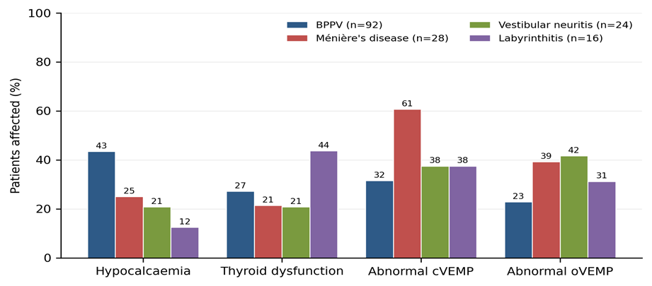
Figure 1. Prevalence of biochemical and VEMP abnormalities within each diagnostic group
Hypocalcaemia was commonest in BPPV (43.5%) and least common in labyrinthitis (12.5%); the difference between BPPV and the other diagnoses was significant (p = 0.002). Abnormal cVEMP was seen most often in Ménière's disease (60.7%) and abnormal oVEMP in vestibular neuritis (41.7%). Thyroid dysfunction occurred in all four groups without any clear preference.
Table 3. Correlation of serum calcium with VEMP parameters (Pearson correlation)
|
VEMP parameter |
n |
r |
95% CI |
p value |
|
cVEMP P13 latency |
155 |
-0.361 |
-0.49 to -0.22 |
<0.001 |
|
cVEMP N23 latency |
155 |
-0.316 |
-0.45 to -0.17 |
<0.001 |
|
cVEMP P13–N23 amplitude |
155 |
+0.337 |
+0.19 to +0.47 |
<0.001 |
|
oVEMP N10 latency |
156 |
-0.317 |
-0.45 to -0.17 |
<0.001 |
|
oVEMP P15 latency |
156 |
-0.360 |
-0.49 to -0.22 |
<0.001 |
|
oVEMP N10–P15 amplitude |
156 |
+0.348 |
+0.20 to +0.48 |
<0.001 |
Serum calcium was significantly correlated with every VEMP parameter (all p < 0.001). The higher the calcium, the larger the cVEMP and oVEMP amplitudes and the shorter the latencies. Patients with an abnormal VEMP had lower mean calcium than those with a normal VEMP (8.70 versus 9.03 mg/dL, p < 0.001).
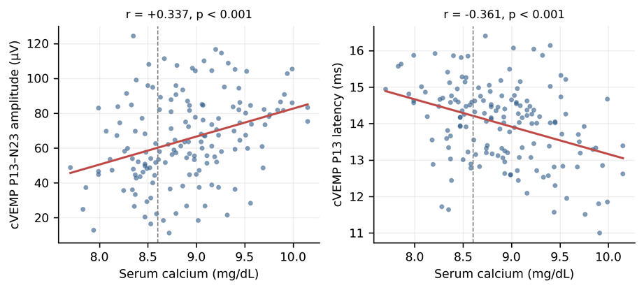
Figure 2. Serum calcium plotted against cVEMP P13–N23 amplitude (left) and cVEMP P13 latency (right); the dashed line marks the lower limit of normal calcium (8.6 mg/dL)
Both plots show a straight-line relationship. cVEMP amplitude increased by about 14 µV for every 1 mg/dL rise in serum calcium, and the P13 latency became correspondingly shorter. Most patients lying below the 8.6 mg/dL line had small amplitudes and long latencies, although the scatter shows that calcium is only one of several factors affecting otolith function.
Table 4. Correlation of thyroid function parameters with VEMP parameters (Pearson correlation)
|
Thyroid parameter |
VEMP parameter |
n |
r |
95% CI |
p value |
|
TSH |
cVEMP P13 latency |
155 |
+0.395 |
+0.25 to +0.52 |
<0.001 |
|
|
|
|
|
|
|
|
TSH |
cVEMP N23 latency |
155 |
+0.406 |
+0.27 to +0.53 |
<0.001 |
|
TSH |
cVEMP P13–N23 amplitude |
155 |
-0.321 |
-0.46 to -0.17 |
<0.001 |
|
TSH |
oVEMP N10 latency |
156 |
+0.308 |
+0.16 to +0.44 |
<0.001 |
|
TSH |
oVEMP P15 latency |
156 |
+0.265 |
+0.11 to +0.41 |
<0.001 |
|
TSH |
oVEMP N10–P15 amplitude |
156 |
-0.359 |
-0.49 to -0.21 |
<0.001 |
|
Free T3 |
cVEMP P13 latency |
155 |
-0.350 |
-0.48 to -0.20 |
<0.001 |
|
Free T3 |
cVEMP N23 latency |
155 |
-0.277 |
-0.42 to -0.12 |
<0.001 |
|
Free T3 |
cVEMP P13–N23 amplitude |
155 |
+0.325 |
+0.18 to +0.46 |
<0.001 |
|
Free T3 |
oVEMP N10 latency |
156 |
-0.246 |
-0.39 to -0.09 |
0.002 |
|
Free T3 |
oVEMP P15 latency |
156 |
-0.242 |
-0.38 to -0.09 |
0.002 |
|
Free T3 |
oVEMP N10–P15 amplitude |
156 |
+0.256 |
+0.10 to +0.40 |
0.001 |
|
Free T4 |
cVEMP P13 latency |
155 |
-0.353 |
-0.48 to -0.21 |
<0.001 |
|
Free T4 |
cVEMP N23 latency |
155 |
-0.315 |
-0.45 to -0.17 |
<0.001 |
|
Free T4 |
cVEMP P13–N23 amplitude |
155 |
+0.360 |
+0.21 to +0.49 |
<0.001 |
|
Free T4 |
oVEMP N10 latency |
156 |
-0.323 |
-0.46 to -0.18 |
<0.001 |
|
Free T4 |
oVEMP P15 latency |
156 |
-0.314 |
-0.45 to -0.17 |
<0.001 |
|
Free T4 |
oVEMP N10–P15 amplitude |
156 |
+0.278 |
+0.13 to +0.42 |
<0.001 |
All three thyroid parameters were significantly correlated with VEMP. A higher TSH went with smaller amplitudes and longer latencies, while a higher free T3 and free T4 went the opposite way. This mirror-image pattern suggests that it is the reduced availability of thyroid hormone that matters. TSH was higher in patients with an abnormal VEMP (3.30 versus 2.16 mIU/L, p < 0.001).
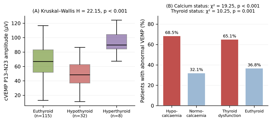
Figure 3. (A) cVEMP P13–N23 amplitude by thyroid status. (B) Proportion of patients with an abnormal VEMP according to calcium status and thyroid status
Panel A shows that the hypothyroid group had the smallest cVEMP amplitude (p < 0.001); only 8 patients were hyperthyroid, too few to interpret, so the finding reflects hypothyroidism. Panel B shows that an abnormal VEMP was about twice as common in hypocalcaemic as in normocalcaemic patients (68.5% versus 32.1%, p < 0.001), and in patients with thyroid dysfunction compared with euthyroid patients (65.1% versus 36.8%, p = 0.001).
On multiple linear regression, serum calcium and TSH each predicted cVEMP amplitude independently of age and sex (both p < 0.001). Age had a small effect (p = 0.038) and sex had none (p = 0.819). Together the four variables explained 21.9% of the variation in cVEMP amplitude (adjusted R² = 0.198, p < 0.001).
DISCUSSION:
This study evaluated, in a single cohort of 160 patients with peripheral vertigo, the relationship between serum calcium, thyroid function and objectively measured otolith function. Both study objectives were met: serum calcium and thyroid parameters were each significantly correlated with cervical and ocular VEMP responses, and both remained independent predictors of VEMP amplitude after adjustment.
The demographic profile of our cohort accords with published series. The mean age of 45.0 years and the female preponderance (57.5%) mirror the age and sex distribution reported by von Brevern et al., who found BPPV to be commonest in middle age with a female-to-male ratio of approximately 2:1.¹ BPPV constituted 57.5% of our peripheral vertigo cases, in keeping with its recognised position as the single commonest cause of peripheral vestibular vertigo.²
With regard to the first objective, we found hypocalcaemia in 54 patients (33.8%) of the whole cohort and in 40/92 (43.5%) of patients with BPPV, significantly more than the 14/68 (20.6%) seen in other peripheral vestibular disorders (p = 0.002), with the lowest mean calcium in the BPPV group (8.72 ± 0.46 mg/dL). This is directionally concordant with Bener et al., who in 177 BPPV patients and 656 controls reported significantly lower serum calcium in cases (1.87 ± 0.90 versus 2.00 ± 0.91 mmol/L, p = 0.004) together with lower 25-hydroxy vitamin D (19.04 ± 8.37 versus 21.19 ± 9.00 ng/mL, p = 0.004) and vitamin D deficiency in 60.5% of cases versus 47.3% of controls (p = 0.007).¹¹
Our findings are, however, at variance with Thomas et al., who in a smaller case-control study of 49 patients and 49 controls found mean serum calcium to be marginally higher in cases than controls (9.34 ± 0.55 versus 8.96 ± 0.82 mg/dL) with no significant difference (p = 0.976), although they did demonstrate a significant negative correlation between vitamin D and the number of BPPV episodes (p = 0.012).¹² The discrepancy is most plausibly explained by sample size, by differences in the nutritional and sunlight-exposure profile of the populations studied, and by the fact that total rather than ionised calcium was measured in both studies.
Supporting evidence for a calcium-dependent mechanism comes from Jeong et al., who found serum 25-hydroxy vitamin D to be significantly lower in 100 patients with idiopathic BPPV than in 192 controls (14.4 ± 8.4 versus 19.1 ± 6.8 ng/mL, p = 0.001), with levels below 20 ng/mL in 80.0% versus 60.1% (p < 0.001),⁴ and from the same group's randomised trial in which vitamin D and calcium supplementation reduced the annual recurrence rate of BPPV from 1.10 to 0.83 recurrences per person-year (incidence rate ratio 0.76, 95% CI 0.66–0.87).¹³ Vibert et al. had earlier demonstrated osteopenia or osteoporosis in 24 of 32 women with BPPV (75%), with significantly lower T-scores than controls (p < 0.026), linking systemic calcium handling to otoconial pathology.¹⁴
The novel contribution of the present study is that this biochemical disturbance was mirrored in objective otolith testing: calcium correlated with cVEMP amplitude (r = +0.337) and inversely with P13 latency (r = -0.361), and abnormal VEMP was more than twice as common in hypocalcaemic patients, 37/54 (68.5%), as in normocalcaemic patients, 34/106 (32.1%) (p < 0.001). This is consistent with the recent work of Zhou et al., who studied 291 elderly patients with idiopathic unilateral BPPV and found osteoporosis in 27.8% and osteopenia in 41.2%; patients with osteoporosis were significantly more likely to show at least unilateral absence of oVEMP (74.1% versus 57.1%, p = 0.008), and osteoporosis remained independently associated with oVEMP absence after adjustment (OR 2.038, p = 0.019), while no such association was seen with cVEMP (p = 0.405).¹⁵ Our cohort showed abnormality of both modalities, which may reflect the younger mean age of our patients and our use of graded latency and amplitude criteria rather than absence of response alone.
With regard to the second objective, thyroid dysfunction was present in 43 patients (26.9%), hypothyroidism accounting for 35 (21.9%). This closely matches Tricarico et al., who studied 797 patients with idiopathic BPPV and found hypothyroidism on hormone replacement in 61 of 250 patients with recurrence (24.4%) compared with 79 of 547 without recurrence (14.4%, p = 0.0006), concluding that hypothyroidism increases the risk of BPPV recurrence, particularly with positive thyroid antibodies.⁵ An association between BPPV and autoimmune chronic thyroiditis had previously been reported by Papi et al.,¹⁶ and Bougerolle et al., in a retrospective review of 422 patients attending for vestibular rehabilitation, showed that hypothyroidism significantly increased the expression of vestibular instability and gait disorders.¹⁷ Our observation that abnormal VEMP occurred in 28/43 (65.1%) patients with thyroid dysfunction versus 43/117 (36.8%) euthyroid patients (p = 0.001) is very close to the findings of Chiarella et al., who documented altered VEMP responses in 52.2% and abnormal caloric responses in 44.7% of 47 euthyroid patients with Hashimoto's thyroiditis, none of whom had a clinical history of vestibular dysfunction.¹⁸ Taken together with our regression analysis, in which TSH independently predicted cVEMP amplitude, these data support a genuine endocrine influence on otolith function rather than a chance association.
The overall VEMP abnormality rate in our cohort (71 (44.4%); cVEMP 61 (38.1%)) is closely comparable with the classical data of Akkuzu et al., who found abnormal VEMP in 30% of affected ears in BPPV and 50% in Ménière's disease, against only 5.9% of control ears (p = 0.012 and p < 0.001 respectively).¹⁹ Our corresponding figures — abnormal cVEMP in 31.5% of BPPV and 60.7% of Ménière's disease — reproduce both the magnitude and the diagnostic ordering of that series. In vestibular neuritis, cVEMP was absent or abnormal in 37.5% of our patients, a lower proportion than the 34% complete absence of p13–n23 reported by Murofushi et al. in 47 patients with acute vestibular neurolabyrinthitis,²⁰ which is expected given that many of our patients were seen beyond the acute phase, when partial recovery of inferior vestibular nerve function may have occurred. The predominance of oVEMP abnormality over cVEMP abnormality in our vestibular neuritis group (41.7% versus 37.5%) is consistent with the recognised predilection of the condition for the superior vestibular nerve.⁹
The mechanistic implication of these findings is coherent. Otoconia are calcium carbonate crystals in continuous exchange with the surrounding endolymph, and a reduction in available calcium favours demineralisation, fragmentation and detachment, reducing the mass loading of the otolithic membrane and hence the amplitude of the myogenic response.³ Thyroid hormone deficiency compounds this by impairing inner-ear microcirculation and slowing neural conduction, which is reflected electrophysiologically as latency prolongation — precisely the pattern observed in our data, where rising TSH tracked with prolonged P13, N23 and N10 latencies. The practical message is that a proportion of patients presenting with peripheral vertigo harbour an inexpensive, readily correctable metabolic or endocrine abnormality, and that VEMP provides an objective means of demonstrating its functional consequence.
SUMMARY
In this cross-sectional study of 160 patients with peripheral vertigo, hypocalcaemia was present in 33.8% and thyroid dysfunction in 26.9%, while VEMP was abnormal in 44.4%. Serum calcium correlated positively with cVEMP and oVEMP amplitudes and negatively with P13, N23, N10 and P15 latencies, and TSH showed the reciprocal pattern, with free T3 and free T4 behaving as expected in the opposite direction; all correlations were significant at p < 0.001. Hypocalcaemia was significantly commoner in BPPV than in other peripheral vestibular disorders, and both hypocalcaemia and thyroid dysfunction approximately doubled the proportion of patients with an abnormal VEMP. Serum calcium and TSH remained independent predictors of cVEMP amplitude after adjustment for age and sex. We therefore conclude that serum calcium estimation and thyroid function testing, interpreted alongside cervical and ocular VEMP, add objective value to the routine evaluation of peripheral vertigo by identifying potentially treatable metabolic and endocrine contributors to otolith dysfunction.
LIMITATIONS
The cross-sectional, single-centre design and absence of healthy controls limit causal inference and comparison with local normative VEMP values.
REFERENCES:
1. von Brevern M, Radtke A, Lezius F, Feldmann M, Ziese T, Lempert T, et al. Epidemiology of benign paroxysmal positional vertigo: a population based study. J Neurol Neurosurg Psychiatry. 2007;78(7):710-5.
2. Kim JS, Zee DS. Clinical practice. Benign paroxysmal positional vertigo. N Engl J Med. 2014;370(12):1138-47.
3. Lundberg YW, Xu Y, Thiessen KD, Kramer KL. Mechanisms of otoconia and otolith development. Dev Dyn. 2015;244(3):239-53.
4. Jeong SH, Kim JS, Shin JW, Kim S, Lee H, Lee AY, et al. Decreased serum vitamin D in idiopathic benign paroxysmal positional vertigo. J Neurol. 2013;260(3):832-8.
5. Tricarico L, Di Cesare T, Galli J, Fetoni AR, Paludetti G, Picciotti PM. Benign paroxysmal positional vertigo: is hypothyroidism a risk factor for recurrence? Acta Otorhinolaryngol Ital. 2022;42(5):465-70.
6. Rosengren SM, Colebatch JG, Young AS, Govender S, Welgampola MS. Vestibular evoked myogenic potentials in practice: methods, pitfalls and clinical applications. Clin Neurophysiol Pract. 2019;4:47-68.
7. Bhattacharyya N, Gubbels SP, Schwartz SR, Edlow JA, El-Kashlan H, Fife T, et al. Clinical practice guideline: benign paroxysmal positional vertigo (update). Otolaryngol Head Neck Surg. 2017;156(3 Suppl):S1-S47.
8. Lopez-Escamez JA, Carey J, Chung WH, Goebel JA, Magnusson M, Mandalà M, et al. Diagnostic criteria for Menière's disease. J Vestib Res. 2015;25(1):1-7.
9. Strupp M, Bisdorff A, Furman J, Hornibrook J, Jahn K, Maire R, et al. Acute unilateral vestibulopathy/vestibular neuritis: diagnostic criteria. J Vestib Res. 2022;32(5):389-406.
10. Colebatch JG, Halmagyi GM, Skuse NF. Myogenic potentials generated by a click-evoked vestibulocollic reflex. J Neurol Neurosurg Psychiatry. 1994;57(2):190-7.
11. Bener A, Erdoğan A, Üstündağ ÜV. The impact of serums calcium 25-hydroxy vitamin D, ferritin, uric acid, and sleeping disorders on benign paroxysmal positional vertigo patients. Audiol Res. 2024;14(4):640-8.
12. Thomas RJ, Goutham MK, Bhat VS, Kamath SD, Aroor R, Bhandary SK. Association of serum calcium and vitamin D with benign paroxysmal positional vertigo. Int Arch Otorhinolaryngol. 2022;26(3):e365-9.
13. Jeong SH, Kim JS, Kim HJ, Choi JY, Koo JW, Choi KD, et al. Prevention of benign paroxysmal positional vertigo with vitamin D supplementation: a randomized trial. Neurology. 2020;95(9):e1117-25.
14. Vibert D, Kompis M, Häusler R. Benign paroxysmal positional vertigo in older women may be related to osteoporosis and osteopenia. Ann Otol Rhinol Laryngol. 2003;112(10):885-9.
15. Zhou CY, Shu L, Wu J, Chen J, Bai YX, Yan R, et al. Osteoporosis correlates with abnormal ocular vestibular evoked myogenic potential in patients with benign paroxysmal positional vertigo. Front Neurol. 2026;17:1785323.
16. Papi G, Corsello SM, Milite MT, Zanni M, Ciardullo AV, Di Donato C, et al. Association between benign paroxysmal positional vertigo and autoimmune chronic thyroiditis. Clin Endocrinol (Oxf). 2009;70(1):169-70.
17. Bougerolle V, El Khiati R, El Ahmadi A, Tighilet B, Besnard S, Chabbert C. Statistical associations between vestibular pathologies and hypothyroidism: a retrospective study. J Clin Med. 2024;13(4):1099.
18. Chiarella G, Tognini S, Nacci A, Sieli R, Costante G, Petrolo C, et al. Vestibular disorders in euthyroid patients with Hashimoto's thyroiditis: role of thyroid autoimmunity. Clin Endocrinol (Oxf). 2014;81(4):600-5.
19. Akkuzu G, Akkuzu B, Ozluoglu LN. Vestibular evoked myogenic potentials in benign paroxysmal positional vertigo and Meniere's disease. Eur Arch Otorhinolaryngol. 2006;263(6):510-7.
20. Murofushi T, Halmagyi GM, Yavor RA, Colebatch JG. Absent vestibular evoked myogenic potentials in vestibular neurolabyrinthitis. An indicator of inferior vestibular nerve involvement? Arch Otolaryngol Head Neck Surg. 1996;122(8):845-8.