Exploring the Cytomorphological Spectrum of Hepatic Space-Occupying Lesions Through Image-Guided FNAC and WHO-Based Reporting: An Observational Study in a Tertiary Care Hospital in Northeast India.
- Shreyoshi Dhar , MD Pathology, Senior Resident, Dept. of Pathology, Agartala Government Medical College & Gobind Vallabh Pant Hospital, Agartala, Tripura, India,
- Tushar Ghosh , MD Pathology, Assistant Professor, Dept. of Pathology, Tripura Santiniketan Medical College, Agartala (Tripura), 799130, India,
- Pallavi Priyadarshini , Post Graduate Trainee, Dept. of Pathology, Agartala Government Medical College & Gobind Ballabh Pant Hospital, Agartala, Tripura, 799006, India,
- Kulashekhar Bhattacharjee , MD Pathology, Associate Professor, Dept. of Pathology, Agartala Government Medical College & Gobind Ballabh Pant Hospital, Agartala (Tripura), 799006,
Article Information:
Abstract:
Background: Fine-needle aspiration cytology (FNAC) is a minimally invasive and cost-effective technique for evaluating focal hepatic lesions. Image-guided FNAC enables accurate targeting of radiologically detected lesions and provides rapid cytological assessment. The World Health Organization (WHO) Reporting System for Liver Cytopathology provides a standardised framework for categorising liver cytology specimens and communicating the risk of malignancy. Objective: To evaluate the cytomorphological spectrum of image-guided FNAC of hepatic space-occupying lesions and classify the cytological diagnoses according to the WHO reporting system for liver cytopathology. Materials and Methods: This cross-sectional observational study was conducted in a tertiary care hospital from April 2025 to March 2026. A total of 60 patients with radiologically confirmed hepatic space-occupying lesions were included using census sampling. Image-guided FNAC was performed, and smears were assessed for adequacy and cytomorphological features. Cases were categorised according to WHO reporting categories: nondiagnostic, benign, atypical, suspicious for malignancy, and malignant. Results: The study demonstrated a broad cytomorphological spectrum of hepatic lesions. Malignant lesions constituted a substantial proportion of cases, with hepatocellular carcinoma and metastatic malignancies being the predominant diagnoses. Among metastatic malignancies, adenocarcinoma was the most frequent subtype, followed by neuroendocrine neoplasms and poorly differentiated carcinoma. Conclusion: Image-guided FNAC is a useful diagnostic modality for hepatic lesions. Application of the WHO reporting system facilitates standardised cytological interpretation and clinically meaningful risk stratification.
Keywords:
Article :
INTRODUCTION:
The liver is a major site for a wide variety of focal and diffuse pathological processes. A hepatic space-occupying lesion (SOL) identified clinically or radiologically may represent a benign lesion, inflammatory process, primary hepatic neoplasm or secondary metastatic malignancy. Distinguishing among these entities is clinically important because their treatment, prognosis, and subsequent diagnostic pathways differ substantially. Fine-needle aspiration cytology (FNAC), particularly when performed under image guidance, provides a minimally invasive way to obtain cellular material for rapid diagnosis and can be especially valuable when a definitive diagnosis is required before initiating systemic therapy.1,2
Primary malignant tumours of the liver include hepatocellular carcinoma (HCC), intrahepatic cholangiocarcinoma and several less common neoplasms. HCC is the predominant primary hepatic malignancy and usually develops in the setting of chronic liver disease or cirrhosis. Hepatitis B and C infection, alcohol-related liver disease and metabolic liver disease constitute important risk factors. Cytological recognition of HCC requires careful assessment because the cytomorphological spectrum varies considerably with the degree of differentiation.3
Metastatic malignancy represents another important component of hepatic SOLs. The liver is a common site of metastatic disease because of its extensive vascular supply and anatomical relationship with the portal and systemic circulations. Gastrointestinal, pancreaticobiliary, breast and lung carcinomas are among the important sources of hepatic metastases. Therefore, the cytological examination of a liver lesion should not be restricted to identifying malignancy; it should also attempt to determine whether the lesion is hepatocellular in origin or represents metastatic disease.4,5
Image-guided FNAC has particular advantages in this setting. Ultrasonography and computed tomography permit precise targeting of focal lesions, including deep-seated and relatively small lesions. Image guidance also permits avoidance of major vascular structures and necrotic areas whenever possible. Several studies have demonstrated that image-guided FNAC can provide a high diagnostic yield in hepatic lesions, with good sensitivity and specificity for distinguishing benign from malignant disease.6,7
The cytological diagnosis of HCC is based on a combination of architectural and cellular features. Tumour cells may occur singly or in cohesive clusters and may demonstrate trabecular, acinar or pseudoglandular arrangements. An increased nuclear-to-cytoplasmic ratio, nuclear enlargement, pleomorphism, prominent nucleoli, atypical naked nuclei, and endothelial cell wrapping may support the diagnosis. Intracytoplasmic bile pigment, although not always present, is an important supportive feature.8,9
Well-differentiated HCC may closely resemble benign hepatocytes, particularly in aspirates obtained from cirrhotic or regenerative nodules. Conversely, poorly differentiated HCC can lose recognisable hepatocellular morphology and may mimic metastatic adenocarcinoma, neuroendocrine neoplasm or other poorly differentiated malignant tumours. Consequently, cytological interpretation must consider the patient's clinical history, radiological findings and, when necessary, ancillary immunocytochemical investigations.8,10
Metastatic adenocarcinoma is frequently encountered in liver aspirates. Cytologically, it may show three-dimensional clusters, acinar or glandular formations, nuclear enlargement, pleomorphism, prominent nucleoli, mucin and necrotic tumour background. In patients with a known extrahepatic primary tumour, the diagnosis may be relatively straightforward; however, in patients presenting with a liver lesion as the initial manifestation of malignancy, identifying the likely primary site may require immunocytochemistry and correlation with imaging.11
Neuroendocrine neoplasms constitute another clinically important metastatic category. Cytological smears may show relatively monotonous cells arranged singly, in loose clusters, rosettes or trabecular formations. The nuclei often demonstrate finely granular chromatin, and nuclear moulding may be present. Because these features may overlap with other poorly differentiated neoplasms, ancillary markers such as synaptophysin, chromogranin and INSM1 may be useful in appropriate cases.12
Specimen adequacy is critical to cytological diagnosis. A technically inadequate specimen can result from insufficient cellularity, sampling of necrotic areas, excessive haemorrhage or inaccurate targeting of the lesion. Barbhuiya et al., in an analysis of 400 liver and gallbladder aspirations, reported 72.2% adequate aspirates, with inconclusive and inadequate samples accounting for 18.7% and 9%, respectively. Metastatic adenocarcinoma was the most frequent positive cytological diagnosis in that study.13
Rapid on-site evaluation (ROSE) has been proposed as an effective way to improve specimen adequacy during image-guided procedures. By allowing immediate assessment of cellularity, providers can request additional passes when the initial aspirate is insufficient. Selhi et al. reported improved diagnostic yield after incorporating ROSE into hepatic FNAC practice, while Walia et al. demonstrated its usefulness in evaluating adequacy in liver mass lesions.14,15
The increasing emphasis on standardised cytopathology terminology has led to the development of the International Academy of Cytology–International Agency for Research on Cancer–WHO reporting systems. These systems aim to promote consistency in terminology, improve communication between cytopathologists and clinicians and provide clinically meaningful categories for cytological diagnoses.16
The WHO Reporting System for Liver Cytopathology uses five diagnostic categories: insufficient/inadequate/nondiagnostic, benign, atypical, suspicious for malignancy and malignant. The system recognises that not every liver aspirate permits a definitive diagnosis and provides a structured framework for conveying the degree of diagnostic certainty.17,18
A recent large institutional study published in 2026 evaluated 976 liver cytology specimens using this WHO framework. The investigators classified cases into nondiagnostic, benign, atypical, suspicious for malignancy and malignant categories and demonstrated that repeat sampling was particularly valuable in initially nondiagnostic and atypical cases.19 This provides contemporary evidence supporting the practical application of the WHO liver cytology framework.
Indian studies have consistently demonstrated the value of liver FNAC. Gatphoh et al., from Northeast India, evaluated 202 liver FNAC cases and reported metastatic carcinoma as the most frequent malignant lesion, followed by HCC.5 More recently, Gupta et al. evaluated 62 image-guided hepatic FNAC specimens and reported 43 malignant lesions, including HCC and metastatic adenocarcinoma, with high diagnostic accuracy when histopathological correlation was available.20
Despite the established utility of FNAC, institution-specific data describing the cytomorphological spectrum of hepatic SOLs and assessing the applicability of standardised WHO reporting terminology remain needed. The present study was therefore undertaken to evaluate the cytomorphological spectrum of image-guided FNAC of radiologically confirmed hepatic SOLs over a one-year period, with particular emphasis on demographic characteristics, anatomical distribution, clinical presentation, specimen adequacy, primary versus metastatic malignancy and WHO-based reporting categories.
OBJECTIVES
1. To evaluate the cytomorphological features of computed tomography (CT)-guided fine needle aspiration (FNA) obtained from liver SOL and categorise them according to the World Health Organization (WHO) reporting system for liver cytology.
2. To determine the frequency of hepatocellular carcinoma and metastatic malignancies.
MATERIALS AND METHODS:
Study Design- Cross-sectional study.
Study Type- Observational study.
Study Setting- The study was carried out in the Department of Pathology, Agartala
Government Medical College (AGMC & GBPH), Agartala, Tripura.
Study Period- 1 year from April 2025 to March 2026.
Study Population- The study population comprised all patients presenting with radiologically detected liver SOL who underwent CT-guided fine needle aspiration (FNA) in the Department of Pathology at a tertiary care hospital in Northeast India during the defined study period.
Exclusion Criteria
1. Patients who were not fit for CT-guided fine needle aspiration due to uncorrected bleeding disorders, severe cardiopulmonary instability, or other contraindications to the procedure.
2. Cases in which the aspirated material was grossly insufficient, and repeat sampling was not performed or not feasible.
Sample Size- 60 cases (determined on the basis of records of the Cytology Section, Department of Pathology, AGMC and GBPH).
Sampling Technique- Census sampling.
Study Tools-
· Patient information proforma.
· 22-23 gauge disposable lumbar puncture needle
· Plunger
· Gloves
· 10ml/20ml disposable syringes.
· Slides
· Giemsa Stain
· Cytology reports
Study Procedure- This observational study was conducted in the Department of Pathology at a tertiary care hospital in Northeast India over a defined study period. Patients presenting with radiologically detected liver SOL were clinically evaluated and subjected to routine haematological and coagulation investigations to assess fitness for CT-guided FNA. Relevant demographic details, clinical history, risk factors, and radiological findings were recorded in a structured proforma.
An experienced radiologist performed CT-guided FNA under aseptic precautions. After localisation of the lesion on CT imaging, a suitable needle (commonly 22–23 gauge) disposable lumbar puncture needle was introduced percutaneously into the targeted liver SOL. Aspirated material was expelled onto clean glass slides. Air-dried slides were prepared. Air-dried smears were stained with May–Grünwald–Giemsa (MGG) stain.
Experienced cytopathologists independently examined all cytology smears. The smears were assessed for cellularity, architectural arrangement, cytoplasmic and nuclear features, background characteristics, and presence of necrosis or inflammatory components. Based on cytomorphological findings, cases were categorised according to the WHO reporting
system for liver cytology into predefined diagnostic categories.
The collected data were compiled and analysed to determine the distribution of diagnostic categories, adequacy rate, and cytomorphological spectrum of liver SOL.
This structured approach ensured uniform sample processing, standardised reporting, and reliable data generation for evaluating the applicability of the WHO liver cytology reporting system in a tertiary care setting.
RESULTS:
Table 1: WHO diagnostic categories for liver cytology
The study applied the five-category WHO Reporting System for Liver Cytopathology:
|
Category |
WHO-based interpretation |
|
I. Insufficient/Inadequate/Nondiagnostic |
Material inadequate or nonrepresentative for a meaningful cytological interpretation |
|
II. Benign |
Findings compatible with a benign/non-neoplastic or benign neoplastic hepatic process |
|
III. Atypical |
Cytological atypia present but insufficient for a definitive malignant diagnosis |
|
IV. Suspicious for malignancy |
Findings strongly suggestive of malignancy but insufficient for definitive categorisation |
|
V. Malignant |
Definite cytological evidence of malignancy |
A total of 60 patients with radiologically confirmed hepatic SOLs underwent image-guided FNAC during the study period.
Age Distribution
The patients ranged from 21 to >80 years. The largest proportion belonged to the 61–70-year age group.
Table 2: Age-wise distribution of patients
|
Age Group |
Number of cases |
|
21-30yrs |
02 |
|
31-40yrs |
04 |
|
41-50yrs |
09 |
|
51-60yrs |
14 |
|
61-70yrs |
17 |
|
71-80yrs |
10 |
|
>80yrs |
04 |
|
Total |
60 |
The highest proportion of patients was observed in the 61–70-year age group (17; 28.3%), followed by the 51–60-year group (14; 23.3%). Patients aged 21–30 years constituted the smallest group.
Gender Distribution
Table 3: Gender Distribution
|
Gender |
Number of cases |
|
Male |
42 |
|
Female |
18 |
|
Total |
60 |
A clear male predominance was observed, with 42 males (70%) and 18 females (30%).
Anatomical Distribution of Lesions
For the purpose of the present analysis, the radiological location was grouped into upper and lower hepatic regions.
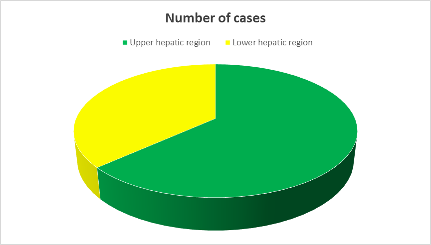
Pie chart 1: Radiological distribution of hepatic lesions
Most lesions were in the upper hepatic region (38; 63.3%), while 17 (28.3%) were in the lower region. Five patients (8.4%) demonstrated multifocal involvement.
Clinical Presentation
Table 4: Clinical presentation of patients
|
Clinical presentation |
Number of cases |
|
Abdominal pain/discomfort |
22 |
|
Weight loss |
14 |
|
Hepatomegaly/abdominal mass |
08 |
|
Jaundice |
05 |
|
Fever |
03 |
|
Known extrahepatic malignancy |
05 |
|
Incidentally detected lesion |
03 |
|
Total |
60 |
Abdominal pain or discomfort was the most common clinical presentation, observed in 22 patients (36.7%), followed by weight loss in 14 patients (23.3%). A small proportion of lesions were detected incidentally.
Adequacy of Sampling
Table 5: Adequacy of image-guided FNAC according to WHO terminology
|
Adequacy category |
Number of cases |
|
Adequate/ satisfactory |
45 |
|
Insufficient /inadequate/nondiagnostic |
15 |
|
Total |
60 |
Adequate cytological material was obtained in 56 cases (93.3%), while four cases (6.7%) were considered insufficient/inadequate/nondiagnostic. The nondiagnostic samples were characterised predominantly by scant cellularity and/or necrotic or haemorrhagic material.
Primary and Metastatic Malignancy
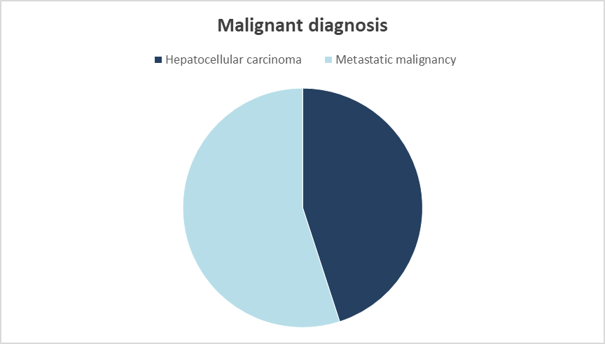
Pie chart 2: Spectrum of primary and metastatic malignant lesions
Among definite malignant lesions, metastatic malignancy was slightly more frequent than HCC. HCC accounted for 18 cases (30.0%), while metastatic malignancies accounted for 21 cases (35.0%).
Cytomorphological Subtypes of Metastatic Malignancy
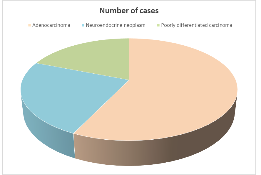
Pie chart 3: Spectrum of metastatic malignancies
Adenocarcinoma represented the predominant metastatic malignancy, accounting for 12 of 21 metastatic cases (57.1%). Neuroendocrine neoplasms accounted for five cases (23.8%), while poorly differentiated carcinoma accounted for four cases (19.1%).
Distribution according to WHO reporting category
Table 6: WHO-based cytological reporting grades
|
WHO category |
Number of cases |
|
Insufficient/ Inadequate/ Nondiagnostic |
15 |
|
Benign |
01 |
|
Atypical |
00 |
|
Suspicious for malignancy |
04 |
|
Malignant |
40 |
|
Total |
60 |
The malignant category was the most frequently assigned WHO category, comprising 39 cases (65.0%), followed by the benign category with 10 cases (16.7%). Four cases (6.7%) were classified as insufficient/inadequate/nondiagnostic, two (3.3%) as atypical and five (8.3%) as suspicious for malignancy.
PHOTOGALLERY
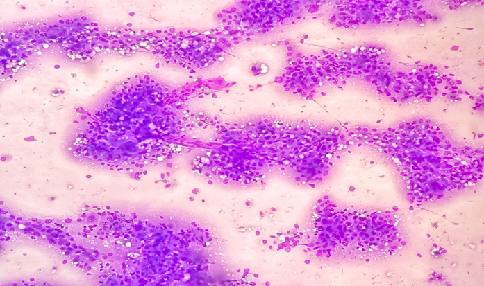
Figure 1: Hepatocellular carcinoma. Photomicrograph showing cells arranged in loosely cohesive, trabecular with occasional dispersed cells. In some areas, endothelial cell wrapping is also noted (Giemsa,100X).
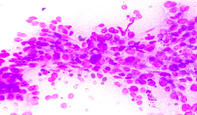
Figure 2: Hepatocellular carcinoma. Photomicrograph showing cells that are polygonal, with moderate to abundant granular cytoplasm. The nuclei are enlarged, round to oval in shape, with a high N:C ratio, coarse chromatin and conspicuous nucleoli (Giemsa 400X).
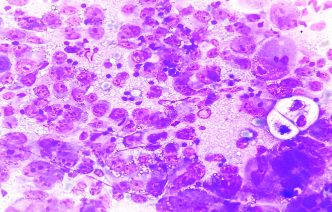
Figure 3: Metastatic Adenocarcinoma. Photomicrograph showing clusters of cells which are round to ovoid with irregular nuclear membranes with a moderate to abundant amount of basophilic cytoplasm and having open chromatin with conspicuous nucleoli (Giemsa, 100X).
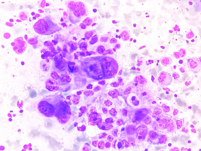
Figure 4: Metastatic high-grade adenocarcinoma. Photomicrograph showing high-grade metastatic adenocarcinoma with large round to oval cells showing a high N:C ratio with conspicuous nucleoli (Giemsa, 400X).
DISCUSSION:
The present study evaluated the cytomorphological spectrum of 60 radiologically confirmed hepatic SOLs undergoing image-guided FNAC over a one-year period. The study also incorporated the recently established WHO Reporting System for Liver Cytopathology to provide a standardised framework for communicating cytological diagnoses. In the present analysis, most patients were in the 61–70-year age group, followed by the 51–60-year group. This predominance of older adults is consistent with the epidemiology of primary and metastatic hepatic malignancies. Gupta et al., in their study of 62 hepatic FNAC cases, also observed that malignant lesions were predominantly encountered among middle-aged and older individuals.20
The present study observed a marked male predominance, with males accounting for 70% of cases. Several liver FNAC series have reported a similar male predominance. In the study by Gatphoh et al., conducted at the Regional Institute of Medical Sciences, Imphal, malignant hepatic lesions showed a substantial male predominance.5 The male predominance is particularly relevant to HCC, which demonstrates a higher incidence among men in many populations. The anatomical distribution in the present study showed a predominance of lesions in the upper hepatic region. However, anatomical localisation should ideally be reported using standardised radiological terminology such as right lobe, left lobe and specific Couinaud segments rather than broad upper and lower categories. A contemporary WHO-based liver cytology study reported that, among cases with documented location, right-lobe lesions were more common than left-lobe lesions.19
Abdominal pain was the most frequent clinical presentation in the present series, followed by weight loss. Hepatic malignancies may remain clinically silent until lesions become large enough to cause discomfort, hepatomegaly, or systemic manifestations. Weight loss and a known history of malignancy were particularly useful clinical clues toward metastatic disease.
Image-guided FNAC adequacy was high in the present study, with 75% of aspirates classified as adequate. This compares favourably with the 72.2% adequate aspiration rate reported by Barbhuiya et al. in their large series of 400 liver and gallbladder aspirates.13 Differences may relate to patient selection, image-guidance techniques, operator experience, availability of ROSE and the nature of the lesions sampled. Selhi et al. showed that incorporating ROSE into hepatic FNAC substantially improved diagnostic yield, with nondiagnostic specimens decreasing from 22 cases among 160 procedures to six cases among 142 procedures after implementing ROSE.14 Walia et al. similarly demonstrated the usefulness of ROSE for assessing adequacy in liver mass lesions.15 These observations support the importance of immediate adequacy assessment wherever facilities are available. Malignant lesions formed the largest diagnostic category in the present study. HCC accounted for 18 cases, while metastatic malignancy accounted for 21 cases. This pattern is comparable to the experience of several published liver FNAC studies in which metastatic disease forms a major component of hepatic malignant lesions. Gatphoh et al. reported 64 metastatic carcinomas and 31 HCCs among 100 malignant cases in their 202-case study.5
A large retrospective study of 755 hepatic SOLs reported metastatic disease in 524 cases and HCC in 148 cases, again demonstrating the substantial contribution of secondary malignancy to hepatic lesions.7 The authors concluded that FNAC was useful for distinguishing primary and metastatic lesions and for clinical decision-making, particularly in advanced malignancy. In another study of 338 FNAs of focal hepatic lesions, metastasis constituted 175 of 245 malignant cases, accounting for 71.4% of malignant lesions. The overall diagnostic accuracy of FNA for categorising lesions as benign or malignant was 97.51%. However, the study also demonstrated that precise tumour subtyping can remain challenging on cytology alone.6
In the present study, adenocarcinoma was the most frequent metastatic tumour, followed by neuroendocrine neoplasm and poorly differentiated carcinoma. The predominance of metastatic adenocarcinoma is consistent with Barbhuiya et al., in which metastatic adenocarcinoma was the most common positive cytological diagnosis, comprising 128 cases (44.2%).13
The cytological distinction between metastatic adenocarcinoma and poorly differentiated HCC remains an important diagnostic challenge. Adenocarcinoma generally shows cohesive glandular or acinar groups, mucin, and marked nuclear atypia, whereas HCC may show trabecular architecture, endothelial wrapping, bile pigment, and characteristic hepatocellular morphology. Nevertheless, poorly differentiated tumours may show overlapping features, and ancillary immunocytochemistry may be necessary.
The present study also identified metastatic neuroendocrine neoplasms. These lesions may demonstrate relatively monotonous tumour cells, finely granular chromatin and variable architectural organisation. In difficult cases, immunocytochemical markers such as synaptophysin, chromogranin, and INSM1 can help establish neuroendocrine differentiation. The WHO reporting system provides a structured approach to handling these diagnostic challenges. The five categories—nondiagnostic, benign, atypical, suspicious for malignancy and malignant—allow the cytopathologist to communicate the degree of certainty rather than forcing an uncertain lesion into a definitive diagnostic category.17-19
The relevance of this framework has been demonstrated in the 2026 study by Geetha et al., which retrospectively classified 976 liver cytology specimens according to the WHO system. The authors reported 79 nondiagnostic, 117 benign, 25 atypical, six suspicious and 749 malignant cases. They also observed that repeat sampling led to malignant upgrades in a substantial proportion of initially nondiagnostic and atypical cases.19 In the present study, 39 cases were assigned to the malignant category, while 10 were classified as benign, 2 as atypical, 5 as suspicious, and 4 as nondiagnostic. The relatively small number of atypical and suspicious cases reflects the deliberately conservative approach adopted for cytological interpretation. This categorisation may be clinically preferable to overdiagnosis because it identifies cases that require further evaluation.
The importance of distinguishing primary from secondary hepatic malignancy cannot be overstated. A diagnosis of metastatic adenocarcinoma may substantially alter clinical management by prompting investigation into an extrahepatic primary tumour, whereas HCC may lead to a completely different therapeutic pathway. FNAC therefore has an important role not merely in detecting malignancy but also in establishing its probable lineage.
Gupta et al. reported 43 malignant neoplastic lesions among 62 hepatic FNAC cases, including 17 HCCs and 23 metastatic adenocarcinomas, and demonstrated high diagnostic concordance between FNAC and histopathology when tissue confirmation was available.²⁰ These findings support the practical diagnostic value of image-guided FNAC in appropriately selected hepatic lesions. The current study is also relevant to the Northeast Indian setting. The earlier study by Gatphoh et al. from Imphal demonstrated the usefulness of liver FNAC in a regional population and established that metastatic carcinoma was a major component of hepatic malignancy.5 The present study extends this diagnostic approach by incorporating the contemporary WHO-based reporting framework.
Strengths of the present study include census sampling, inclusion of all eligible cases encountered over a defined one-year period, image-guided sampling, and systematic application of standardised WHO categories. The study also evaluates not only malignant diagnoses but the complete reporting spectrum, including nondiagnostic, benign, atypical and suspicious cases.
Limitations: The sample size of 60 cases is relatively small, and the study was conducted at a single tertiary-care centre. Histopathological confirmation and long-term follow-up may not be available for every patient. Furthermore, grouping lesions broadly into upper and lower hepatic regions provides less anatomical precision than right/left lobe or segment-based reporting. Future studies should incorporate standardised segmental radiological localisation, systematic histopathological correlation, immunocytochemistry and follow-up to determine the diagnostic accuracy and risk of malignancy associated with individual WHO categories.
Overall, the findings support the continued role of image-guided FNAC in evaluating hepatic SOLs. The WHO reporting framework provides a clinically meaningful structure for reporting and may be particularly valuable for borderline or limited specimens.
CONCLUSION:
Image-guided FNAC is a useful minimally invasive technique for evaluating radiologically detected hepatic space-occupying lesions. In the present study, malignant lesions constituted the major cytological diagnostic group, with HCC and metastatic malignancies forming the principal malignant categories.
Among metastatic malignancies, adenocarcinoma was the predominant subtype, followed by neuroendocrine neoplasm and poorly differentiated carcinoma. The high adequacy rate demonstrates the usefulness of image-guided sampling in obtaining diagnostically valuable material. The WHO Reporting System for Liver Cytopathology provides a structured, reproducible method for communicating cytological findings. The five-category system allows appropriate separation of nondiagnostic, benign, atypical, suspicious and malignant specimens and may help guide repeat sampling and ancillary investigations.
Image-guided FNAC, when interpreted with clinical and radiological information and supplemented by ancillary testing when required, can contribute substantially to the diagnosis and management of hepatic SOLs. Larger multicentric studies with histopathological correlation and follow-up are recommended to validate the diagnostic performance and malignancy risk associated with individual WHO categories.
REFERENCES:
1. Field AS, Pitman M, Cree IA, Canberk S, Bubendorf L, Mehrotra R, et al. The rationale for the development and publication of the World Health Organization reporting systems for cytopathology and a brief overview of the first editions of the lung and pancreaticobiliary systems. Cancer Cytopathol. 2023;131(12):751-61.
2. Gao Y, Zhang YH. Navigating the horizon: the World Health Organization reporting system for liver cytopathology. Hum Pathol Rep. 2024;36:300737.
3. International Agency for Research on Cancer. WHO Classification of Tumours Editorial Board. Digestive System Tumours. 5th ed. Lyon: IARC; 2019.
4. Jin Z, Li Y, Yi H, Wang M, Wang C, Du S, et al. Pathogenetic development, diagnosis and clinical therapeutic approaches for liver metastasis from colorectal cancer (Review). Int J Oncol. 2025;66(3):22.
5. Gatphoh ED, Gaytri S, Babina S, Singh AM. Fine needle aspiration cytology of liver: a study of 202 cases. Indian J Med Sci. 2003;57(1):22-5.
6. Rai SP, Kumar VK, Basavaiah SH, Sreeram S, Gopal S, Tantry BV. Efficacy and validity of image-guided percutaneous fine needle aspiration and core biopsy of liver pathologies: saga of focal hepatic lesions from the nodule to the needle to the slide. J Cytol. 2021;38:21-30.
7. Reddy CV, Goud YG, Poornima R, Deshmane V, Madhusudhana BA, Gayathridevi M. Role of FNAC in hepatic lesions: risk of track metastases. South Asian J Cancer. 2015;4(1):35-7.
8. Das DK. Cytodiagnosis of hepatocellular carcinoma in fine-needle aspirates of the liver: its differentiation from reactive hepatocytes and metastatic adenocarcinoma. Diagn Cytopathol. 1999;21(6):370-7.
9. Devi VL, Hazarika D. Fine needle aspiration cytology of hepatocellular carcinoma. Indian J Pathol Microbiol. 1995;38(4):389-92.
10. Bottles K, Cohen MB. An approach to fine-needle aspiration biopsy diagnosis of hepatic masses. Diagn Cytopathol. 1991;7(2):204-10.
11. Cohen MB, Haber MM, Holly EA, Ahn DK, Bottles K, Stoloff K. Cytologic criteria to distinguish hepatocellular carcinoma from nonneoplastic liver. Am J Clin Pathol. 1991;95(2):125-30.
12. Saeed OAM, Cramer H, Wng X, Wu HH. Fine needle aspiration cytology of hepatic metastases of neuroendocrine tumors: a 20-year retrospective, single institutional study. Diagn Cytopathol. 2018;46(1):35-9.
13. Barbhuiya M, Bhunia S, Kakkar M, Shrivastava B, Tiwari PK, Gupta S. Fine needle aspiration cytology of lesions of liver and gallbladder: an analysis of 400 consecutive aspirations. J Cytol. 2014;31(1):20-4.
14. Selhi PK, Tyagi R, Bansal P, Kaur H, Sood N. Hepatic fine-needle aspiration cytology: The role of rapid on-site evaluation in the assessment of hepatic lesions. Turk J Gastroenterol. 2018;29(4):442-7.
15. Walia S, Aron M, Hu E, Chopra S. Utility of rapid on-site evaluation for needle core biopsies and fine-needle aspiration cytology done for diagnosis of mass lesions of the liver. J Am Soc Cytopathol. 2019;8(2):69-77.
16. Nigam JS, Bharti JN, Rath A, Pradeep I. A comprehensive review of the newest World Health Organization cytopathology reporting systems. Adv Anat Pathol. 2023 Sep 1;30(5):301-06.
17. International Academy of Cytology-IARC-WHO Joint Editorial Board. WHO Reporting System for Cytopathology. Lyon: International Agency for Research on Cancer; 2022.
18. Kupati S, Siddiqui I, Ingin R, SS. Image guided fine needle aspiration cytology study of Liver lesions. Vascular & Endovascular Review; 2025; 8(9):73-78.
19. Geetha SD, Ali A, Kashi M, Burke A, Ort P, Chiu A, et al. Classifying liver cytology using the WHO reporting system: institutional experience and malignancy risk assessment. Diagn Cytopathol. 2026;54(6):436-43.
20. Gupta SK, Ashu T, Bharti M, Paswan MK, Sindhu VS, Venkatesh N. Cytomorphological spectrum of hepatic lesions on ultrasonography or CT-guided fine-needle aspiration cytology in a tertiary care centre, Rajendra Institute of Medical Sciences, Ranchi, Jharkhand, India. Cureus. 2024;16(9).