Correlation Of Serum Vitamin B12 And Ldh Levels With Peripheral Blood Smear Findings In Macrocytic Anaemia.
- Sonali Chaurasiya , Post Graduate Resident, Department of Pathology, Gandhi Medical College, Bhopal, Madhya Pradesh, India
- Maneesh Sulya , Professor & Head, Department of Pathology, Gandhi Medical College, Bhopal, Madhya Pradesh, India
- Archana Shrivastava , Associate Professor, Department of Pathology, Gandhi Medical College, Bhopal, Madhya Pradesh, India.
Article Information:
Abstract:
Background: Macrocytic anemia requires integration of peripheral smear morphology, MCV, RDW and biochemical markers. Objective: To assess the correlation between serum vitamin B12, LDH levels and peripheral blood smear findings in macrocytic anaemia. Materials and Methods: This prospective observational study was conducted in the Department of Pathology, Gandhi Medical College, Bhopal, Madhya Pradesh, over 18 months and included 200 patients with macrocytic anemia. Peripheral blood smear morphology, serum vitamin B12, and LDH levels were evaluated. Results: Vitamin B12 deficiency was present in 59%, while LDH was elevated in 82%. Mean MCV was 109.33 fL and mean RDW was 21.65%. Vitamin B12 showed a weak negative correlation with LDH (r = −0.136), whereas MCV and RDW showed weak positive correlations (r = 0.124 and 0.055). Conclusion: Integrated assessment of peripheral smear, vitamin B12, and LDH provides complementary information in macrocytic anemia.
Keywords:
Article :
INTRODUCTION:
Macrocytic anaemia is characterized by enlarged red blood cells, generally reflected by a mean corpuscular volume (MCV) greater than 100 fL. It may result from megaloblastic processes, particularly vitamin B12 or folate deficiency, or from non-megaloblastic conditions such as liver disease, alcohol use, hypothyroidism, medications, and bone marrow disorders [1,2].
Vitamin B12 deficiency interferes with DNA synthesis and nuclear maturation, resulting in ineffective erythropoiesis and characteristic megaloblastic changes. Peripheral blood smear may demonstrate macro-ovalocytes and other morphological abnormalities, while associated cytopenias may occur in more severe disease [2,3].
Automated MCV and RDW, particularly MCV and RDW, provide quantitative information about macrocytosis and anisocytosis, whereas peripheral blood smear examination provides direct morphological assessment. Therefore, combining these parameters with biochemical evaluation of vitamin B12 may improve recognition of the underlying mechanism [1,4].
Serum LDH may also be increased in megaloblastic states because of ineffective erythropoiesis and intramedullary destruction. However, the relationship between biochemical abnormalities, MCV and RDW, and peripheral smear morphology is not necessarily uniform. Hence, the present study was undertaken to assess the correlation between serum vitamin B12 and LDH levels and to evaluate the relationship between MCV and RDW and peripheral blood smear findings in patients with macrocytic anemia.
Peripheral blood smear examination remains an indispensable component of haematological evaluation. Morphological features such as macro-ovalocytes, hyper segmented neutrophils, polychromasia, Howell–Jolly bodies, Cabot rings and basophilic stippling strongly suggest megaloblastic anaemia, whereas round macrocytes and target cells are more characteristic of non-megaloblastic disorders.
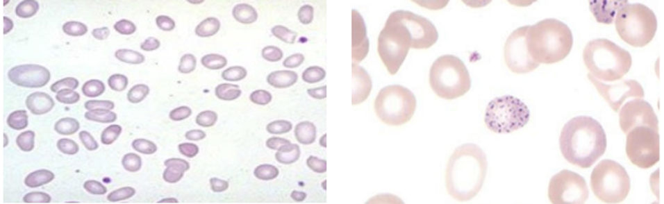
Image1- Macro-ovalocytes and macrocytes Image 2- Basophilic Stippling
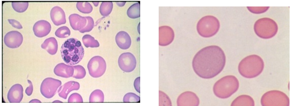
Image 3- Hyper segmented neutrophils Image 4- Polychromatic red cell
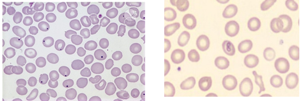
Image 5-Howell-Jolly bodies Image 6- Cabot ring
MATERIALS AND METHODS:
Study design and setting: This prospective observational study was conducted over a period of 18 months in the Department of Pathology, Gandhi Medical College, Bhopal, Madhya Pradesh, at the Central Clinical Laboratory and associated Hamidia Hospital. Samples received from various clinical departments, including Medicine, Surgery, Obstetrics and Gynaecology, Paediatrics, and Emergency Services, were evaluated. Complete blood counts were performed using fully automated haematology analysers. Peripheral blood smears were prepared in the haematology section, stained with Leishman stain, and examined by light microscopy. Serum LDH and vitamin B12 were subsequently evaluated in eligible patients.
Study population and sample size: Patients showing macrocytosis on complete blood count and peripheral blood smear were considered for inclusion. The sample size was calculated using the formula n = Z²PQ/D², taking Z = 1.96, prevalence (P) = 2.1%, Q = 97.9%, and allowable error (D) = 2%. The calculated sample size was approximately 200, and a total of 200 patients were included.
Inclusion criteria: Patients with MCV >100 fL on complete blood count and a macrocytic anaemia picture on peripheral blood smear were included. Exclusion criteria: Patients with MCV <100 fL or normocytic/microcytic morphology on peripheral smear, anaemia attributable to other conditions such as iron-deficiency or haemolytic anaemia, women in the third trimester of pregnancy, and patients receiving chemotherapy or radiotherapy were excluded.
Haematological evaluation: Complete blood count with particular attention to mean corpuscular volume (MCV), mean corpuscular haemoglobin (MCH) and red cell distribution width (RDW). The reference ranges documented in the thesis were MCV 80–100 fL, MCH 27–32 pg and RDW 11.5–14.5%.
Peripheral blood smear examination: A tongue-shaped peripheral blood smear was prepared on a clean glass slide, stained with Leishman stain, and examined microscopically for red-cell morphology.
Biochemical investigations: Serum vitamin B12 and LDH levels were measured in patients fulfilling the haematological and morphological criteria for macrocytosis.
Study procedure: CBC samples showing MCV >100 fL were initially identified. Peripheral blood smears were subsequently prepared, stained with Leishman stain, and examined microscopically. In patients whose smears demonstrated macrocytosis or macro-ovalocytosis, venous blood samples were processed for serum vitamin B12 and LDH estimation. Relevant clinical information, including nutritional status, alcohol consumption, drug intake, and associated medical conditions, was recorded whenever available. Laboratory and clinical findings were cross-checked and entered into structured case-record forms for analysis.
Statistical analysis: Data were analysed using appropriate statistical software. Continuous variables were summarized as mean and standard deviation, while categorical variables were expressed as frequencies and percentages. Normality of continuous variables was assessed before selection of statistical tests. Pearson's or Spearman's correlation coefficient, as appropriate, was used for evaluation. Comparative analyses were performed using parametric or corresponding non-parametric tests where applicable. A two-sided p value <0.05 was considered statistically significant.
Ethical considerations: The study was initiated after approval from the Institutional Ethics Committee. Routine diagnostic samples were utilized, and no additional procedures were undertaken solely for research purposes. Patient confidentiality and privacy were maintained, and the study was conducted according to institutional ethical requirements and the principles of the Declaration of Helsinki.
RESULTS:
A total of 200 patients with macrocytic anaemia were evaluated. The age distribution showed the highest proportion in the 21–30-year group, while males constituted a slightly larger proportion of the study population. MCV and RDW demonstrated marked macrocytosis and anisocytosis. Vitamin B12 deficiency and elevated LDH were frequent biochemical findings.
Table 1: Age Group Distribution of Study Participants (n = 200)
|
Age group (years) |
Frequency (n=200) |
Percentage |
|
0-10 |
14 |
7.0% |
|
11-20 |
28 |
14.0% |
|
21-30 |
47 |
23.5% |
|
31-40 |
40 |
20.0% |
|
41-50 |
28 |
14.0% |
|
51-60 |
22 |
11.0% |
|
>60 |
21 |
10.5% |
The largest proportion of patients belonged to the 21–30-year age group (23.5%), followed by the 31–40-year group (20.0%). Cases were represented across all age groups, indicating that macrocytic anaemia occurred throughout the studied age range.
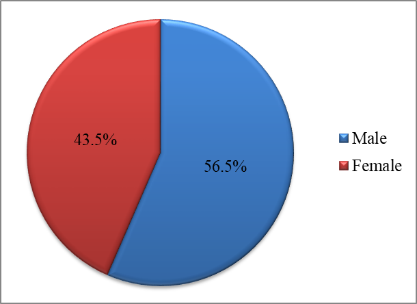
Figure 1: Gender Distribution of Study Participants
Males constituted 56.5% of the study population, whereas females accounted for 43.5%, demonstrating a slight male predominance.
Table 2: Distribution of MCV and RDW
|
Parameter |
Mean ± SD |
|
MCV (fL) |
109.33 ± 11.86 |
|
RDW (%) |
21.65 ± 6.89 |
The mean MCV was 109.33 fL, consistent with macrocytosis, while the mean RDW was 21.65%, indicating considerable variation in red cell size and anisocytosis.
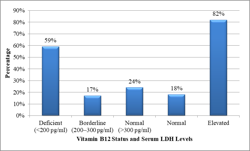
Figure 2: Distribution of Vitamin B12 Status and Serum LDH Levels
Vitamin B12 deficiency was identified in 59.0% of patients, while 17.0% had borderline and 24.0% had normal levels. Serum LDH was elevated in 82.0% of patients and normal in 18.0%.
Table 3: Peripheral Blood Smear Findings in Macrocytic Anaemia
|
Peripheral blood smear finding |
Frequency (n=200) |
Percentage |
|
Macrocytic anaemia |
36 |
18.0% |
|
Macrocytic anaemia with thrombocytopenia |
17 |
8.5% |
|
Macrocytic blood picture |
10 |
5.0% |
|
Macrocytic anaemia with pancytopenia |
7 |
3.5% |
|
Macrocytic anaemia with leucopenia |
5 |
2.5% |
|
Other/remaining macrocytic findings |
125 |
62.5% |
Peripheral blood smear examination showed isolated macrocytic anaemia in 36 (18.0%) patients, while macrocytic anaemia associated with thrombocytopenia was observed in 17 (8.5%), pancytopenia in 7 (3.5%), and leucopenia in 5 (2.5%) patients. A macrocytic blood picture without the specified associated cytopenias was documented in 10 (5.0%) patients. The remaining 125 (62.5%) patients had other macrocytic smear findings that were not further categorized in the available dataset.
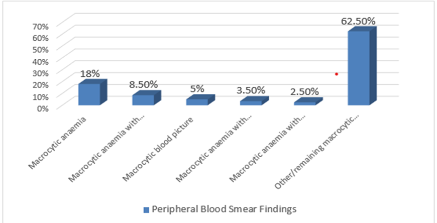
Figure 3: Peripheral Blood Smear Findings among study patients
Isolated macrocytic anemia accounted for 18.0% of cases, followed by macrocytic anaemia with thrombocytopenia (8.5%), a macrocytic blood picture (5.0%), macrocytic anaemia with pancytopenia (3.5%), and macrocytic anaemia with leucopenia (2.5%). Other/remaining macrocytic findings constituted 62.5% of cases.
Table 4: Correlation of Vitamin B12 and MCV and RDW with Serum LDH
|
Variables |
Correlation coefficient (r) |
P value |
|
Vitamin B12 vs LDH |
−0.136 |
0.055 |
|
MCV vs LDH |
0.124 |
0.080 |
|
RDW vs LDH |
0.055 |
0.439 |
Serum vitamin B12 showed a weak inverse correlation with LDH (r = −0.136), whereas MCV and RDW showed weak positive correlations (r = 0.124 and r = 0.055, respectively); none of these correlations reached statistical significance.
Table 5: Relationship Between MCV,RDW and Peripheral Blood Smear Morphology
|
RBC index |
Megaloblastic morphology on PBS (n=42), Median (IQR) |
Other macrocytic morphology (n=158), Median (IQR) |
Mann–Whitney U |
p value |
|
MCV (fL) |
110.25 (104.03–117.30) |
108.10 (102.03–117.00) |
3607.0 |
0.387 |
|
RDW (%) |
24.50 (20.38–30.23) |
18.45 (16.23–22.48) |
4845.5 |
<0.001 |
Peripheral smear findings suggestive of megaloblastic morphology were identified in 42 (21.0%) patients, while 158 (79.0%) showed other macrocytic morphological patterns. The median MCV was comparable between patients with megaloblastic morphology and those with other macrocytic morphology [110.25 (IQR 104.03–117.30) vs 108.10 (102.03–117.00) fL; p=0.387]. In contrast, RDW was significantly higher among patients demonstrating megaloblastic morphology [24.50% (IQR 20.38–30.23)] than among those with other macrocytic morphology [18.45% (IQR 16.23–22.48); p<0.001]. These findings indicate that RDW showed a significant association with megaloblastic peripheral smear morphology, whereas MCV did not significantly discriminate between the two morphological groups.
DISCUSSION:
The present study observed Vitamin B12 deficiency in 59% of patients, while serum LDH was elevated in 82% along with mean MCV 109.33±11.86 fL and the mean RDW was 21.65±6.89%, indicating substantial macrocytosis accompanied by marked variation in red cell size. These findings reinforce the value of combining quantitative haematological indices with morphological and biochemical parameters rather than relying on a single laboratory marker [1-4].
Vitamin B12 status
Vitamin B12 deficiency was present in 59% of the patients in the current study, while 17% had borderline concentrations and 24% had values within the normal range. The high frequency of deficiency suggests that vitamin B12 deficiency constitutes an important contributor to macrocytic anaemia in this population. The findings are broadly consistent with Indian studies demonstrating a substantial burden of vitamin B12 deficiency and its haematological manifestations. Singh et al. reported anaemia in 87.7% of vitamin B12-deficient patients, macrocytosis in 88.6%, macro-ovalocytes in 20%, and hyper segmented neutrophils in 60%. Another Indian study reported that vitamin B12 deficiency accounted for 40% of megaloblastic anaemia, whereas folate deficiency and combined B12-folate deficiency accounted for 25% and 35%, respectively [6, 7]. Vitamin B12 is essential for normal DNA synthesis, particularly in rapidly proliferating hematopoietic cells. Deficiency leads to nuclear-cytoplasmic asynchrony, ineffective erythropoiesis and abnormal maturation of erythroid and myeloid precursors. Consequently, peripheral blood may demonstrate macro-ovalocytes, anisopoikilocytosis and hyper segmented neutrophils, with leukopenia, thrombocytopenia or pancytopenia in more advanced deficiency. Recent reviews continue to identify macro-ovalocytes and hyper segmented neutrophils as characteristic morphological manifestations of megaloblastic erythropoiesis.
Serum LDH and ineffective erythropoiesis
One of the prominent observations of the present study was the high frequency of elevated serum LDH, which was detected in 82% of patients. This is remarkably similar to the northern Indian study by Singh et al., where LDH was elevated in 84.15% of tested vitamin B12-deficient patients. In another Indian study of megaloblastic anaemia, LDH was elevated in all affected patients, with a mean value of 3208 IU/L, and lower hemoglobin concentrations were associated with higher LDH levels [5, 6]. The increase in LDH in megaloblastic anaemia can largely be explained by ineffective erythropoiesis and intramedullary destruction of abnormal erythroid precursors. Because erythroid precursors fail to mature normally, many undergo destruction within the marrow before entering the peripheral circulation. This releases intracellular LDH and may be accompanied by indirect hyperbilirubinemia and reduced haptoglobin. Contemporary reviews recognize elevated LDH as an important biochemical manifestation of this intramedullary cell destruction. Severe vitamin B12 deficiency can occasionally produce extremely high LDH concentrations together with thrombocytopenia, fragmented erythrocytes and biochemical evidence resembling peripheral haemolysis or thrombotic microangiopathy.
Relationship between vitamin B12 and LDH
In the present study, serum vitamin B12 demonstrated a weak negative correlation with LDH (r = −0.136). The direction of this relationship is biologically plausible: greater vitamin B12 deficiency may result in more severe ineffective erythropoiesis and consequently greater intracellular enzyme release. However, the weak magnitude of the observed correlation indicates considerable interindividual variability. Importantly, the present findings should not be interpreted as demonstrating a strong inverse relationship between vitamin B12 and LDH. LDH is a nonspecific marker that can be affected by numerous haematological and nonhematological conditions. Moreover, serum vitamin B12 concentration alone does not always reflect the functional severity of cobalamin deficiency. Therefore, LDH should be regarded as a supportive marker of ineffective erythropoiesis rather than a substitute for vitamin B12 measurement.
This interpretation is consistent with previous observations that the haematological severity of megaloblastic anaemia does not necessarily show a direct linear relationship with serum vitamin concentrations [7, 9]. The weak correlation found in our study therefore supports a multimodal diagnostic approach rather than dependence on either vitamin B12 or LDH in isolation.
Correlation of MCV and RDW with LDH
MCV showed only a weak positive correlation with LDH (r = 0.124), while RDW showed a minimal positive correlation (r = 0.055). These findings indicate that increasing macrocytosis or anisocytosis does not necessarily parallel the biochemical severity of ineffective erythropoiesis.
This distinction is clinically relevant because MCV represents the average erythrocyte volume in peripheral blood, RDW reflects variation in erythrocyte size, whereas LDH predominantly reflects cellular injury and turnover. Although all three abnormalities may occur simultaneously in megaloblastic anaemia, they represent different biological aspects of the disease.
Furthermore, MCV may be influenced by concurrent iron deficiency, thalassemia trait, liver disease, alcohol consumption, hypothyroidism and other conditions. Recent literature has emphasized that coexisting iron deficiency or hemoglobinopathies can mask macrocytosis in a proportion of patients with vitamin B12 deficiency [8,10].
Peripheral blood smear findings and cytopenias
Peripheral smear examination in the current study demonstrated macrocytic morphological changes, with thrombocytopenia, leukopenia and pancytopenia occurring in subsets of patients.
Such findings are compatible with ineffective haematopoiesis extending beyond the erythroid lineage. Singh et al. reported leukopenia in 14.1%, thrombocytopenia in 7.27% and pancytopenia in 12.73% of vitamin B12-deficient patients. Other Indian data have also demonstrated pancytopenia in patients with megaloblastic anaemia. Severe cobalamin deficiency can impair maturation across multiple hematopoietic lineages, explaining the occurrence of bicytopenia and pancytopenia. Recent studies have further shown that these abnormalities are particularly associated with severe vitamin B12 deficiency [6, 11]. Therefore, vitamin B12 deficiency should remain an important and readily treatable differential diagnosis when macrocytosis occurs together with unexplained thrombocytopenia, leukopenia or pancytopenia.
Clinical implications
The major clinical implication of the present study is that no single parameter adequately characterizes macrocytic anaemia. MCV and RDW identify macrocytosis and anisocytosis, peripheral smear demonstrates morphological abnormalities, serum vitamin B12 identifies an important nutritional cause, and LDH provides supportive evidence of ineffective erythropoiesis.
CONCLUSION:
The present study demonstrates that macrocytic anaemia is best evaluated through an integrated assessment of haematological, morphological, and biochemical parameters.
The observed concordance between elevated MCV, increased RDW, and peripheral smear features such as macrocytes, macro-ovalocytes, and anisocytosis further emphasizes the continued diagnostic value of peripheral smear examination alongside automated MCV and RDW. Therefore, a combined approach incorporating MCV, RDW, peripheral blood smear morphology, serum vitamin B12, and LDH may facilitate a more comprehensive and practical evaluation of patients with macrocytic anaemia, particularly in routine laboratory diagnosis.
REFERENCES:
1. Nagao T, Hirokawa M. Diagnosis and treatment of macrocytic anemias in adults. J Gen Fam Med. 2017 Apr 13;18(5):200-204. doi: 10.1002/jgf2.31.
2. Socha DS, DeSouza SI, Flagg A, Sekeres M, Rogers HJ. Severe megaloblastic anemia: Vitamin deficiency and other causes. Cleve Clin J Med. 2020 Mar;87(3):153-164. doi: 10.3949/ccjm.87a.19072.
3. Green R, Miller JW. Vitamin B12 deficiency. Vitam Horm. 2022; 119:405-439. doi: 10.1016/bs.vh.2022.02.003.
4. Langan RC, Goodbred AJ. Vitamin B12 Deficiency: Recognition and Management. Am Fam Physician. 2017 Sep 15;96(6):384-389.
5. Goel S, Gajendra S, Sahni T, Jha B, Dang M, Rawat HS, Sharif MS, Raina V, Sachdev R. Role of NEUT-X & NEUT-Y in picking up megaloblastic anaemia on peripheral blood & in differentiating from other macrocytic anaemia. Indian J Med Res. 2017 Mar;145(3):395-398. doi: 10.4103/ijmr.IJMR_201_15.
6. Singh J, Dinkar A, Gupta P, Atam V. Vitamin B12 deficiency in northern India tertiary care: Prevalence, risk factors and clinical characteristics. J Family Med Prim Care. 2022 Jun;11(6):2381-2388. doi: 10.4103/jfmpc.jfmpc_650_21.
7. Hall JA, Mason J, Choi J, Holguin M. B12 deficiency leading to marked poikilocytosis versus true schistocytosis, a pernicious problem. Transfus Apher Sci. 2017 Aug;56(4):576-577. doi: 10.1016/j.transci.2017.06.003.
8. Teixeira AM, Macedo B, Fontes CP, Manuel M. Macrocytic Anaemia: Not Always a Straightforward Diagnosis. Cureus. 2022 Mar 14;14(3):e23152. doi: 10.7759/cureus.23152.
9. Andrès E, Serraj K, Zhu J, Vermorken AJ. The pathophysiology of elevated vitamin B12 in clinical practice. QJM. 2013 Jun;106(6):505-15. doi: 10.1093/qjmed/hct051. Epub 2013 Feb 27. PMID: 23447660.
10. Shilpy, Mishra M, Saha SS, Kumar A. Clinical and Hematological Parameters and Their Correlation with the Severity of Vitamin B12 Deficiency: A Hospital-Based Study. Cureus. 2025 Sep 9;17(9): e91907. doi: 10.7759/cureus.91907. PMID: 41080292
11. Khanduri U, Sharma A. Megaloblastic anaemia: prevalence and causative factors. Natl Med J India. 2007 Jul-Aug;20(4):172-5. PMID: 18085121.