King Vision Video Laryngoscope versus Macintosh Laryngoscope for Endotracheal Intubation in Elective Surgery: A Prospective Randomized Comparative Study.
- Manjula Bestha , Assistant professor, Department of Anaesthesiology, MNR medical college and hospital, Sangareddy, Telangana, India.
- Asha Sharma , Assistant professor, Department of Anaesthesiology, MNR medical college and hospital, Sangareddy, Telangana, India.
Article Information:
Abstract:
Background: Laryngoscopy and tracheal intubation trigger sympathetic stimulation that may produce transient tachycardia and hypertension. Video laryngoscopy improves laryngeal visualization without requiring the same degree of alignment of the oral, pharyngeal, and laryngeal axes as direct laryngoscopy, but its effect on intubation difficulty and peri-intubation haemodynamics remains variable across studies. The study is designed to compare the King Vision Video Laryngoscope with the Macintosh laryngoscope for ease of tracheal intubation, assessed by the Intubation Difficulty Score, and for changes in haemodynamic variables during elective general anaesthesia. Methods: This prospective randomized comparative study included 101 adults aged 18-60 years with American Society of Anesthesiologists physical status I or II and Mallampati class I or II who underwent elective surgery requiring endotracheal intubation. Patients were randomized to Macintosh laryngoscopy (MCL, n=51) or King Vision video laryngoscopy (KVVL, n=50). A standardized anaesthetic technique was used. Intubation difficulty was graded using the seven-component Intubation Difficulty Score. Heart rate, systolic blood pressure, diastolic blood pressure, mean arterial pressure, and oxygen saturation were recorded before induction, after induction, and at 1, 3, and 5 minutes. Between-group p values are reported as presented in the source study. Results: The groups were comparable in sex, age distribution, body mass index, ASA class, and Mallampati grade. The mean total Intubation Difficulty Score was lower with KVVL than with MCL (0.36 ± 0.80 vs 0.84 ± 1.28; p=0.027). Better glottic exposure contributed to this difference (N4: 0.08 ± 0.27 vs 0.38 ± 0.53; p=0.001), and external laryngeal pressure was required less often with KVVL (N6: 0 vs 0.16 ± 0.37; p=0.003). Heart rate, systolic blood pressure, and oxygen saturation did not differ significantly between groups at the reported time points. Diastolic blood pressure and mean arterial pressure were higher in the KVVL group at baseline and remained higher during follow-up. Conclusion: King Vision video laryngoscopy reduced overall intubation difficulty, mainly through improved glottic visualization and less need for external laryngeal manipulation. It did not demonstrate a clear advantage over Macintosh laryngoscopy in attenuating the cardiovascular response to intubation in this study.
Keywords:
Article :
INTRODUCTION:
Endotracheal intubation is a routine component of general anaesthesia, but the act of laryngoscopy and passage of the tracheal tube is physiologically active rather than neutral. Mechanical stimulation of the tongue base, supraglottic tissues, larynx, and trachea activates autonomic reflexes and is commonly followed by a short-lived increase in heart rate and arterial pressure [1-5]. These changes are usually well tolerated in healthy adults, although they can be clinically important in patients with limited cardiovascular or cerebrovascular reserve [3-5,24].
The Macintosh laryngoscope remains a familiar and widely used device for direct laryngoscopy. Its use depends on obtaining a direct line of sight to the glottis, which generally requires positioning of the head and neck and application of lifting force to displace the tongue and related soft tissues [1,2,23]. Difficult or failed laryngoscopy can increase the number of airway manipulations and the possibility of dental, mucosal, and hypoxaemic complications [13,18,19,22,25,26].
Video laryngoscopes were developed to improve laryngeal visualization by transferring the image from a camera near the blade tip to an external display. This design reduces dependence on direct alignment of the airway axes and may improve the glottic view, particularly when conventional direct laryngoscopy is difficult [8,13,19-22,30]. However, a better view does not always translate into faster tube delivery or a smaller haemodynamic response. Differences in blade geometry, tube channel design, operator familiarity, and the stimulation produced by tracheal tube passage can influence the overall response [9,10,12,15-17,21].
The King Vision Video Laryngoscope is an indirect video laryngoscope with a reusable display and disposable blades, including a channelled blade that guides the endotracheal tube toward the glottis. Previous investigations have reported favourable laryngeal views and useful intubating conditions with this device, although results differ according to airway difficulty, comparator device, and clinical setting [6,7,14,27-29]. Evidence from larger comparative studies and systematic reviews also supports a general advantage of video laryngoscopy for glottic visualization and first-attempt success, while showing considerable variation in intubation time and haemodynamic outcomes [27,30].
The present study was therefore undertaken in adults with an apparently normal airway who were scheduled for elective surgery. The primary practical question was whether King Vision video laryngoscopy made intubation easier than conventional Macintosh laryngoscopy when difficulty was assessed with a structured Intubation Difficulty Score. A second objective was to compare peri-intubation heart rate, blood pressure, mean arterial pressure, and oxygen saturation between the two techniques.
MATERIALS AND METHODS:
Study design and setting
A prospective randomized comparative study was conducted at Narayana Hrudayalaya, Bommasandra, Bangalore. Institutional review and ethics approval were obtained before recruitment, and written informed consent was taken from each participant. The study compared tracheal intubation performed with a Macintosh laryngoscope and with the King Vision Video Laryngoscope in patients undergoing elective surgery under general anaesthesia.
Participants and eligibility
Adults aged 18-60 years of either sex were eligible when they had American Society of Anesthesiologists physical status I or II, Mallampati class I or II, and were scheduled for elective surgery requiring general anaesthesia with endotracheal intubation. Patients were excluded for upper airway pathology such as maxillofacial fracture or tumour, cervical spine disease, anticipated difficult intubation including Mallampati class III or IV, emergency surgery, obesity, pregnancy, or ASA physical status III or IV. The discussion section of the source thesis specified obesity as body mass index above 30 kg/m².
Sample size and randomization
The sample size was derived from an earlier comparison in which first-attempt success was 97% with King Vision and 86% with Macintosh laryngoscopy [6]. Using a two-proportion calculation with a two-sided alpha of 0.05 and 80% power, a total sample of 101 patients was selected. One day before surgery, patients were allocated by a computer-generated random number table to the Macintosh group (MCL) or the King Vision group (KVVL). The final groups contained 51 and 50 patients, respectively.
Preoperative assessment and anaesthetic technique
A preanaesthetic evaluation was completed on the day before surgery. Clinical history, physical examination, airway assessment, height, weight, and body mass index were recorded, together with a history of previous difficult intubation when present. Patients received standardized premedication and remained fasting overnight. In the operating room, intravenous access was established and routine monitoring included electrocardiography, non-invasive blood pressure, heart rate, and pulse oximetry.
After preoxygenation, anaesthesia was induced with fentanyl 2 micrograms/kg, midazolam 0.03 mg/kg, propofol 2 mg/kg, and vecuronium 0.1 mg/kg. Mask ventilation was continued for three minutes with oxygen and isoflurane at approximately 1% end-tidal concentration. Laryngoscopy was then performed with the allocated device. In the Macintosh group, direct laryngoscopy and intubation were performed with an appropriate blade and tracheal tube. In the King Vision group, the video laryngoscope blade was introduced in the midline, advanced over the tongue with the tip positioned in the vallecula, and lifted until the glottic view was optimized. The endotracheal tube was then advanced through the vocal cords. Tube position was confirmed by bilateral air entry and a sustained end-tidal carbon dioxide waveform.
Outcome measures
Ease of intubation was assessed with the Intubation Difficulty Score, a composite measure that captures the number of supplementary attempts and operators, use of alternative techniques, grade of glottic exposure, lifting force, need for external laryngeal pressure, and position of the vocal cords. Glottic exposure was graded using the Cormack and Lehane system [26]. An IDS of 0 represented easy intubation, a score greater than 0 but below 5 indicated slight difficulty, a score above 5 indicated moderate or major difficulty, and an unsuccessful intubation was classified as impossible. The components of the score were recorded as N1 through N7, consistent with the study proforma.
Haemodynamic assessment included heart rate, systolic blood pressure, diastolic blood pressure, mean arterial pressure, and oxygen saturation. Measurements were documented before induction, after induction, and at 1, 3, and 5 minutes during the peri-intubation observation period. The study also planned to record intubation time, but a numerical comparative intubation-time table was not available in the thesis results and was therefore not reconstructed for this manuscript.
Statistical analysis
The source study used SPSS version 22.0. Continuous variables were summarized as mean ± standard deviation and categorical variables as counts and percentages. Independent-samples t tests were used for continuous between-group comparisons, while chi-square or Fisher exact tests were used for categorical data as appropriate. A p value below 0.05 was considered statistically significant. The protocol stated that serial haemodynamic variables would be assessed with repeated-measures analysis of variance; however, the available results tables reported unpaired t tests at individual time points. Because patient-level data were not available for reanalysis, this manuscript reports the pointwise p values presented in the thesis rather than generating unreported repeated-measures statistics.
Ethical considerations
The study documents were reviewed and approved by the Narayana Health Academic Ethics Committee. The thesis includes the committee approval letter and records written informed consent from participants. Participation was voluntary, and the study information sheet stated that withdrawal would not affect subsequent clinical care.
RESULTS:
Participant profile and baseline comparability
A total of 101 patients were analyzed, with 51 patients in the Macintosh group and 50 in the King Vision group. Men accounted for 52.94% of MCL and 58.0% of KVVL participants, and the sex distribution was not significantly different (p=0.687). Mean age was 43.80 ± 10.84 years in MCL and 43.38 ± 12.70 years in KVVL. The groups were also comparable in the reported distributions of age, body mass index, ASA physical status, and Mallampati grade. Baseline characteristics are summarized in Table 1.
Table 1: Baseline demographic and airway characteristics
|
Characteristic |
MCL (n=51) |
KVVL (n=50) |
P value |
|
Male sex, n (%) |
27 (52.94) |
29 (58.0) |
0.687 |
|
Female sex, n (%) |
24 (47.06) |
21 (42.0) |
|
|
Age <=30 years, n (%) |
6 (11.77) |
9 (18.0) |
0.600 |
|
Age 31-45 years, n (%) |
23 (45.09) |
18 (36.0) |
|
|
Age >=46 years, n (%) |
22 (43.14) |
23 (46.0) |
|
|
Mean age, years |
43.80 ± 10.835 |
43.38 ± 12.697 |
|
|
Height, cm |
163.62 ± 3.61 |
165.08 ± 15.79 |
0.506 |
|
Weight, kg |
59.10 ± 8.299 |
60.96 ± 1.323 |
0.995 |
|
BMI, kg/m² |
22.09 ± 3.112 |
22.36 ± 3.799 |
0.337 |
|
ASA I, n (%) |
29 (56.86) |
32 (64.0) |
0.539 |
|
ASA II, n (%) |
22 (43.14) |
18 (36.0) |
|
|
Mallampati I, n (%) |
24 (47.06) |
22 (44.0) |
0.688 |
|
Mallampati II, n (%) |
27 (52.94) |
28 (56.0) |
|
Data are n (%) or mean ± SD. P values are those reported in the source thesis. Categorical comparisons used chi-square or Fisher exact tests as appropriate; continuous variables used independent-samples t tests. ASA, American Society of Anesthesiologists; BMI, body mass index; MCL, Macintosh laryngoscope; KVVL, King Vision Video Laryngoscope
Reported comorbidities were similarly distributed between groups. Hypertension was present in 10 MCL and 9 KVVL patients (p=0.799), diabetes mellitus in 10 and 6 (p=0.275), chronic obstructive pulmonary disease in 1 and 0 (p=0.315), bronchial asthma in 2 and 3 (p=0.646), smoking history in 3 and 4 (p=0.695), and hypothyroidism in 2 and 1 patients (p=0.558). None of these comparisons reached statistical significance.
Intubation difficulty
The total Intubation Difficulty Score was significantly lower in the King Vision group. Mean total IDS was 0.84 ± 1.28 with Macintosh and 0.36 ± 0.80 with King Vision (p=0.027). The largest contribution to this difference came from glottic exposure. The N4 component was 0.38 ± 0.53 in MCL and 0.08 ± 0.27 in KVVL (p=0.001), indicating a better laryngoscopic view with King Vision. External laryngeal pressure was also required less often with the video laryngoscope, reflected by an N6 score of 0.16 ± 0.37 in MCL and 0 in KVVL (p=0.003). Other IDS components did not differ significantly (Table 2 and Figure 1).
Table 2: Intubation Difficulty Score components
|
IDS component |
MCL |
KVVL |
P value |
|
N1: supplementary attempts |
0.06 ± 0.23 |
0.06 ± 0.23 |
1.000 |
|
N2: supplementary operators |
0.02 ± 0.14 |
0.06 ± 0.23 |
0.312 |
|
N3: alternative techniques |
0.00 ± 0.00 |
0.00 ± 0.00 |
NA |
|
N4: glottic exposure |
0.38 ± 0.53 |
0.08 ± 0.27 |
0.001 |
|
N5: lifting force |
0.18 ± 0.38 |
0.16 ± 0.37 |
0.793 |
|
N6: external laryngeal pressure |
0.16 ± 0.37 |
0.00 ± 0.00 |
0.003 |
|
N7: vocal cord position |
0.04 ± 0.28 |
0.00 ± 0.00 |
0.320 |
|
Total IDS |
0.84 ± 1.28 |
0.36 ± 0.80 |
0.027 |
Values are mean ± SD. IDS is the sum of N1-N7. P values are those reported in the thesis. NA, not applicable because both groups had a zero value for N3. Statistically significant comparisons are N4, N6, and total IDS.
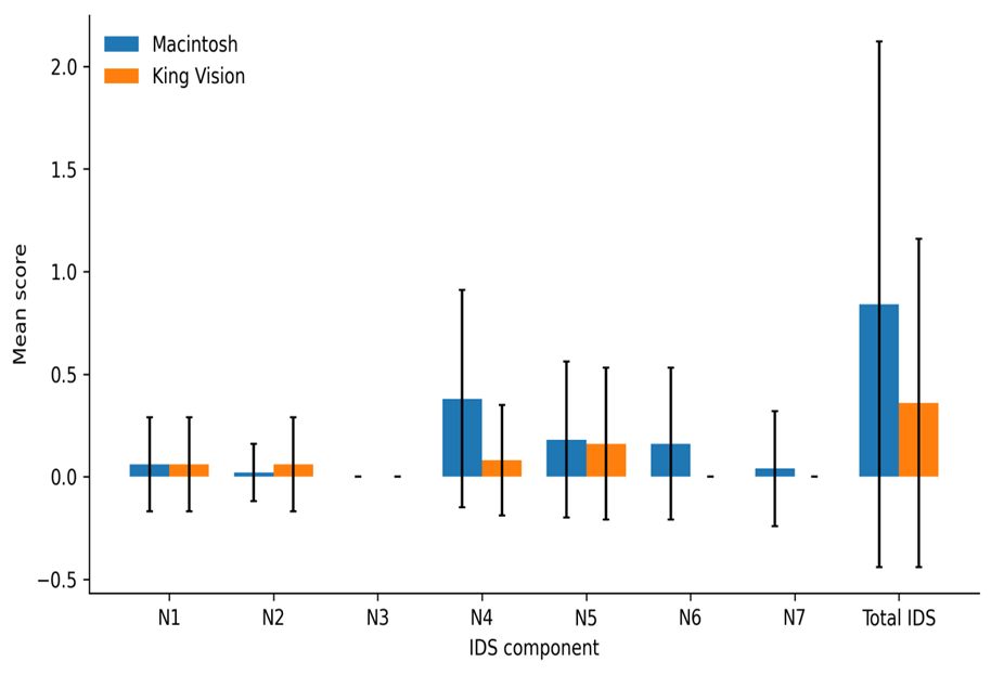
Figure 1: Mean Intubation Difficulty Score components in the Macintosh and King Vision groups. Error bars represent standard deviation. The largest between-group differences were seen for glottic exposure (N4), external laryngeal pressure (N6), and total IDS
Haemodynamic response
Heart rate
Baseline heart rate was 79.98 ± 11.89 beats/min in the Macintosh group and 82.02 ± 9.40 beats/min in the King Vision group (p=0.344). Both groups showed their highest mean heart rate after induction, followed by a gradual return toward baseline. No between-group heart-rate comparison reached statistical significance at the reported time points, although the difference at 1 minute approached significance (84.74 ± 11.57 vs 88.60 ± 8.42 beats/min; p=0.059). The time course is shown in Figure 2.
Table 3: Heart rate at measured time points
|
Time point |
MCL, beats/min |
KVVL, beats/min |
P value |
|
Before induction |
79.98 ± 11.886 |
82.02 ± 9.401 |
0.344 |
|
After induction |
89.24 ± 11.754 |
90.32 ± 8.849 |
0.605 |
|
1 min |
84.74 ± 11.568 |
88.60 ± 8.420 |
0.059 |
|
3 min |
80.68 ± 11.316 |
81.40 ± 8.091 |
0.715 |
|
5 min |
79.26 ± 11.593 |
80.40 ± 17.565 |
0.703 |
Values are mean ± SD. P values are from the unpaired t tests reported in the source results. MCL, Macintosh laryngoscope; KVVL, King Vision Video Laryngoscope
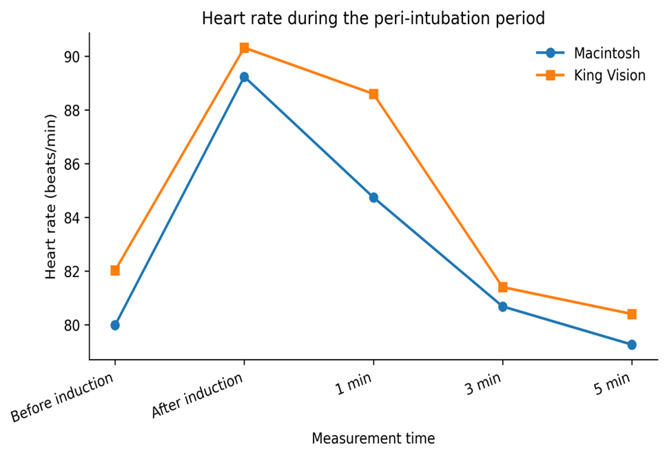
Figure 2: Mean heart rate before induction, after induction, and at 1, 3, and 5 minutes in the two study groups
Blood pressure
Systolic blood pressure was similar between groups at baseline and throughout the observation period. Baseline values were 128.32 ± 8.26 mmHg in MCL and 127.88 ± 7.56 mmHg in KVVL (p=0.782). Both groups reached their highest mean systolic pressure after induction and showed lower values by 5 minutes, with no significant between-group difference at any reported time point (Figure 3).
In contrast, diastolic blood pressure and mean arterial pressure were already higher in the KVVL group before induction and remained statistically higher at all subsequent reported measurements. Baseline diastolic pressure was 76.10 ± 5.70 mmHg in MCL and 81.22 ± 5.78 mmHg in KVVL (p<0.001), while baseline mean arterial pressure was 93.20 ± 5.84 and 96.76 ± 6.02 mmHg, respectively (p=0.003). Because these differences were present before induction, the absolute between-group values cannot be attributed solely to the laryngoscopy technique. The detailed measurements are provided in Table 4 and Figures 4 and 5.
Table 4: Systolic, diastolic, and mean arterial pressure
|
Parameter and time |
MCL, mmHg |
KVVL, mmHg |
P value |
|
SBP: before induction |
128.32 ± 8.257 |
127.88 ± 7.561 |
0.782 |
|
SBP: after induction |
137.14 ± 9.483 |
135.46 ± 7.696 |
0.333 |
|
SBP: 1 min |
133.80 ± 9.470 |
132.92 ± 7.693 |
0.611 |
|
SBP: 3 min |
125.18 ± 8.528 |
122.94 ± 6.756 |
0.149 |
|
SBP: 5 min |
120.24 ± 8.620 |
118.34 ± 6.598 |
0.219 |
|
DBP: before induction |
76.10 ± 5.704 |
81.22 ± 5.783 |
<0.001 |
|
DBP: after induction |
79.38 ± 7.082 |
85.00 ± 5.739 |
<0.001 |
|
DBP: 1 min |
77.30 ± 7.377 |
83.06 ± 5.836 |
<0.001 |
|
DBP: 3 min |
72.66 ± 6.297 |
77.38 ± 5.182 |
<0.001 |
|
DBP: 5 min |
69.86 ± 5.599 |
74.50 ± 5.108 |
<0.001 |
|
MAP: before induction |
93.20 ± 5.841 |
96.76 ± 6.019 |
0.003 |
|
MAP: after induction |
98.40 ± 7.100 |
101.84 ± 5.905 |
0.010 |
|
MAP: 1 min |
96.10 ± 7.284 |
99.64 ± 6.134 |
0.010 |
|
MAP: 3 min |
90.21 ± 6.246 |
92.52 ± 5.281 |
0.048 |
|
MAP: 5 min |
86.42 ± 5.693 |
89.10 ± 5.250 |
0.016 |
Values are mean ± SD. P values are from the unpaired t tests reported in the source results. SBP, systolic blood pressure; DBP, diastolic blood pressure; MAP, mean arterial pressure. Baseline DBP and MAP differed significantly between groups, which should be considered when interpreting later absolute values
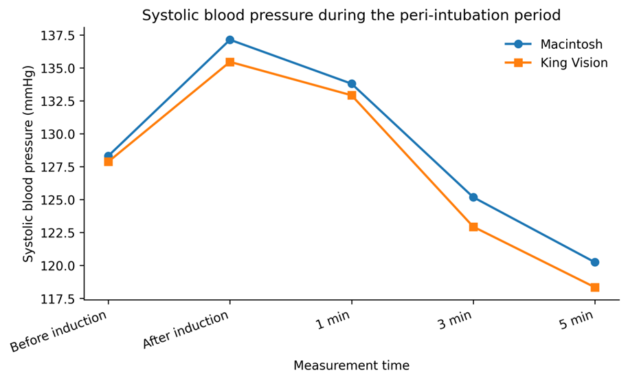
Figure 3: Mean systolic blood pressure during the peri-intubation observation period
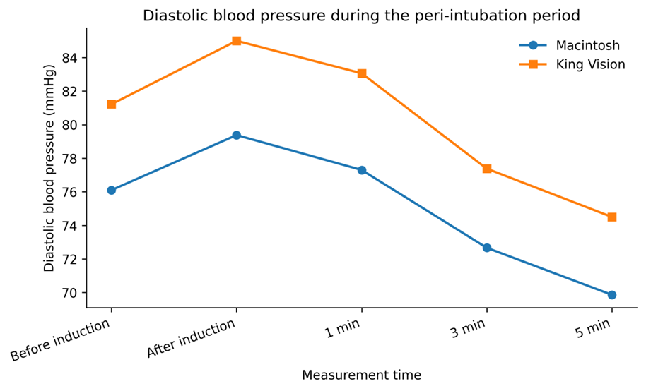
Figure 4: Mean diastolic blood pressure during the peri-intubation observation period. The KVVL group had a higher baseline DBP before induction
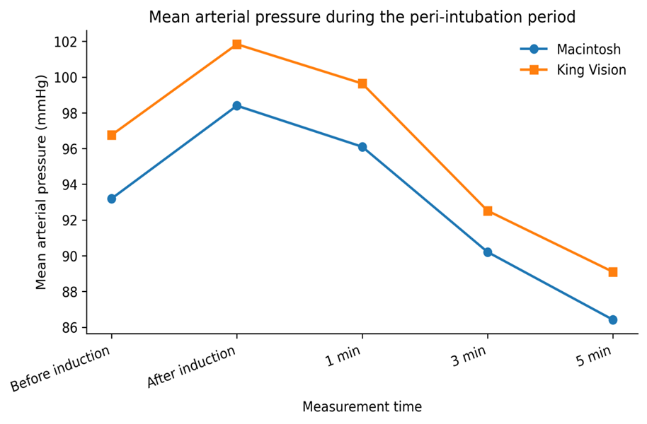
Figure 5: Mean arterial pressure during the peri-intubation observation period. Baseline MAP was higher in the KVVL group
Oxygen saturation
Oxygen saturation remained stable in both groups. Mean SpO2 ranged from 97.74% to 98.26% in MCL and from 97.94% to 98.48% in KVVL across the reported measurements. No comparison was statistically significant, suggesting that both techniques maintained adequate oxygenation during the observed peri-intubation period.
Table 5: Oxygen saturation at measured time points
|
Time point |
MCL, % |
KVVL, % |
P value |
|
Before induction |
97.74 ± 1.306 |
98.14 ± 1.443 |
0.149 |
|
After induction |
98.26 ± 1.411 |
98.20 ± 1.340 |
0.828 |
|
1 min |
98.00 ± 1.471 |
97.96 ± 1.498 |
0.893 |
|
3 min |
97.84 ± 1.517 |
97.94 ± 1.531 |
0.744 |
|
5 min |
98.26 ± 1.226 |
98.48 ± 1.282 |
0.383 |
Values are mean ± SD. P values are from the unpaired t tests reported in the thesis. SpO2, peripheral oxygen saturation
DISCUSSION:
This randomized comparative study found a practical advantage of the King Vision Video Laryngoscope in ease of intubation. The total Intubation Difficulty Score was lower with King Vision, and the difference was mainly explained by better glottic exposure and less need for external laryngeal pressure. These are clinically relevant components of the intubation process because they reflect how much additional manipulation is required to obtain a usable view and pass the tracheal tube. The haemodynamic findings were less persuasive. Heart rate, systolic pressure, and oxygen saturation were similar between groups, while diastolic pressure and mean arterial pressure were higher in the King Vision group from baseline onward. Taken together, the data support easier visualization and intubation conditions with King Vision but do not show clear attenuation of the cardiovascular response.
The reduction in IDS is consistent with earlier work involving King Vision and other video laryngoscopes. Ali and colleagues reported a lower IDS with King Vision in patients with cervical spine immobilization [6]. Elhadi and colleagues also observed favourable intubating conditions and reduced need for assisting manoeuvres with King Vision compared with conventional laryngoscopy [7]. Studies of C-MAC and other indirect devices have similarly shown improved glottic visualization, fewer adjunctive manoeuvres, or higher first-attempt success in difficult or non-standard airway settings [8,13,18-22]. Maharaj and colleagues reported a modest improvement in IDS with the Airtraq compared with Macintosh laryngoscopy [11]. These findings suggest that improved visualization can reduce some of the mechanical steps that contribute to intubation difficulty, even when the device itself requires a distinct tube-delivery technique.
The present glottic-view result also agrees with more recent King Vision data. Erdivanli and colleagues found that King Vision and Macintosh laryngoscopy produced similar first-pass success in experienced hands, although King Vision required more time for glottic visualization and intubation [27]. Reena reported a higher first-attempt success rate and less need for optimization manoeuvres with a channelled King Vision blade when armoured tracheal tubes were used [29]. Schoettker and Corniche observed good laryngeal visualization with King Vision in a simulated difficult-airway comparison [14]. These studies reinforce an important distinction: visualization may be excellent while tube advancement can still depend on blade channel geometry, operator familiarity, and alignment of the tube with the glottic opening.
The haemodynamic literature is more heterogeneous. Pournajafian and colleagues found no significant overall difference in cardiovascular response between GlideScope and Macintosh laryngoscopy despite a longer intubation time with the video device [9]. Siddiqui and colleagues similarly reported no clear haemodynamic benefit among direct laryngoscopy, GlideScope, and Trachlight techniques [10]. Kanchi and colleagues found comparable haemodynamic responses in patients with coronary artery disease despite a longer procedure with video laryngoscopy [12]. Xue and colleagues also reported similar responses between GlideScope and Macintosh laryngoscopy [17]. In contrast, Tsai and colleagues suggested that an indirect device could attenuate the stress response under specific conditions [15], and Mahjoubifar and Boroojeny observed a smaller rise in mean arterial pressure with GlideScope than with direct laryngoscopy [16]. These differences are likely related to anaesthetic depth, operator technique, intubation duration, degree of airway manipulation, and the timing of haemodynamic measurements.
A later comparison of King Vision with GlideScope found no significant difference in haemodynamic response between the two video devices, although both were considered effective alternatives for limiting the stress response [28]. The current study does not provide the same evidence of attenuation. Heart rate and systolic pressure were not significantly different between groups, and the statistically higher diastolic and mean arterial pressures in the King Vision group were already present before induction. This baseline imbalance makes it difficult to interpret the later absolute differences as an effect of the laryngoscope itself. A change-from-baseline analysis or a mixed-effects model would have been more informative, but individual patient data were not available for such reanalysis.
The broader evidence base increasingly favours video laryngoscopy for airway success rather than for a predictable reduction in sympathetic response. Hypes and colleagues found higher first-attempt success and fewer desaturation events with video laryngoscopy in the intensive care unit [13]. Noppens and colleagues reported improved laryngeal visualization and first-attempt success in ICU patients with predictors of difficulty [19], while Sakles and colleagues observed improved Cormack-Lehane views and higher success in emergency department practice [22]. A large Cochrane review including 222 trials and more than 26,000 participants concluded that video laryngoscopes generally reduce failed intubation and improve first-attempt success and glottic views compared with direct laryngoscopy [30]. The same evidence base shows substantial heterogeneity in intubation time, which helps explain why superior visualization does not always produce a smaller haemodynamic response.
The study has several limitations. It included mainly ASA I and II patients with Mallampati class I or II, so the results should not be generalized directly to anticipated difficult airways, emergency intubation, critical illness, or patients with marked cardiovascular instability. Blinding of the laryngoscopist was not possible, and operator experience with each device was not quantified in the available thesis. The protocol planned repeated-measures analysis for serial haemodynamic variables, but the reported results provided separate unpaired t tests at each time point. Baseline DBP and MAP were also significantly different between groups, which limits causal interpretation of later absolute values. Finally, although intubation time was planned as a recorded variable, a numerical comparative result was not available in the thesis. These issues do not alter the observed IDS difference, but they do reduce confidence in any claim that one device produces a superior haemodynamic profile.
From a clinical perspective, King Vision appears useful when the goal is to obtain a clearer glottic view with less external laryngeal manipulation. This may be particularly valuable when conventional alignment is difficult or when repeated direct-laryngoscopy manoeuvres are undesirable. The findings should not, however, be interpreted as evidence that video laryngoscopy alone reliably prevents the sympathetic response to tracheal intubation. Anaesthetic depth, opioid administration, duration of airway instrumentation, and the stimulation caused by the endotracheal tube remain important determinants of the cardiovascular response [3-5,9,10,12,24].
CONCLUSION:
In adults undergoing elective surgery with an apparently normal airway, the King Vision Video Laryngoscope produced a lower Intubation Difficulty Score than the Macintosh laryngoscope. The advantage was driven mainly by improved glottic exposure and a reduced need for external laryngeal pressure. These findings support King Vision as an effective device for facilitating laryngeal visualization and reducing selected components of intubation difficulty.
The study did not demonstrate a clear haemodynamic advantage for King Vision. Heart rate, systolic blood pressure, and oxygen saturation were similar between groups, while diastolic blood pressure and mean arterial pressure differed from baseline onward. Larger studies using prespecified repeated-measures or change-from-baseline analyses are required to determine whether the mechanical advantages of King Vision translate into meaningful cardiovascular benefit.
REFERENCES:
1. Hagberg CA. Benumof’s Airway Management. 2nd ed. Mosby Elsevier; 2007. p. 422-429.
2. Miller DR. Airway management of anaesthesia. In: Miller’s Anesthesia. 8th ed. Churchill Livingstone; 2015. p. 1667.
3. Kaur S, Gupta A, Ranjana, Rita. Intubating conditions and stress response to laryngoscopy: comparison between Macintosh and levering (McCoy type) laryngoscope. J Anaesthesiol Clin Pharmacol. 2009;25(3):333-336.
4. Singhal S, Neha. Hemodynamic response to laryngoscopy and intubation: comparison of McCoy and Macintosh laryngoscope. Internet J Anesthesiol. 2007;17(1).
5. King BD, Harris LC, Griefenstein FE, Elder JD, Dripps RD. Reflex circulatory responses to direct laryngoscopy with or without prior lidocaine. Anesthesiology. 1977;47(4):381-384.
6. Ali Q, Amir SH, Ahmed S. A comparative evaluation of King Vision video laryngoscope (channelled blade), McCoy, and Macintosh laryngoscopes for tracheal intubation in patients with immobilized cervical spine. Sri Lankan J Anaesthesiol. 2017;25(2).
7. Elhadi SM, Rady WK, Elfadly AM. A comparative study between the Macintosh laryngoscope and the King Vision video laryngoscope in endotracheal intubation. Res Opin Anesth Intensive Care. 2016;3(4):168.
8. Aziz MF, Dillman D, Fu R, Brambrink AM. Comparative effectiveness of the C-MAC video laryngoscope versus direct laryngoscopy in the setting of the predicted difficult airway. Anesthesiology. 2012;116(3):629-636.
9. Pournajafian AR, Ghodraty MR, Faiz SHR, Rahimzadeh P, Goodarzynejad H, Dogmehchi E. Comparing GlideScope video laryngoscope and Macintosh laryngoscope regarding hemodynamic responses during orotracheal intubation: a randomized controlled trial. Iran Red Crescent Med J. 2014;16(4):e12334.
10. Siddiqui N, Katznelson R, Friedman Z. Heart rate/blood pressure response and airway morbidity following tracheal intubation with direct laryngoscopy, GlideScope and Trachlight: a randomized control trial. Eur J Anaesthesiol. 2009;26(9):740-745.
11. Maharaj CH, O’Croinin D, Curley G, Harte BH, Laffey JG. A comparison of tracheal intubation using the Airtraq or the Macintosh laryngoscope in routine airway management: a randomised, controlled clinical trial. Anaesthesia. 2006;61(11):1093-1099.
12. Kanchi M, Nair HC, Banakal S, Murthy K, Murugesan C. Haemodynamic response to endotracheal intubation in coronary artery disease: direct versus video laryngoscopy. Indian J Anaesth. 2011;55(3):260.
13. Hypes CD, Stolz U, Sakles JC, Joshi RR, Natt B, Malo J, et al. Video laryngoscopy improves odds of first-attempt success at intubation in the intensive care unit: a propensity-matched analysis. Ann Am Thorac Soc. 2016;13(3):382-390.
14. Schoettker P, Corniche J. The AirView study: comparison of intubation conditions and ease between the Airtraq-AirView and the King Vision. Biomed Res Int. 2015;2015:284142.
15. Tsai P, Chen B. Haemodynamic responses to endotracheal intubation comparing the Airway Scope, GlideScope and Macintosh laryngoscope. Internet J Anesthesiol. 2009;4(2).
16. Mahjoubifar M, Boroojeny SB. Hemodynamic changes during orotracheal intubation with the GlideScope and direct laryngoscope. Iran Red Crescent Med J. 2010;12(4):406-408.
17. Xue FS, Zhang GH, Li XY, Sun HT, Li P, Li CW, et al. Comparison of hemodynamic responses to orotracheal intubation with the GlideScope videolaryngoscope and the Macintosh direct laryngoscope. J Clin Anesth. 2007;19:245-250.
18. Mosier J, Chiu S, Patanwala AE, Sakles JC. A comparison of the GlideScope video laryngoscope to the C-MAC video laryngoscope for intubation in the emergency department. Ann Emerg Med. 2013;61(4).
19. Noppens RR, Geimer S, Eisel N, David M, Piepho T. Endotracheal intubation using the C-MAC videolaryngoscope or the Macintosh laryngoscope: a prospective, comparative study in the ICU. Crit Care. 2012;16(3):R103.
20. Bhat R, Sanickop CS, Patil MC, Umrani VS, Dhorigol MG. Comparison of Macintosh laryngoscope and C-MAC video laryngoscope for intubation in lateral position. J Anaesthesiol Clin Pharmacol. 2015;31(2):226-229.
21. Hodgetts V, Danha RF, Mendonca C, Hillerman C. A randomized comparison of C-MAC videolaryngoscope versus Macintosh laryngoscope for tracheal intubation. J Anesth Clin Res. 2011;2(9):1-3.
22. Sakles JC, Mosier J, Chiu S, Cosentino M, Kalin L. Comparison of the C-MAC video laryngoscope to the Macintosh direct laryngoscope for intubation in the emergency department. Ann Emerg Med. 2012;60(6):739-748.
23. Hagberg CA, Artime CA. Airway management. In: Miller’s Anesthesia. 8th ed. Philadelphia: Saunders Elsevier; 2015. p. 1647-1651.
24. Joffe AM, Deem SA. Physiologic response to airway manipulation. In: Benumof and Hagberg’s Airway Management. 3rd ed. Philadelphia: Saunders Elsevier; 2013. p. 185-188.
25. Samsoon GL, Young JR. Difficult tracheal intubation: a retrospective study. Anaesthesia. 1987;42(5):487-490.
26. Cormack RS, Lehane J. Difficult tracheal intubation in obstetrics. Anaesthesia. 1984;39(11):1105-1111.
27. Erdivanli B, Sen A, Batcik S, Koyuncu T, Kazdal H. Comparison of King Vision video laryngoscope and Macintosh laryngoscope: a prospective randomized controlled clinical trial. Braz J Anesthesiol. 2018;68(5):499-506.
28. El-Shmaa NS. Evaluation of King’s Vision videolaryngoscope and GlideScope on hemodynamic stress response to laryngoscopy and endotracheal intubation. J Anaesthesiol Clin Pharmacol. 2020;36(2):233-237.
29. Reena. Comparison of King Vision video laryngoscope (channeled blade) with Macintosh laryngoscope for tracheal intubation using armored endotracheal tubes. J Anaesthesiol Clin Pharmacol. 2019;35(3):359-362.
30. Hansel J, Rogers AM, Lewis SR, Cook TM, Smith AF. Videolaryngoscopy versus direct laryngoscopy for adults undergoing tracheal intubation: a Cochrane systematic review and meta-analysis update. Br J Anaesth. 2022;129(4):612-623.