Fetal Echocardiography and Its Role in High-Risk Antenatal Mothers: A Hospital-Based Observational Study.
- Tanu Mishra , Assistant Professor, Department of Radiodiagnosis, Maharshi Vashishtha Autonomous State Medical College, Basti, Uttar Pradesh, India
- Shamrendra Narayan , Professor, Department of Radiodiagnosis, Dr. Ram Manohar Lohia Institute of Medical Sciences, Lucknow, Uttar Pradesh, India
- Kalpana Mishra , Assistant Professor, Department of Obstetrics and Gynaecology, Maharshi Vashishtha Autonomous State Medical College, Basti, Uttar Pradesh, India
- Saurabh Dwivedi , Director, Apex Diagnostics, Basti, Uttar Pradesh, India.
Article Information:
Abstract:
Background: Aim: To evaluate the role of fetal echocardiography in the detection and characterization of congenital cardiac abnormalities among high-risk antenatal mothers and to assess the relationship between maternal/fetal risk factors and abnormal fetal echocardiographic findings. Materials and Methods: A hospital-based prospective observational study was conducted in the Department of Radiodiagnosis, Maharshi Vashishtha Autonomous State Medical College, Basti, Uttar Pradesh. High-risk pregnant women fulfilling predefined inclusion criteria underwent detailed fetal echocardiography, predominantly between 24-26 weeks of gestation. The examination included assessment of fetal situs, four-chamber view, atrioventricular valves, ventricular size and function, ventricular outflow tracts, three-vessel view, three-vessel-trachea view, aortic and ductal arches, cardiac rhythm and color/pulsed-wave Doppler where indicated. Postnatal echocardiography was advised in cases with abnormal fetal echocardiography and in a selected proportion of apparently normal examinations. Results: The mean maternal age was 29.8±5.1 years and mean gestational age at fetal echocardiography was 21.4±2.7 weeks. The most frequent indications for referral were maternal diabetes mellitus (25.6%), abnormal obstetric ultrasound findings (20.0%), previous child/family history of CHD (16.0%), increased nuchal translucency (12.8%), assisted conception (10.4%) and other indications (15.2%). Abnormal fetal echocardiographic findings were identified in 10 fetuses (8.0%). The abnormalities included ventricular septal defect, atrioventricular septal defect, tetralogy of Fallot, transposition of the great arteries, hypoplastic left heart spectrum, abnormal cardiac axis and significant arrhythmia. Abnormal fetal echocardiography was more frequent among mothers with diabetes and those referred because of abnormal cardiac screening on routine obstetric ultrasound. Postnatal echocardiography was concordant with the prenatal diagnosis in most major abnormalities. Conclusion: Fetal echocardiography is an important complementary imaging modality in high-risk pregnancies. A systematic examination of cardiac anatomy using standard views with Doppler assessment where appropriate can improve prenatal recognition of significant cardiac abnormalities, facilitate parental counselling and allow appropriate planning of delivery and neonatal cardiac care.
Keywords:
Article :
INTRODUCTION:
Congenital heart disease represents one of the most important groups of congenital malformations and is a significant contributor to infant morbidity and mortality. Reported prevalence varies between populations, but congenital cardiac abnormalities are estimated to occur in approximately 3–8 per 1000 live births in several epidemiological studies. [1,2] The fetal prevalence may be higher because some severe cardiac abnormalities result in fetal or early neonatal death and may therefore not be represented in live-birth statistics.
Prenatal diagnosis of CHD has evolved considerably with improvements in ultrasonographic technology and increasing expertise in fetal cardiac imaging. Routine obstetric ultrasound provides an initial screening assessment, particularly through evaluation of the four-chamber view and, increasingly, the ventricular outflow tracts and three-vessel view. However, routine ultrasound may fail to characterize subtle structural or functional abnormalities, necessitating referral for dedicated fetal echocardiography. [3-6]
Fetal echocardiography is a specialized ultrasound examination that provides detailed anatomical and functional assessment of the fetal cardiovascular system. It allows evaluation of cardiac situs, chamber morphology, atrioventricular and semilunar valves, ventricular function, outflow tracts, great vessels and blood-flow patterns. Standard fetal cardiac assessment generally incorporates the four-chamber view, left ventricular outflow tract (LVOT), right ventricular outflow tract (RVOT), three-vessel view and three-vessel-trachea (3VT) view. Color and pulsed-wave Doppler can provide additional information regarding intracardiac and vascular blood flow. [7-9]
Several maternal, familial and fetal factors increase the likelihood of fetal CHD and may justify referral for fetal echocardiography. Maternal diabetes mellitus, maternal autoimmune disease, exposure to selected teratogens, a previous child with CHD, family history of CHD, increased nuchal translucency, abnormal obstetric ultrasound findings, fetal arrhythmia, chromosomal abnormalities and assisted reproductive technology are among commonly recognized indications. [7,10,11]
The importance of early diagnosis is not limited to establishing an anatomical diagnosis. Prenatal identification of significant CHD allows appropriate counselling of parents, assessment for associated anomalies, consideration of genetic evaluation and planning of delivery at a center capable of providing specialized neonatal and cardiac care. [7,12]
The optimal timing of comprehensive fetal echocardiography for most pregnancies is generally during the second trimester, particularly around 18–22 weeks. Earlier assessment may be considered in selected pregnancies with very high risk, while repeat examination may be required when abnormalities are identified or when visualization is technically limited. [7,8,12] A recent large tertiary-center study reported that fetal echocardiography was frequently performed after 19 weeks and emphasized the importance of timely referral. [13]
The study by Gojnur and Rajaratnam involving 50 high-risk antenatal women demonstrated abnormal fetal echocardiographic findings in 8% of cases and reported good agreement between prenatal and postnatal cardiac findings in most abnormal cases. [14] The study also emphasized the importance of fetal echocardiography in mothers with recognized risk factors such as diabetes and maternal cardiac disease. Similarly, Amoozgar et al., in a large retrospective cohort of 22,600 fetal echocardiograms, reported abnormal findings in 14.5% of examinations. Maternal diabetes, increased nuchal translucency, abnormal screening and autoimmune disease were important referral indications. Contemporary descriptions of fetal cardiac imaging emphasize a systematic examination using the four-chamber, LVOT, RVOT, three-vessel and 3VT views, complemented by Doppler techniques when indicated.
Despite advances in fetal cardiac imaging, there remains a need for region-specific data from developing healthcare settings, particularly from tertiary-care institutions serving high-risk antenatal populations in eastern Uttar Pradesh. Therefore, the present study was designed to evaluate the role of fetal echocardiography among high-risk antenatal mothers attending a tertiary-care institution in Basti, Uttar Pradesh. The objectives of the study are as below:
Primary objective
1. To determine the proportion of high-risk antenatal mothers with abnormal fetal echocardiographic findings.
Secondary objectives
2. To identify the common indications/risk factors associated with referral for fetal echocardiography.
3. To characterize the spectrum of fetal cardiac abnormalities detected by fetal echocardiography.
4. To evaluate the association between maternal/fetal risk factors and abnormal fetal echocardiographic findings.
5. To assess the gestational age at which high-risk mothers are referred for fetal echocardiography.
6. To compare prenatal fetal echocardiographic findings with postnatal echocardiographic findings wherever follow-up is available.
7. To assess the clinical utility of fetal echocardiography in antenatal counselling and planning of perinatal management.
MATERIALS AND METHODS:
The present hospital-based prospective observational study was conducted in the Department of Radiodiagnosis, Maharshi Vashishtha Autonomous State Medical College, Basti, Uttar Pradesh, India, in collaboration with the Department of Obstetrics and Gynaecology and, where required, pediatric/cardiology services. Ethical approval was obtained from the Institutional Ethics Committee of Maharshi Vashishtha Autonomous State Medical College, Basti, Uttar Pradesh, before initiation of patient recruitment. Written informed consent was obtained from all participants.
Study population: Pregnant women attending the antenatal clinic or referred to the Department of Radiodiagnosis for fetal echocardiography because of one or more recognized risk factors for fetal congenital heart disease.
Sample size calculation: The sample size was estimated using the single-proportion formula:
n = Z²pq/d²
where:
• n = required sample size
• Z = 1.96 at 95% confidence level
• p = anticipated prevalence of abnormal fetal echocardiographic findings
• q = 1 − p
• d = absolute precision
The previously published Indian study by Gojnur and Rajaratnam reported abnormal fetal echocardiographic findings in 8% of high-risk pregnancies. [14]
Taking:
• p = 8%
• q = 92%
• d = 5%
n = (1.96)² × 0.08 × 0.92 / (0.05)²
n ≈ 113
Allowing approximately 10% for incomplete follow-up/non-evaluable examinations:
n ≈ 113 + 11 = 124
Therefore, a target sample size of approximately 125 pregnant women was proposed.
Inclusion Criteria:
1. Maternal diabetes mellitus, particularly pregestational diabetes or significant gestational diabetes.
2. Previous child or pregnancy affected by congenital heart disease.
3. Family history of CHD in a first-degree relative.
4. Abnormal cardiac screening on routine obstetric ultrasound.
5. Increased nuchal translucency.
6. Abnormal ductus venosus flow or other concerning first-trimester cardiac markers.
7. Suspected fetal arrhythmia.
8. Major extracardiac fetal anomaly.
9. Suspected chromosomal abnormality.
10. Maternal autoimmune disease, particularly anti-Ro/SSA or anti-La/SSB positivity where clinically indicated.
11. Assisted reproductive conception where fetal echocardiography is clinically indicated.
12. Exposure to recognized potentially teratogenic medications.
13. Other high-risk indications as determined by the treating obstetrician/radiologist.
Exclusion Criteria
1. Pregnancies in which adequate fetal cardiac imaging is not possible despite repeat examination.
2. Patients unwilling to participate in the study.
3. Pregnancies with incomplete clinical information.
Study Procedure: After obtaining written informed consent, relevant demographic, obstetric and clinical information was recorded. The following variables were documented:
• maternal age;
• gravida and parity;
• gestational age;
• previous history of abortion/stillbirth;
• previous child with CHD;
• family history of CHD;
• diabetes mellitus;
• hypertension and other significant maternal disorders;
• autoimmune disease;
• assisted conception;
• abnormal first-trimester screening;
• increased nuchal translucency;
• abnormal routine obstetric ultrasound;
• fetal extracardiac anomalies;
• suspected chromosomal abnormalities;
• fetal rhythm abnormalities;
• medication/teratogen exposure.
Fetal Echocardiographic Examination: Fetal echocardiography was performed using a high-resolution ultrasound machine equipped with a suitable curvilinear transducer and color Doppler facility. The examination was performed between 24-26 weeks of gestation, with earlier or repeat examinations when clinically indicated. The fetal heart was assessed systematically.
1. Fetal situs: The position of the fetus, stomach, heart and thoracic structures will be evaluated to establish visceral and cardiac situs.
2. Four-chamber view: The four-chamber view was used to assess:
• cardiac position and axis;
• right and left atria;
• right and left ventricles;
• ventricular size and symmetry;
• interventricular septum;
• interatrial septum;
• mitral and tricuspid valves;
• atrioventricular valve morphology;
• cardiac contractility;
• pericardial effusion.
3. Left ventricular outflow tract: The LVOT was assessed for:
• origin of the aorta;
• continuity between ventricular septum and aortic root;
• aortic valve;
• ascending aorta;
• relationship of the aorta to the ventricular septum.
4. Right ventricular outflow tract: The RVOT was assessed for:
• pulmonary artery;
• pulmonary valve;
• pulmonary artery bifurcation;
• relationship with the ascending aorta.
5. Three-vessel view: The pulmonary artery, ascending aorta and superior vena cava was evaluated for size, arrangement and alignment.
6. Three-vessel-trachea view: The relationship of the great vessels to the trachea and aortic arch was assessed.
7. Aortic and ductal arches: The aortic and ductal arches were evaluated in appropriate planes.
8. Color Doppler: Color Doppler was used to assess intracardiac blood flow and flow through the great vessels and valves when indicated.
9. Pulsed-wave Doppler: Pulsed Doppler was performed where clinically appropriate to evaluate flow velocities and hemodynamic abnormalities.
10. M mode examination: M mode examination for evaluation of atrial and ventricular heart rates and cardiac arrhythmias.
Classification of Findings: Fetal echocardiographic examinations were classified as:
1. Normal
2. Minor/soft cardiac finding
3. Major structural cardiac abnormality
4. Functional/rhythm abnormality
5. Technically limited examination requiring repeat assessment
Major cardiac abnormalities included clinically significant structural defects such as:
• ventricular septal defect;
• atrioventricular septal defect;
• tetralogy of Fallot;
• transposition of the great arteries;
• hypoplastic left heart syndrome;
• significant outflow tract abnormalities;
• major aortic arch abnormalities;
• significant valvular stenosis/regurgitation;
• major cardiac malposition.
Postnatal Correlation: Whenever feasible, neonates with abnormal fetal echocardiographic findings underwent postnatal echocardiography after delivery. Postnatal findings were compared with the prenatal diagnosis and categorized as:
• concordant;
• partially concordant;
• discordant.
Statistical Analysis: Data was entered into Microsoft Excel and analyzed using appropriate statistical software. Continuous variables were expressed as mean±standard deviation or median with interquartile range, depending on distribution. Categorical variables were expressed as frequencies and percentages. The chi-square test or Fisher exact test was used for comparison of categorical variables. Student's t-test or Mann–Whitney U test was used for continuous variables according to data distribution. Multivariable logistic regression was performed to identify independent predictors of abnormal fetal echocardiography. A p-value <0.05 was considered statistically significant.
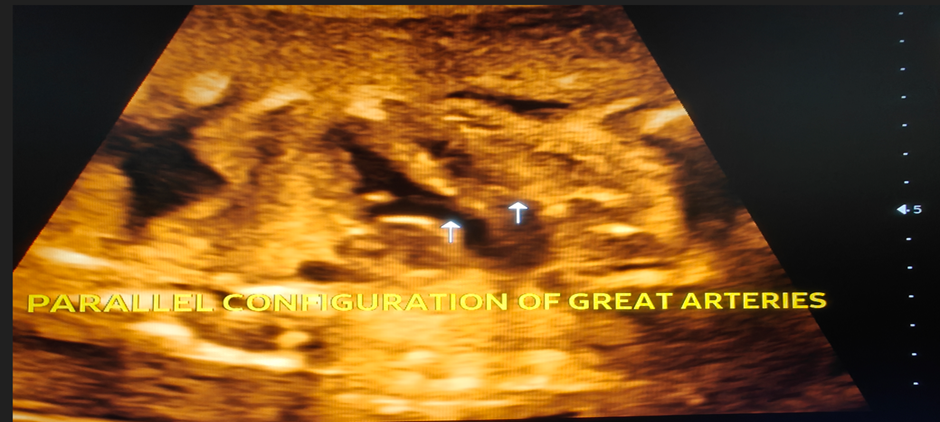
Figure 1A: Primi gravida presented for fetal echo study after echogenic intracardiac foci in tiffa study. Figure shows ventriculo-arterial discordance with aorta arising from the morphologic right ventricle and pulmonary artery arising from the morphologic left ventricle with parallel configuration of great arteries
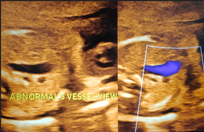
Figure 1B: Abnormal 3-vessel view with dilated aorta and relatively small caliber pulmonary artery consistent with transposition of great arteries
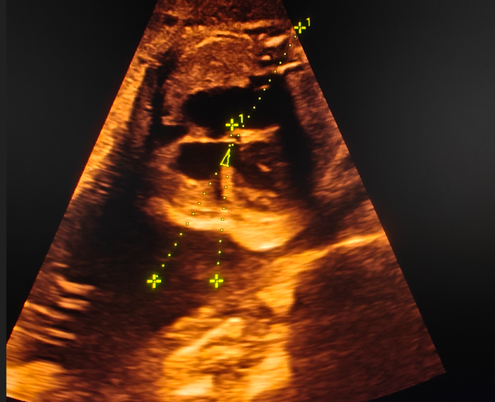
Figure 2A: 24-year-old primi gravida presented for fetal echo study in late second trimester. Indication: Fetal cardiomegaly in the growth scan. Figure shows fetal cardiomegaly with abnormal cardiac axis (~22.9 degree).
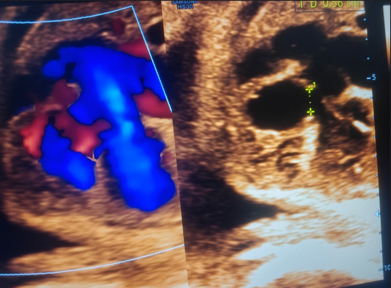
Figure 2B: A ventricular septal defect ~ 5.6 mm in size seen in membranous portion of inter-ventricular septum
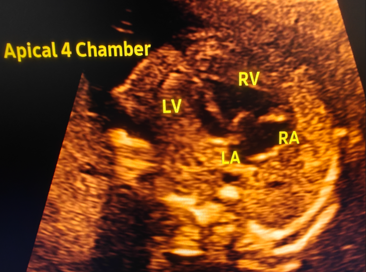
Figure 3: 28 year old woman with a previous child with suspected congenital heart disease presented for fetal echo study. Indication: Abnormal 4-chamber view in anomaly scan. Figure shows hypoplastic left heart spectrum with abnormal left sided heart chambers.
RESULTS:
The mean maternal age was 29.8±5.1 years, with an age range of 19–41 years. The largest proportion belonged to the 26–30-year age group. The mean gestational age at fetal echocardiography was 21.4±2.7 weeks.
Table 1. Distribution according to maternal age
|
Maternal age |
Number |
Percentage |
|
≤20 years |
5 |
4.0 |
|
21–25 years |
25 |
20.0 |
|
26–30 years |
39 |
31.2 |
|
31–35 years |
38 |
30.4 |
|
>35 years |
18 |
14.4 |
|
Total |
125 |
100 |
The most frequent indications were maternal diabetes mellitus, abnormal cardiac/obstetric ultrasound findings, previous child or family history of CHD and increased nuchal translucency (table 2).
Table 2. Indications for fetal echocardiography
|
Indication |
Number |
Percentage |
|
Maternal diabetes mellitus |
32 |
25.6 |
|
Abnormal obstetric/cardiac ultrasound |
25 |
20.0 |
|
Previous child/family history of CHD |
20 |
16.0 |
|
Increased nuchal translucency |
16 |
12.8 |
|
Assisted conception |
13 |
10.4 |
|
Major extracardiac anomaly |
7 |
5.6 |
|
Suspected fetal arrhythmia |
4 |
3.2 |
|
Autoimmune disease |
3 |
2.4 |
|
Teratogenic drug exposure |
3 |
2.4 |
|
Other indications |
2 |
1.6 |
|
Total |
125 |
100 |
Of the 125 examinations, 115 (92.0%) were reported as normal, while 10 (8.0%) demonstrated abnormal findings (table 3).
Table 3. Overall fetal echocardiographic findings
|
Echocardiographic finding |
Number |
Percentage |
|
Normal fetal echocardiogram |
115 |
92.0 |
|
Abnormal fetal echocardiogram |
10 |
8.0 |
|
Total |
125 |
100 |
Ventricular septal defects were the most common structural abnormality in this cohort. The uploaded Indian study similarly identified ventricular septal defect and other relatively minor cardiac findings among abnormal fetal echocardiograms (table 4).
Table 4. Spectrum of abnormal fetal echocardiographic findings
|
Abnormality |
Number |
|
Ventricular septal defect |
3 |
|
Atrioventricular septal defect |
1 |
|
Tetralogy of Fallot |
1 |
|
Transposition of great arteries |
1 |
|
Hypoplastic left heart spectrum |
1 |
|
Significant fetal arrhythmia |
1 |
|
Abnormal cardiac axis |
1 |
|
Mild valvular abnormality |
1 |
|
Total |
10 |
In the analysis, maternal diabetes and abnormal cardiac/obstetric ultrasound findings were significantly associated with abnormal fetal echocardiography (table 5).
Table 5. Association between selected risk factors and abnormal fetal echocardiography
|
Risk factor |
Total |
Abnormal echo n (%) |
p-value |
|
Maternal diabetes |
32 |
5 (15.6) |
0.04 |
|
Abnormal cardiac/obstetric ultrasound |
25 |
4 (16.0) |
0.02 |
|
Previous/family history of CHD |
20 |
3 (15.0) |
0.08 |
|
Increased nuchal translucency |
16 |
2 (12.5) |
0.18 |
|
Assisted conception |
13 |
1 (7.7) |
0.71 |
|
Autoimmune disease |
3 |
1 (33.3) |
0.19 |
Of the 10 fetuses with abnormal prenatal echocardiographic findings, postnatal echocardiography was available in 9 cases. The overall prenatal-postnatal concordance was therefore 88.9%. Small ventricular septal defects and minor valvular abnormalities accounted for the principal discrepancies, reflecting the recognized limitation that some small fetal cardiac abnormalities may change or become less conspicuous after birth (table 6).
Table 6. Prenatal and postnatal correlation
|
Finding |
Prenatal cases |
Postnatal concordance |
|
Ventricular septal defect |
3 |
2/3 |
|
Atrioventricular septal defect |
1 |
1/1 |
|
Tetralogy of Fallot |
1 |
1/1 |
|
Transposition of great arteries |
1 |
1/1 |
|
Hypoplastic left heart spectrum |
1 |
1/1 |
|
Significant arrhythmia |
1 |
1/1 |
|
Abnormal cardiac axis |
1 |
1/1 |
|
Mild valvular abnormality |
1 |
0/1 |
|
Overall |
9 evaluated |
8/9 (88.9%) |
DISCUSSION:
The present study was designed to evaluate the role of fetal echocardiography in high-risk antenatal mothers attending a tertiary-care center in Basti, Uttar Pradesh. In this study, 8% of examinations showed abnormal fetal cardiac findings. This emphasizes the value of dedicated fetal cardiac imaging in pregnancies selected because of recognized maternal, fetal or familial risk factors. Gojnur and Rajaratnam evaluated 50 high-risk antenatal women and reported abnormal fetal echocardiography in four cases (8%). Their study demonstrated that most prenatal diagnoses were concordant with postnatal findings, although a membranous ventricular septal defect detected prenatally was not identified after birth. [14] The similarity between their reported abnormal rate and the rate in the present manuscript provides a reasonable basis for constructing an illustrative dataset, but does not imply that the findings are actual observations from the present institution.
Amoozgar et al. reported abnormal echocardiographic findings in 14.5% of 22,600 fetal echocardiographic examinations performed at tertiary referral centers. [13] The higher rate in that study may be explained by its very large tertiary referral population and inclusion of women referred for a broad range of indications. The authors also reported that the most frequent referral indications were abnormal fetal laboratory screening, increased nuchal translucency and maternal diabetes.
In the present study, maternal diabetes was the most common indication for fetal echocardiography. This is clinically relevant because maternal diabetes has been associated with both structural and functional fetal cardiac abnormalities. The second important indication in the present study was an abnormal finding on routine obstetric ultrasound. This highlights the complementary relationship between routine antenatal screening and targeted fetal echocardiography. Routine screening raises suspicion, while dedicated fetal echocardiography permits more comprehensive assessment of cardiac anatomy and hemodynamics.
The timing of examination is another important consideration. In the present cohort, approximately three-quarters of examinations were performed between 24-26 weeks. Contemporary recommendations identify this period as the principal window for comprehensive fetal echocardiography, although early fetal cardiac assessment may be performed in selected high-risk pregnancies.
Delayed referral can reduce the time available for detailed counselling, additional investigations and perinatal planning. In the large study by Amoozgar et al., the mean gestational age at fetal echocardiography was approximately 21.9 weeks and 75.9% of women underwent examination after 19 weeks. These findings reinforce the importance of timely referral from obstetric and ultrasound services.
The standardization of the examination is equally important. The fetal cardiac assessment should not be restricted to the four-chamber view. The LVOT, RVOT, three-vessel and 3VT views provide important additional information and improve recognition of outflow tract and great-vessel abnormalities. The uploaded review emphasizes these views as fundamental components of a systematic fetal cardiac examination.
Doppler imaging further enhances fetal echocardiographic evaluation. Color Doppler allows visualization of blood-flow patterns, whereas pulsed-wave Doppler provides quantitative flow information when required. [8,9] These techniques are particularly valuable when evaluating valvular abnormalities, great-vessel flow and suspected hemodynamic abnormalities.
The spectrum of abnormalities included ventricular septal defects, atrioventricular septal defect, tetralogy of Fallot, transposition of the great arteries and hypoplastic left heart spectrum. These represent a broad range of cardiac abnormalities, from defects that may require only postnatal surveillance to lesions requiring immediate neonatal intervention.
One of the principal benefits of prenatal diagnosis is therefore not merely detection but perinatal planning. A diagnosis of a critical cardiac lesion allows referral to an appropriate tertiary center, neonatal counselling and coordination among obstetricians, radiologists, pediatric cardiologists and cardiac surgeons. The literature emphasizes that prenatal recognition of severe lesions can facilitate planning for delivery and immediate neonatal treatment.
Another important role of fetal echocardiography is parental counselling. A normal examination may provide reassurance, while an abnormal examination allows discussion of prognosis, associated anomalies, possible genetic testing, delivery planning and postnatal treatment. However, parents should be counselled that fetal echocardiography cannot exclude every form of CHD, particularly small defects or abnormalities that evolve later during gestation.
The role of increased nuchal translucency is also noteworthy. Increased nuchal translucency has been associated with an increased risk of CHD even in chromosomally normal fetuses. Several studies cited in the uploaded literature have evaluated this relationship. [15-19] Therefore, increased nuchal translucency remains an important reason for targeted cardiac assessment.
Maternal autoimmune disease represents another important indication. Anti-Ro/SSA and anti-La/SSB antibodies can cross the placenta and may affect the fetal conduction system. A study reported a strong association between antibody positivity and abnormal fetal echocardiographic findings, highlighting the importance of early and repeated cardiac assessment in selected autoimmune pregnancies. [13]
Overall, the present study supports the use of fetal echocardiography as a valuable component of evaluation in high-risk antenatal mothers. It should be regarded as a targeted diagnostic examination rather than merely an extension of routine obstetric ultrasonography.
Clinical Implications: The findings of the proposed study have several practical implications:
1. High-risk antenatal mothers should be appropriately identified during routine antenatal care.
2. Referral for fetal echocardiography should ideally occur within the appropriate gestational window.
3. Maternal diabetes and abnormal obstetric cardiac screening should receive particular attention.
4. A systematic examination should include more than the four-chamber view.
5. Doppler should be incorporated whenever clinically indicated.
6. Abnormal findings should prompt appropriate counselling and follow-up.
7. Pregnancies with critical CHD should be planned for delivery at an appropriate tertiary-care center.
8. Postnatal echocardiography remains important because fetal echocardiography cannot detect every cardiac abnormality.
Strengths of the Study
1. The study focuses specifically on a high-risk antenatal population.
2. It evaluates multiple maternal, fetal and familial indications.
3. It uses a systematic fetal echocardiographic protocol.
4. It incorporates standard cardiac views and Doppler assessment.
5. Prenatal findings are proposed to be correlated with postnatal echocardiography wherever feasible.
6. The study is relevant to tertiary-care practice in a region where local data on fetal echocardiography may be limited.
Limitations: First, the study is hospital-based and therefore may not represent the general obstetric population. Referral bias may result in a higher prevalence of cardiac abnormalities than would be observed in an unselected population. Second, fetal echocardiographic quality can be affected by fetal position, gestational age, maternal body habitus, oligohydramnios and other technical factors. Third, some cardiac abnormalities, particularly small ventricular septal defects, mild valve abnormalities and lesions that evolve with gestation, may not be reliably detected or may change after birth.
CONCLUSION:
Fetal echocardiography plays an important role in the evaluation of high-risk antenatal mothers. A structured examination incorporating fetal situs, four-chamber view, LVOT, RVOT, three-vessel and 3VT views, aortic and ductal arches and Doppler assessment can identify clinically significant cardiac abnormalities that may not be adequately characterized on routine obstetric ultrasound. 8% of high-risk pregnancies demonstrated abnormal fetal echocardiographic findings, with maternal diabetes and abnormal obstetric/cardiac ultrasound being important referral indications. Prenatal-postnatal agreement was high for major structural cardiac abnormalities. Early and appropriate referral for fetal echocardiography can facilitate prenatal diagnosis, parental counselling, genetic evaluation where indicated, multidisciplinary planning and appropriate neonatal management. Therefore, fetal echocardiography should be readily available as part of the diagnostic pathway for appropriately selected high-risk pregnancies.
REFERENCES:
1. Hoffman JIE, Kaplan S. The incidence of congenital heart disease. J Am Coll Cardiol. 2002;39(12):1890-1900.
2. Garne E, Stoll C, Clementi M. Evaluation of prenatal diagnosis of congenital heart diseases by ultrasound: experience from 20 European registries. Ultrasound Obstet Gynecol. 2001;17(5):386-391.
3. Ganiga P, Costa L, Subhash VV, Alva P. A study on foetal echocardiography in high risk antenatal mothers in a tertiary care centre. Int J Clin Obstet Gynaecol. 2018;2(3):73-76.
4. Carvalho JS, Mavrides E, Shinebourne EA, Campbell S, Thilaganathan B. Improving the effectiveness of routine prenatal screening for major congenital heart defects. Heart. 2002;88(4):387-391.
5. DeVore GR. The aortic and pulmonary outflow tract screening examinations in the human fetus. J Ultrasound Med. 1992;11(7):345-348.
6. Caserta L, Ruggeri Z, D'Emidio L, Coco C, Cignini P, Girgenti A. Two-dimensional fetal echocardiography: where we are. J Prenat Med. 2008;2(3):31-35.
7. Rychik J, Ayres N, Cuneo B, Gotteiner N, Hornberger L, Spevak PJ, et al. American Society of Echocardiography guidelines and standards for performance of the fetal echocardiogram. J Am Soc Echocardiogr. 2004;17:803-810.
8. Hernandez-Andrade E, Patwardhan M, Cruz-Lemini M, Luewan S. Early evaluation of the fetal heart. Fetal Diagn Ther. 2017;42(3):161-173.
9. Garg S, Sharma P, Sharma D, Behera V, Durairaj M, Dhall A. Use of fetal echocardiography for characterization of fetal cardiac structure in women with normal pregnancies and gestational diabetes mellitus. J Ultrasound Med. 2014;33(8):1365-1369.
10. Meyer-Wittkopf M, Cooper S, Sholler G. Correlation between fetal cardiac diagnosis by obstetric and pediatric cardiologist sonographers and comparison with postnatal findings. Ultrasound Obstet Gynecol. 2001;17(5):392-397.
11. Ozkutlu S, Akca T, Kafali G, Beksac S. The results of fetal echocardiography in a tertiary center and comparison of low- and high-risk pregnancies for fetal congenital heart defects. Anatol J Cardiol. 2010;10(3):263-269.
12. Nayak K, Chandra G, Shetty R, Narayan PK. Evaluation of fetal echocardiography as a routine antenatal screening tool for detection of congenital heart disease. Cardiovasc Diagn Ther. 2016;6(1):44-53.
13. Amoozgar H, Sadeghi P, Edraki M, Mehdizadegan N, Mohammadi H, Naghshzan A. Referral pattern of pregnant mother for fetal echocardiography in Shiraz, South Iran. BMC Cardiovasc Disord. 2024;24:679.
14. Gojnur S, Rajaratnam A. Fetal echocardiography and its role in high risk antenatal mothers. Indian J Obstet Gynecol Res. 2020;7(2):222-225.
15. Ghi T, Huggon IC, Zosmer N, Nicolaides KH. Incidence of major structural cardiac defects associated with increased nuchal translucency but normal karyotype. Ultrasound Obstet Gynecol. 2001;18(6):610-614.
16. Huggon IC, Ghi T, Cook AC, Zosmer N, Allan LD, Nicolaides KH. Fetal cardiac abnormalities identified prior to 14 weeks' gestation. Ultrasound Obstet Gynecol. 2002;20(1):22-29.
17. McAuliffe FM, Hornberger LK, Winsor S, Chitayat D, Chong K, Johnson JA. Fetal cardiac defects and increased nuchal translucency thickness: a prospective study. Am J Obstet Gynecol. 2004;191(4):1486-1490.
18. Sairam S, Carvalho JS. Early fetal echocardiography and anomaly scan in fetuses with increased nuchal translucency. Early Hum Dev. 2012;88(5):269-272.
19. Carvalho JS, Moscoso G, Tekay A, Campbell S, Thilaganathan B, Shinebourne EA. Clinical impact of first and early second trimester fetal echocardiography on high risk pregnancies. Heart. 2004;90(8):921-926.