Cytological Evaluation of Women Presenting with Leucorrhea Using Pap Smear: A Tertiary Care Hospital-Based Study.
- Keerti Sahi , Assistant Professor, Department of Pathology, Navodaya Medical College, Raichur
- Madhavi Ajit Kulkarni , Professor, Department of Pathology, Navodaya Medical College, Raichur
- A S Aanand , Professor and Head, Department of pathology, Navodaya Medical College, Raichur
- Ashraf Ahmed Zubair , Professor, Department of Pathology, Navodaya Medical College, Raichur.
Article Information:
Abstract:
Background: Vaginal discharge is a common clinical complaint and can be related to inflammatory and infectious conditions of the genital tract. Pap smear examination can provide cytological information about inflammatory changes, selected infectious organisms, and abnormalities of the cervical epithelium. Aims: To identify the etiological factors of vaginal discharge, to determine the prevalence of pathogenic microorganisms in leucorrhea and to study the pattern of cervical smear cytology in women presenting with vaginal discharge. Methods: A prospective study was conducted for a period of one year from September 2024 to September 2025 in the Cytology Section of the Department of Pathology, Navodaya Medical College, Raichur. 111 women with vaginal discharge attending the Obstetrics and Gynecology OPD were evaluated. Demographic details, clinical history and per-speculum finding were recorded. Two cytopathologists reviewed conventional cervical smears prepared and fixed in absolute alcohol and stained by the Papanicolaou method according to the Bethesda system. Results: 111 women were included in the analysis. The age ranged from 17 to 60 years with a mean age of 33.0 ± 8.2 years and a median age of 33 years. Most were in the age group of 30-39 years (55; 49.5%). The most common cytological finding was nilm with inflammatory changes (65; 58.6%). The most common specific infectious diagnosis was bacterial vaginosis (27; 24.3%) followed by trichomoniasis (9; 8.1%) and fungal infection/candidiasis (9; 8.1%). One case of ASC-US (0.9%) was observed. Specific infectious diagnoses were found in 45 of 111 women (40.5%). Conclusion: Pap smear examination in women presenting with leucorrhea provides useful information regarding inflammatory changes and selected infectious organisms and may also identify cervical epithelial abnormalities that require further evaluation.
Keywords:
Article :
INTRODUCTION:
Vaginal discharge may arise from any part of upper and lower genital tract. The vaginal microbial environment is dynamic and shaped by factors such as pregnancy, the menstrual cycle, sexual activity, age, and contraceptive use[1]. Vaginal dischargecan be normal physiological or pathological but its presence can be a source of concern for women.[2] Physiological discharge is seen during ovulation and secretory phase of menstrual cycle.[3]In many women, Lactobacilli dominate the vaginal flora. These beneficial bacteria help protect against harmful pathogens, thereby lowering the risk of urinary tract infections and sexually transmitted infections (STIs)[4].Changes in balance of normal vaginal flora can cause overgrowth of some organisms, when one class of them dominates leading to vaginitis or vaginosis.[5]
Inadequate genital hygiene is a significant factor underlying the high rates of excessive vaginal discharge observed among Indian women.[6] The most common infections commonly encountered with vaginal discharge include Bacterial Vaginosis (BV), Vaginal Candidiasis (VC) and Trichomonas Vaginalis (TV).[5][ Hence, excessive vaginal discharge called as Leucorrhoea is the clinical evidence of genital infection.[4]Untreated or poorly managed vaginitis is associated with a range of adverse outcomes. Pelvic inflammatory disease may occur as a consequence of untreated bacterial vaginosis or STIs like trichomoniasis, leading to long-term sequelae such as chronic pelvic pain, ectopic pregnancy, and infertility. Furthermore, vaginitis heightens the risk of STI acquisition and transmission, including HIV, due to disruption of mucosal integrity and immune protection.
Pap test is a simple and inexpensive method to screen in women for squamous dysplasia and carcinoma of uterine cervix. Cervical carcinoma which may present as leucorrhoea is a leading cause of morbidity and mortality among women worldwide [7]. It is the second most common cancer among women after breast cancer [8]. It is presented by spectrum of precancerous lesions called cervical intraepithelial neoplasia (CIN) [9]. Therefore, evaluating women presenting with leucorrhea through Pap smear screening is essential, as it provides valuable insights into the underlying etiology and guides appropriate management. The present study was undertaken to identify various causes of vaginal discharge, frequency of various pathogenic microorganisms in cervical smear and to study various cytopathological changes of cervix in cases with reproductive tract infection.
METHODOLOGY:
This is a prospective study conducted for a duration of one year from September 2024 to September 2025. The study iscarried out in cytology section of pathology department at Navodaya Medical College, Raichur. It included 111 female patients aged between 18 years to 75 years who attended an obstetrics and gynaecology OPD at a tertiary care hospital with complaint of vaginal discharge. Study participants were explained about the objectives of the study and informed consent was obtained from them. Demographic variables and detailed history was obtained. Per speculum examination was done and any abnormality noted was recorded. Characteristics of vaginal discharge were also noted. Cervical smear samples were collected by gynaecologists and conventional smears were prepared by trained technical staff and were sent to cytology section after fixation with absolute alcohol. The slides were stained with Papanicolaou stain, examined and reported by two cytopathologists according to the Bethesda system. Bacterial vaginosis was reported based on the presence of clue cells and coccobacilli. Candidiasis was diagnosed as a characteristic organism with spore, budding yeast or fungal hyphae.Trichomonas vaginalis was identified as a pear-shaped blue or grey organism with bright red granules and a proteinaceous background, often with severe acute inflammation.
With objective
1) To identify causes of vaginal discharge
2) Frequency of pathogenic microorganisms which are responsible for infective causes of leucorrhoea.
3) To evaluate the pattern of cervical smear cytology
Inclusion Criteria: patients attending obstetrics and gynaecology clinic at tertiary care hospital with complaints of vaginal discharge.
Exclusion Criteria: 1) Patients with history of medication in past 14 daysm
2) Pregnant woman
Sample Size Calculation
n= (zα/2)2*p*(1-P)/d2
The formula is used to calculate the sample size.
*n=sample size
*zα/2 =Z value corresponding to the desired confidence level (e.g.1.96 for a 95% confidence level)
Prevalence=83% or 0.83(taken from the parental article) 10
Non prevalence, 1-p=13% or 0.13
Allowable error=7% =0.07
Zα/2=1.96 at 95% confidence interval
|
P |
Prevalence or sensitivity |
837% or 0.83 |
|
1-α |
Confidence interval |
0.95 |
|
Zα/2 |
Z-value associated with confidence |
1.96 |
|
D |
Allowable Error |
0.07 |
|
N |
Minimum sample size |
111 |
By substituting the above values in the formula, the calculated sample size is 111.
Data will be analysed using SPSSv22 software, all categorical data will be presented using frequencies and percentages. The continuous data will be described using mean (or median) and standard deviationto study the association or significance of clinical and demographic factors with poor clinical outcome, chi-square (χ2-test) will be used. T-test will be used for continuous variables as test of significance. p<0.05 is considered to be significant.
RESULTS:
A total of 111 women presenting with leucorrhea were included in the study. The age of the participants ranged from 17 to 60 years, with a mean age of 33.0 ± 8.2 years and a median age of 33 years. The majority of participants belonged to the 30–39-year age group (55; 49.5%), followed by the 20–29-year age group (32; 28.8%).On Pap-smear examination, the most common finding was NILM with inflammatory changes, observed in 65 women (58.6%). Among the specific infectious diagnoses, bacterial vaginosis was the most frequent, identified in 27 women (24.3%). Trichomoniasis was identified in 9 women (8.1%), while fungal infection/candidiasis was identified in 9 women (8.1%). One woman (0.9%) showed ASC-US.
Thus, specific infectious diagnoses were identified in 45 of 111 women (40.5%), with bacterial vaginosis constituting the predominant infectious finding. The remaining majority showed NILM with inflammatory changes. The single case of ASC-US represented 0.9% of the study population.On age-wise analysis, the 30–39-year age group contributed the largest number of inflammatory as well as infectious Pap-smear findings, corresponding with its predominance in the study population. Among women aged 30–39 years, 31 had NILM with inflammatory changes, 13 had bacterial vaginosis, 6 had trichomoniasis, 4 had fungal infection and 1 had ASC-US.Per-speculum examination demonstrated findings including cervical erosion, bleeding on touch, thick/curdy white discharge and foul-smelling discharge in different participants. These clinical findings were variably associated with inflammatory and infectious cytological findings.Overall, the findings demonstrate that Pap smear can provide useful cytological information regarding inflammatory changes and selected infectious etiologies among women presenting with leucorrhea, while also identifying occasional epithelial abnormalities such as ASC-US.
Table 1. Overall Pap-smear diagnostic profile of Study participants (n=111)
|
Pap-smear diagnosis |
No. |
Percentage |
|
NILM – inflammatory |
65 |
58.6% |
|
Bacterial vaginosis (BV) |
27 |
24.3% |
|
Trichomoniasis (TV) |
9 |
8.1% |
|
Fungal infection/candidiasis |
9 |
8.1% |
|
ASC-US |
1 |
0.9% |
|
Total |
111 |
100% |
The most frequent Pap-smear finding was NILM with inflammatory changes (58.6%). This indicates that although no epithelial lesion was identified, a substantial proportion of women showed inflammatory cytological changes.Among specific infectious diagnoses, bacterial vaginosis was the predominant infection (24.3%), followed by trichomoniasis (8.1%) and fungal infection (8.1%).Thus, Pap smear provided clinically relevant information regarding theinflammatory and infectious causes associated with leucorrhea, in addition to screening for epithelial abnormalities.
Table 2. Age distribution of Study participants (n=111)
The age of women ranged from 17 to 60 years. The majority belonged to the 30–39-year age group (55/111; 49.5%), followed by 20–29. years (32/111; 28.8%).
|
Age group |
No. |
% |
|
≤19 years |
3 |
2.7% |
|
20–29 years |
32 |
28.8% |
|
30–39 years |
55 |
49.5% |
|
40–49 years |
17 |
15.3% |
|
50–59 years |
2 |
1.8% |
|
≥60 years |
2 |
1.8% |
For interpretation, the Pap-smear diagnosis can also be grouped into:
· NILM with inflammatory changes:65 (58.6%)
· Infectious diagnoses: 45 (40.5%)
· BV: 27
· Trichomoniasis: 9
· Fungal infection: 9
· ASC-US: 1 (0.9%)
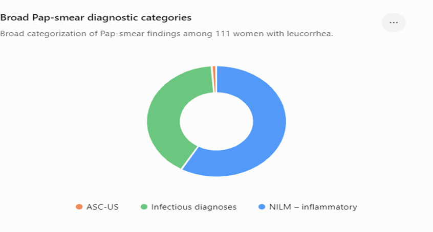
Fig 1. Infectious versus non-infectious/inflammatory findings of Study participants(n=111)
Approximately two-fifths (40.5%) of women had a specific infectious diagnosis identifiable on Pap smear. This is an important finding because leucorrhea is a symptom with multiple possible etiologies, and cytological identification of organisms or characteristic changes can contribute to etiological assessment.At the same time, 58.6% had NILM with inflammatory changes, emphasizing that inflammatory cytology is much more common than a specific identifiable infection in this dataset.
Table 4. Age-wise distribution of Pap-smear findings of study participants (n=111)
|
Age group |
NILM-inflammatory |
BV |
TV |
Fungal |
ASC-US |
Total |
|
≤19 |
1 |
1 |
0 |
1 |
0 |
3 |
|
20–29 |
18 |
7 |
3 |
4 |
0 |
32 |
|
30–39 |
31 |
13 |
6 |
4 |
1 |
55 |
|
40–49 |
12 |
5 |
0 |
0 |
0 |
17 |
|
50–59 |
1 |
1 |
0 |
0 |
0 |
2 |
|
≥60 |
2 |
0 |
0 |
0 |
0 |
2 |
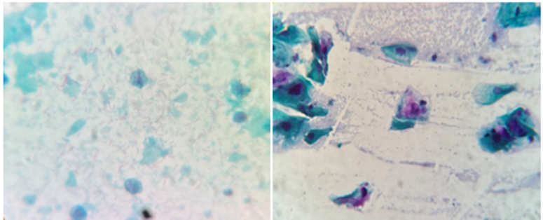
Fig 2. Trichomonas Vaginalis
Fig 3 ASCUS (Atypical squamous cell of undetermined significance)
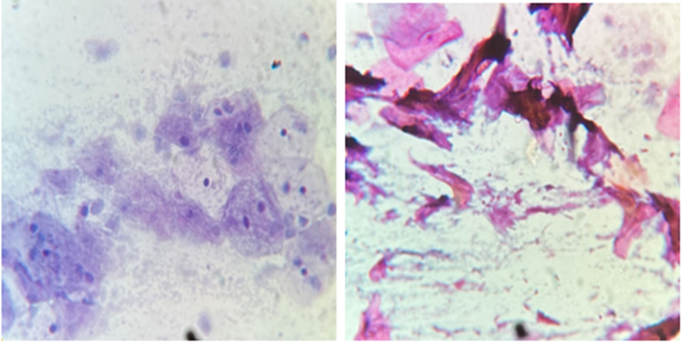
Fig 4. Bacterial Vaginosis Fig 5. Candidiasis
DISCUSSION:
Among the 111 women with vaginal discharge attending obstetrics and gynaecology OPD evaluated for Pap smear examination, specific infectious diagnoses were identified in 45 of 111 women (40.5%). The results are correlating with the studies done by Salih et al11, and Sivaranjini et al12, who found 53% and 44.5%, respectively. However, a study done by Priyadarshi et al, found that76% of cases in their study were found to have infectious causes for vaginal discharge.Among the specific infectious diagnoses, bacterial vaginosis was the most frequent, identified in 27 women (24.3%). Trichomoniasis was identified in 9 women (8.1%), while fungal infection/candidiasis was identified in 9 women (8.1%). The most common organism found in the study conducted by Priyadarshi et al, was BV (71%), followed by vaginal candidiasis (3%) and T. vaginalis (2%) - (total 76%)10. Salih et al11,has found 33.5% cases of candidiasis and 19.5% cases of bacterial vaginosis (Total 53%), Barautiet al13, found 17.2% cases of bacterial vaginosis, followed by 10.6% candidiasis and 0.4% T. vaginalis, and R. Sivaranjini et al12, concluded BV in 26.25% cases, candidiasis in 15.25% of cases, and T.
vaginalis in 3% cases (total 44.5%). An article published by Hildebrand has mentioned that Bacterial vaginosis is the most common cause of vaginitis, accounting for 40% to 50% of cases, followed by yeast infections, which affect about 20% to 25% of women at least once in their lifetime.14On age-wise analysis, the 30–39-year age group contributed the largest number of inflammatory as well as infectious Pap-smear findings, corresponding with its predominance in the study population. Study done by Priyadarshi et al has shown the presence of pathogenic organisms greater among the age group of 26–35 years, followed by 36–45 years.1058.6% had NILM with inflammatory changes. Inflammation was detected in 83% of cases and 77% of cases in the studies done by Priyadarshi et al10 and Barautiet al13 respectively.In the present study, Onlyone patient (0.9%) had ASC-US on Pap smear. Study done by Priyadarshi et al10 showed 3 cases (3%) of ASCUS, 2 case of (2%) of LSIL and I case (1%) of HSIL whereas Salih et al, showed 1% of cases of LSIL and 2% of cases of HSIL presented with vaginal discharge.11 This is important because the primary purpose of the study is not merely identification of infections. Pap smear also helps to identify epithelial abnormalities in symptomatic women.
This observational study has several limitations. The small sample size (n=111) from a single tertiary center restricts generalizability and limits the detection of rare cytological abnormalities. Furthermore, because the cohort exclusively comprised symptomatic women (vaginal discharge), the findings do not represent asymptomatic screening populations, nor can causal relationships be established. Finally, the absence of confirmatory microbiological testing and histopathological or colposcopic follow-up for the ASC-US case precluded the calculation of diagnostic accuracy metrics such as sensitivity and specificity.
CONCLUSION:
Infectious causes contribute substantially to abnormal vaginal discharge. Pap smear examination, particularly in symptomatic women, can provide useful information on vaginal microorganisms and cervical premalignant lesions. Improving awareness of cervical screening and incorporating Pap smear into routine screening of sexually active women may facilitate early detection of clinically significant cervical abnormalities.
REFERENCES:
1. Chee WJY, Chew SY, Than LTL. Vaginal microbiome: considerations for reproductive health. Microorganisms. 2022;10(7):1301
2. Patel V, Pednekar S, Weiss H, Rodrigues M, Barros P, Nayak B, et al. Why do women complain of vaginal discharge? A population survey of infectious and pyschosocial risk factors in a South Asian community. Int J Epidemiol. 2005 Apr 15;34(4):853-62.
3. Rani S, Qamarunissa, Fakharunissa, Waqarunissa, Frequency of Abnormal Cervical Smear in Women Presenting with Vaginal Discharge; Imperial J Interdisci Res (IJIR). 2016;2(4):722-6.
4. Saraf VS, Sheikh SA, Ahmad A, Gillevet PM, Bokhari H, Javed S. Vaginal microbiome: normalcy vs dysbiosis. Arch Microbiol. 2021 Sep;203(7):3793-3802
5. Sujatha P, Indira V, Kumar KM. Study of PAP smear examination in patients complaining of leucorrhoea-A 2 years prospective study in a teaching hospital. IAIM. 2016;3(5):106-12.
6. Koteswari K, Rao N et al. A Study of Pap smear Examination in Women Complaining of Leucorrhea, DOI: 10.9790/0853-14143742, e-ISSN: 2279-0853, p-ISSN: 2279-0861 Ver. IV. 2015; 14(1):37-42.
7. Patel M, Pandya A, Modi J. Cervical Pap Smear Study And Its Utility In Cancer Screening, To Specify, The Strategy For Cervical Cancer Control. National Journal of Community Medicine. 2011; 2(1). ISSN: 0976 3325
8. Bengtsson E, Malm P, Screening for Cervical Cancer Using Automated Analysis of PAP-Smears, 2014, Article ID 842037
9. Warpe B, Warpe S, Sawant S. An institution-based cervical PAP smear study, correlation with clinical findings & histopathology in the Konkan region of Maharashtra state, India. Walawalkar International Medical Journal. 2016; 3(1).
10. Priyadarshi A, Halder NR, Halder B, Singh N, Sinha A. Evaluation of Pap smear examination in patients with complaint of vaginal discharge in a tertiary care hospital. Int J Pharm Clin Res. 2023;15(5):1265-1270
11. Salih MM, Alhag FT, Khalifa MA, El Nabi AH. Cervical cytopathological changes among women with vaginal discharge attending teaching hospital. J Cytol. 2017 Apr;34(2):90-4.
12. Sivaranjini R, Jaisankar TJ, Thappa DM, Kumari R, Chandrasekhar L, Malathi M, et al. Spectrum of vaginal discharge in a tertiary care setting. Trop Parasitol. 2013 Jul; 3(2):135.
13. Barouti E, Farzaneh F, Sene AA, Tajik Z, Jafari B. The pathogenic microorganisms in Papanicolaou vaginal smears and correlation with inflammation. J Family Repr Health. 2013 Mar;7(1):23.
14. Hildebrand JP, Carlson K, Kansagor AT. Vaginitis. [Updated 2025 Jan 19]. In: StatPearls [Internet]. Treasure Island (FL): StatPearls Publishing; 2026 Jan.