Cytomorphological Spectrum of Thyroid Lesions on Fine-Needle Aspiration Cytology with Cytoradiological Correlation.
- Saara Neeha , Assistant Professor, Dept of Pathology, Navodaya Medical College Hospital And Research Center, Raichur.
- Seemant Kumar Saini , Professor, Department of Pathology Ananta Institute of Medical Sciences Rajsamand Rajasthan.
- Pranshu Sharma , Professor, Department of Pathology Ananta Institute of Medical Sciences Rajsamand Rajasthan.
- Mohammed Ismail , Associate Professor, Dept of Radiology, Raichur Institute of Medical Sciences, Raichur
Article Information:
Abstract:
Background: Fine-needle aspiration cytology (FNAC) is an established minimally invasive investigation for thyroid nodules and is central to distinguishing benign from potentially malignant lesions. Standardized reporting using the Bethesda System improves diagnostic communication and links cytological categories to clinically relevant management pathways. Ultrasonography provides complementary structural risk assessment, with ACR TI-RADS offering a standardized approach to thyroid nodule stratification. Aim: To evaluate the cytomorphological spectrum of thyroid lesions diagnosed by FNAC, describe their distribution according to the Bethesda System, and assess the descriptive relationship between cytological diagnoses and available ACR TI-RADS categories. Materials and Methods:This retrospective descriptive study included 50 thyroid FNAC cases. Demographic, clinical, nodule, procedural, adequacy, cytomorphological, Bethesda, and available ACR TI-RADS data were analyzed. Histopathological follow-up was not available; therefore, diagnostic performance parameters were not calculated. Results: Patients were 19–57 years old (mean 34.98 years); 41 (82%) were female and 9 (18%) were male. Palpation-guided FNAC was performed in 31 (62%) cases and ultrasound-guided FNAC in 19 (38%). Forty-five (90%) specimens were recorded as adequate. Bethesda II was predominant (41/50, 82%), followed by Bethesda I (6/50, 12%), Bethesda VI (2/50, 4%), and Bethesda IV (1/50, 2%); no Bethesda III or V cases were recorded. Colloid nodule (38%) and Hashimoto/chronic lymphocytic thyroiditis (34%) were the leading diagnoses. Macrophages were present in 48%, lymphocytes in 40%, and Hurthle cells in 34%. ACR TI-RADS categories were TR1 70%, TR2 20%, TR3 4%, and TR4 6%; no TR5 cases were recorded. All Bethesda IV and VI lesions occurred in TR3/TR4 nodules, although benign Bethesda II lesions were also present in these categories. Conclusion: The series showed a predominantly benign cytological spectrum, with colloid nodules and chronic lymphocytic/Hashimoto thyroiditis as the major diagnostic groups. Bethesda reporting provides a consistent framework for thyroid FNAC, while ACR TI-RADS offers complementary radiological risk stratification. The small sample size, incomplete radiological variables, and absence of histopathological follow-up limit conclusions regarding diagnostic performance or a statistically validated cytoradiological association.
Keywords:
Article :
INTRODUCTION:
Thyroid nodules are common in clinical practice and most of them are benign. The challenge for diagnosis is to detect the small minority of nodules that represent clinically significant malignancy, while avoiding unnecessary invasive procedures. FNAC is an important first-line cytological investigation because of its minimally invasive nature, wide availability and ability to distinguish many benign lesions from those requiring further investigation [1,2].
The Bethesda System for Reporting Thyroid Cytopathology was established to standardize terminology of thyroid FNA and to serve as a communication tool between cytopathologists and clinicians. The 2023 edition maintains the six diagnostic categories of nondiagnostic, benign, atypia of undetermined significance, follicular neoplasm, suspicious for malignancy, and malignant and revises the terminology and risk estimates to align with contemporary thyroid pathology [3].
Cytologic interpretation is an integrated assessment of cellularity, architecture, colloid, background inflammatory cells, Hurthle-cell change, macrophages and nuclear features. This is especially true in chronic lymphocytic/Hashimoto thyroiditis, in which lymphocytes and Hurthle cells may be prominent and may occasionally cause diagnostic problems [4,5]. Ultrasonography provides additional information about nodule composition, echogenicity, shape, margins and echogenic foci compared with cytology.
The ACR TI-RADS then uses these standardized features to assign risk categories and to guide biopsy or follow-up decisions [6]. It has been shown in studies that in selected settings ultrasound-guided FNA can improve diagnostic yield over palpation-guided sampling, although the magnitude of benefit varies with case mix and operator factors [7].
Studies in India have demonstrated a wide variability in the distribution of Bethesda categories, reflecting variation in referral patterns, iodine status, case selection and availability of histopathological follow-up [2,8-12]. In the present study, we have looked at the cytomorphological spectrum of thyroid lesions in a series of 50 cases with Bethesda categorization as the primary reporting framework and ACR TI-RADS as a secondary descriptive radiological variable.
Aim
To study the cytomorphological spectrum of thyroid lesions diagnosed by FNAC and to evaluate their Bethesda category distribution with descriptive correlation with available ACR TI-RADS categories.
MATERIALS AND METHODS:
Study design and population
This was a retrospective descriptive study of 50 patients who underwent FNAC for thyroid nodules over a period of one year. Demographic, clinical, cytological, procedural, and available radiological data were retrieved from private diagnostic center in Raichur after informed consent.
Variables assessed
The following variables were evaluated: age, sex, clinical presentation, solitary or multinodular involvement, nodule site, maximum nodule size, FNAC guidance, specimen adequacy, Bethesda category, final cytological diagnosis, colloid quantity, lymphocytes, Hurthle cells, macrophages, follicular pattern, necrosis, calcification, granulomatous/giant-cell reaction, and ACR TI-RADS category.
Cytological classification
FNAC specimens were categorized using the Bethesda System for Reporting Thyroid Cytopathology [3]. The recorded dataset included Bethesda I (nondiagnostic), II (benign), IV (follicular neoplasm), and VI (malignant), with no Bethesda III or V cases.
Radiological classification
Where available, nodules were assigned an ACR TI-RADS category using the recorded ultrasound classification [6]. Because the source dataset contained the final TI-RADS category but not the complete component-level ultrasound scores, the radiological analysis was limited to category-level descriptive comparison.
Statistical analysis
Descriptive statistics were used. Continuous variables were summarized using mean, median, and range, and categorical variables using frequencies and percentages. Because histopathological follow-up was unavailable, sensitivity, specificity, positive predictive value, negative predictive value, diagnostic accuracy, and cytohistological concordance were not calculated. Inferential testing of the Bethesda–TI-RADS relationship was not performed because of the small number of observations in the higher-risk categories.
RESULTS:
Demographic, clinical, and procedural characteristics
The study comprised 50 patients aged 19–57 years, with a mean age of 34.98 years and median age of 35 years. There was a marked female predominance (41, 82%). All patients were recorded as presenting with swelling in front of the neck. Solitary and multinodular thyroid involvement were equally represented (25 cases each). Bilateral involvement was recorded in 21 cases (42%), right-lobe involvement in 15 (30%), and left-lobe involvement in 14 (28%). The maximum nodule size ranged from 10 to 30 mm, with a mean of 20.08 mm and median of 20 mm.
Palpation-guided FNAC was performed in 31 cases (62%) and ultrasound-guided FNAC in 19 cases (38%). Forty-five specimens (90%) were recorded as adequate and five (10%) as unsatisfactory. The adequacy field and final Bethesda classification were not completely concordant in the source dataset: five specimens were recorded as unsatisfactory, whereas six cases were classified as Bethesda I. This discrepancy should be reconciled against the original cytology reports before submission.
Table 1. Baseline clinical, nodule, and FNAC characteristics
|
Variable |
Number |
% |
|
Total cases |
50 |
100 |
|
Female |
41 |
82 |
|
Male |
9 |
18 |
|
Solitary nodule |
25 |
50 |
|
Multinodular |
25 |
50 |
|
Bilateral involvement |
21 |
42 |
|
Right-lobe involvement |
15 |
30 |
|
Left-lobe involvement |
14 |
28 |
|
Palpation-guided FNAC |
31 |
62 |
|
Ultrasound-guided FNAC |
19 |
38 |
|
Adequate specimen |
45 |
90 |
|
Unsatisfactory specimen |
5 |
10 |
Bethesda distribution and final cytological diagnoses
Bethesda II was the predominant category, accounting for 41 cases (82%), followed by Bethesda I in 6 cases (12%), Bethesda VI in 2 cases (4%), and Bethesda IV in 1 case (2%). No cases were classified as Bethesda III or Bethesda V. The predominance of benign Bethesda-category lesions is consistent with several Indian series, although the exact proportions vary across institutions.
The most frequent final cytological diagnosis was colloid nodule (19 cases, 38%), followed by Hashimoto/chronic lymphocytic thyroiditis (17 cases, 34%). Cyst fluid only, unsatisfactory, and nodular goitre each accounted for 3 cases (6%); multinodular goitre accounted for 2 cases (4%); papillary thyroid carcinoma for 2 cases (4%); and suspicious for follicular neoplasm for 1 case (2%).
Table 2. Bethesda categories and final cytological diagnoses
|
Category/diagnosis |
Number |
% |
Comment |
|
Bethesda I – Nondiagnostic |
6 |
12 |
No Bethesda III or V cases |
|
Bethesda II – Benign |
41 |
82 |
Predominant category |
|
Bethesda IV – Follicular neoplasm |
1 |
2 |
One case |
|
Bethesda VI – Malignant |
2 |
4 |
Both diagnosed cytologically as papillary thyroid carcinoma |
|
Colloid nodule |
19 |
38 |
Most frequent specific diagnosis |
|
Hashimoto/chronic lymphocytic thyroiditis |
17 |
34 |
Second most frequent diagnosis |
|
Cyst fluid only |
3 |
6 |
|
|
Unsatisfactory |
3 |
6 |
Final cytological diagnosis field |
|
Nodular goitre |
3 |
6 |
|
|
Multinodular goitre |
2 |
4 |
|
|
Papillary thyroid carcinoma |
2 |
4 |
|
|
Suspicious for follicular neoplasm |
1 |
2 |
|
Cytomorphological findings
Colloid was moderate in 18 cases (36%), abundant in 16 (32%), scanty in 8 (16%), and absent in 8 (16%). Lymphocytes were present in 20 cases (40%), Hurthle cells in 17 (34%), and macrophages in 24 (48%). No necrosis, calcification, or granulomatous/giant-cell reaction was recorded. Where follicular pattern was documented, 13 cases showed a macrofollicular pattern and 8 a mixed pattern; because this variable was missing in a substantial proportion of cases, it was not used for further analysis.
The combination of lymphocytes and Hurthle cells is particularly relevant in chronic lymphocytic/Hashimoto thyroiditis, in which cytological appearances may vary with disease stage and may occasionally mimic a Hurthle-cell-rich neoplasm [10,13]. Macrophages are commonly encountered in cystic or degenerative thyroid lesions and should be interpreted in the context of the overall smear pattern.
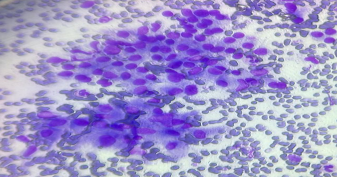
Figure 1. Thyroid FNAC smear showing hurthle cells and epithelioid like cells in a case of Hashimoto’s thyroiditis.(40x magnification, MGG Stain)
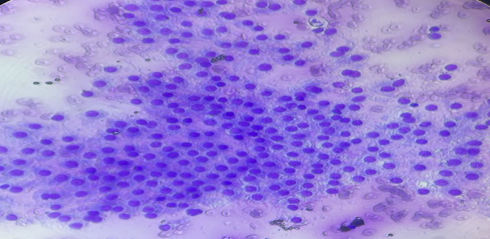
Figure 2. Thyroid FNAC smear demonstrating a cellular aspirate composed of relatively uniform round-to-oval cells arranged predominantly in cohesive groups, case of colloid nodule.( 40X,MGG Stain).
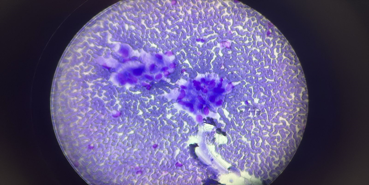
Figure 3. Thyroid FNAC smear showing hurthle cells and epithelioid like cells in a case of Hashimoto’s thyroiditis.(40x magnification, MGG Stain).
ACR TI-RADS distribution and cytoradiological correlation
TR1 was the most frequent ACR TI-RADS category (35 cases, 70%), followed by TR2 (10, 20%), TR4 (3, 6%), and TR3 (2, 4%). No TR5 cases were recorded. The predominance of lower TI-RADS categories parallels the predominantly benign cytological spectrum in this series, although category-level concordance cannot be interpreted as diagnostic validation .
All three Bethesda IV/VI cases occurred in TR3/TR4 nodules: the single Bethesda IV case was TR4, and the two Bethesda VI cases were TR3 and TR4, respectively. Two Bethesda II lesions were also present among TR3/TR4 nodules. Thus, the observed relationship is descriptive rather than definitive. The small number of TR3/TR4 cases and absence of TR5 cases preclude robust statistical assessment.
Table 3. Bethesda category distribution across ACR TI-RADS categories
|
ACR TI-RADS |
Bethesda I |
Bethesda II |
Bethesda IV |
Bethesda VI |
Total |
|
TR1 |
6 |
29 |
0 |
0 |
35 |
|
TR2 |
0 |
10 |
0 |
0 |
10 |
|
TR3 |
0 |
1 |
0 |
1 |
2 |
|
TR4 |
0 |
1 |
1 |
1 |
3 |
|
TR5 |
0 |
0 |
0 |
0 |
0 |
|
Total |
6 |
41 |
1 |
2 |
50 |
DISCUSSION:
The present series of 50 cases showed a predominantly benign spectrum of thyroid cytology with 82% cases being Bethesda II. This is generally similar to Indian studies and reviews where benign lesions form the largest Bethesda group although the reported distributions vary with institutional case mix and referral patterns [2,8–12]. The extensive use of Bethesda terminology and the difference in category frequencies among centers were noted by Agarwal and Jain in their review of Indian thyroid cytology [2]. The predominance of women in the present study (82%) also corresponds to the predominance of women reported in several cohorts of thyroid FNAC [8–11].
The mean age of 34.98 years indicates that the thyroid lesions in this series were mostly encountered in young and middle-aged adults, however the retrospective dataset does not allow broader epidemiological inference. The most common specific diagnosis was colloid nodule (38%) followed by chronic lymphocytic/Hashimoto thyroiditis (34%). The high frequency of colloid-rich lesions is consistent with the frequent occurrence of benign hyperplastic/nodular thyroid disease in routine FNAC practice. The cytological features observed in the current series, that is, lymphocytes, Hurthle cells and variable colloid, are also typical of Hashimoto thyroiditis [4,5].
Because Hurthle-cell-rich aspirates can mimic a neoplasm, it is important to assess the inflammatory background and the relationship of Hurthle cells to lymphocytes carefully [5]. The Bethesda IV case illustrates the inherent limitation of cytology in follicular patterned lesions. Cytology cannot assess capsular or vascular invasion, histological criteria necessary to differentiate follicular carcinoma from follicular adenoma. Follicular neoplasm remains a distinct entity in the 2023 Bethesda System with a well-defined management algorithm [3].
The two Bethesda VI cases were diagnosed cytologically as papillary thyroid carcinoma. The diagnosis of papillary carcinoma still rests on recognition of characteristic nuclear and architectural changes. No histopathological follow-up was available in this data set, therefore the cytological diagnoses cannot be used to calculate sensitivity, specificity or cytohistological concordance. Worth noting is that 12% Bethesda I rating. Nondiagnostic aspirates may result from inadequate follicular-cell material, cystic lesions, or sampling problems. The literature emphasizes the importance of specimen adequacy and repeat sampling when indicated [3,10]. The difference in the figures for unsatisfactory specimens (5) and for Bethesda I cases (6) as separately tabulated in the present study should be explained by examination of the original reports prior to submission.
Guidance for FNAC was palpation in 62% and ultrasound in 38%. Although contemporary evidence suggests that ultrasound guidance may improve diagnostic performance and decrease nondiagnostic sampling in selected populations, real world studies show that nodule characteristics and selection effects influence differences [7]. There was not enough paired or outcome data in the present study to statistically compare the two techniques.The distribution of ACR TI-RADS was heavily skewed toward TR1 and TR2 with only five cases in TR3/TR4 and none in TR5. ACR TI-RADS was designed to standardize the ultrasound evaluation and to recommend decisions on biopsy or follow-up [6]. All Bethesda IV and VI lesions in this series were identified in TR3/TR4 nodules, although benign Bethesda II lesions were also identified in those categories. This supports the complementary nature of ultrasound and cytology rather than establishes either alone in determining malignancy.
Comparisons with published Indian series should be interpreted with caution. Nandedkar et al. described 606 thyroid FNACs with histopathological correlation in a subset, whereas, Kamboj et al. and Sen et al. reported larger cohorts with surgical follow-up [8–10]. Bethesda classification has also been demonstrated to be useful by Mondal et al. in a large retrospective series with histological follow-up [11]. The main limitation of the present dataset is that it does not allow to calculate the diagnostic performance as for other studies.
CONCLUSION:
Most thyroid lesions in this study were benign, with colloid nodules and chronic lymphocytic/Hashimoto thyroiditis as the main diagnoses. The most common category was Bethesda II (82%), followed by I (12%), VI (4%) and IV (2%). The cytomorphological findings emphasize the importance of colloid, lymphocytes, Hurthle cells, macrophages, and follicular architecture as a whole.
According to ACR TI-RADS, all Bethesda IV and VI lesions were in TR3/TR4 nodules, but benign II lesions were too. This suggests using standardized ultrasound risk stratification and cytological assessment together. Larger studies with complete ultrasound variables and uniform histopathological follow-up are needed to evaluate diagnostic performance and establish statistically significant cytoradiological correlations.
REFERENCES:
1. Bongiovanni M, Spitale A, Faquin WC, Mazzucchelli L, Baloch ZW. The Bethesda System for Reporting Thyroid Cytopathology: a meta-analysis. Acta Cytol. 2012;56(4):333–339. doi:10.1159/000339959.
2. Agarwal S, Jain D. Thyroid cytology in India: contemporary review and meta-analysis. J Pathol Transl Med. 2017;51(6):533–547. doi:10.4132/jptm.2017.08.04.
3. Ali SZ, Baloch ZW, Cochand-Priollet B, Schmitt FC, Vielh P, VanderLaan PA. The 2023 Bethesda System for Reporting Thyroid Cytopathology. Thyroid. 2023;33(9):1039–1044. doi:10.1089/thy.2023.0141.
4. Kumar N, Ray C, Jain S. Aspiration cytology of Hashimoto's thyroiditis in an endemic area. Cytopathology. 2002;13(1):31–39. doi:10.1046/j.0956-5507.2002.00366.x.
5. Chandanwale SS, Kulkarni TV, Patel RJ, Thakkar D. A focal nodular Hürthle cell hyperplasia in Hashimoto's thyroiditis: a diagnostic dilemma on fine needle aspiration. J Cytol. 2014;31(4):236–238. doi:10.4103/0970-9371.151145.
6. Tessler FN, Middleton WD, Grant EG, Hoang JK, Berland LL, Teefey SA, et al. ACR Thyroid Imaging, Reporting and Data System (TI-RADS): White Paper of the ACR TI-RADS Committee. J Am Coll Radiol. 2017;14(5):587–595. doi:10.1016/j.jacr.2017.01.046.
7. Tarigan TJE, Anwar BS, Sinto R, Wisnu W. Diagnostic accuracy of palpation versus ultrasound-guided fine needle aspiration biopsy for diagnosis of malignancy in thyroid nodules: a systematic review and meta-analysis. BMC Endocr Disord. 2022;22(1):181. doi:10.1186/s12902-022-01085-5.
8. Nandedkar SS, Dixit M, Malukani K, Varma AV, Gambhir S. Evaluation of thyroid lesions by fine-needle aspiration cytology according to Bethesda System and its histopathological correlation. Int J Appl Basic Med Res. 2018;8(2):76–82. doi:10.4103/ijabmr.IJABMR_169_17.
9. Kamboj M, Mehta A, Pasricha S, Gupta G, Sharma A, Durga G. Cytomorphological categorization of thyroid lesions according to the Bethesda System for Reporting Thyroid Cytology and correlation with their histological outcome: an Indian Oncology Centre experience. J Cytol. 2022;39(1):44–50. doi:10.4103/JOC.JOC_79_20.
10. Sen A, Sarkar M, Das RN, Paul SS, Chatterjee U. The Bethesda system for reporting thyroid fine needle aspirates: a study of 1763 patients; with histopathological correlations, in eastern India. Diagn Cytopathol. 2023;51(3):174–181. doi:10.1002/dc.25080.
11. Mondal SK, Sinha S, Basak B, Roy DN, Sinha SK. The Bethesda system for reporting thyroid fine needle aspirates: a cytologic study with histologic follow-up. J Cytol. 2013;30(2):94–99. doi:10.4103/0970-9371.112650.
12. Jain D, Sharma R, Pachori G, Bayla T. Evaluation of thyroid lesions by fine needle aspiration cytology according to Bethesda System. Indian J Otolaryngol Head Neck Surg. 2023;75(2):457–462. doi:10.1007/s12070-022-03292-x.