Histopathological Insights from Medicolegal Autopsies in Undetermined and Suspected Deaths: An Observational and Descriptive study.
- Shaik Riyaz Parveen , Professor, Department of Pathology, Mamata Medical College, Khammam, Telangana.
- Sandeep Kumar Lakkarasu , Assistant Professor, Department of Pathology, Government Medical College, Mahabubabad, Telangana.
- Kalyani Sharmila , Assistant Professor, Department of Pathology, Government Medical College, Khammam, Telangana.
- C. Saritha , Professor and Head, Department of Pathology, Government Medical College, Khammam, Telangana.
- Arun Kumar Voruganti , Assistant Professor, Department of Pathology, Rajarajeswari Institute of Medical Sciences, Hyderabad, Telangana.
- Nishkala. K , Assistant Professor, Department of Pathology, Sri Manakula Vinayagar Medical College, Pondicherry.
Article Information:
Abstract:
Introduction: Medico-legal autopsies often encounter cases in which the cause of death remains undetermined or is based on a provisional clinical diagnosis. In such scenarios, histopathological examination plays a pivotal role in uncovering occult disease processes that may not be evident on gross examination alone. Aim of the study was to evaluate the diagnostic value of histopathological findings in ascertaining the final cause of death in undetermined and suspected medico-legal autopsies, and to assess concordance and discordance between provisional clinical diagnoses and histopathological findings. Materials and Methods: A prospective cross-sectional descriptive study was conducted on 69 medico-legal autopsies over a two-year period (January 2024 to December 2025) at a tertiary care center. Histopathological examination of major organs was performed in cases with undetermined cause of death or suspected deaths with provisional clinical diagnoses. Findings were analyzed with respect to age, sex, organ involvement, concordance and discordance with clinical diagnoses. Results: Of the 69 cases studied,. The most common histopathological finding was diffuse pulmonary edema in 22 cases (32%), followed by steatohepatitis in 16 cases (23%) and myocardial infarction in 12 cases (17%) out of total 69 cases. Histopathology independently established the cause of death in 28 cases (40%) without provisional clinical diagnoses. Among 41 suspected deaths, concordance between clinical and histopathological diagnoses was observed in 13 (32%) cases, while 28 cases (68%) showed discordance. Incidental and unexpected findings included metastatic malignancy, myocarditis, fungal pneumonitis, miliary tuberculosis, and early myocardial ischemia. males constituted 39 (56%) cases Conclusion: Histopathological examination significantly enhances diagnostic accuracy in medico-legal autopsies, particularly in undetermined and suspected deaths. The high rate of discordance between clinical and histopathological findings underscores the limitations of gross examination and provisional diagnoses alone. Routine histopathological analysis of major organs should be considered as an essential component of medico-legal autopsy practice.
Keywords:
Article :
INTRODUCTION:
The term “autopsy” is derived from Greek word ‘Autopsia’ meaning “to see for oneself” and refers to examination of human body to discover diseases and Cause of Death (COD)[1]. Medicolegal autopsies are performed to determine the cause and manner of death. Examination of cellular details and tissue architecture at the microscopic level in forensic autopsies can reveal pathological findings that contribute to mortality assessment. Cases where COD is either not readily apparent or obvious on gross autopsy are sent for histopathological examination to: a) to evaluate the COD; b) to confirm preexisting disease that may contribute to death; and c) to form the basis of COD[2].
In forensic autopsy practice, microscopic tissue examination plays a pivotal role in confirming specific pathological conditions , including myocarditis, amniotic fluid embolism and fat embolism, which may not be reliably identified on gross examination alone [3,4]. Histopathological analysis has a very significant role to play in autopsies, whereby incidental and interesting lesions, otherwise unsuspected in the life of an individual, become apparent [5,6].
Therefore this study aims to analyze the histopathological findings in cases of undetermined and suspected deaths, to evaluate their contribution in establishing the cause of death, emphasize the indispensable role of histopathological examination in modern forensic practice and identify patterns or correlations between specific microscopic lesions and demographic or circumstantial factors associated with the deaths.
OBJECTIVES
• To investigate the role of histopathology in ascertaining the final cause of Death.
• To study the concordant and discordant Histopathological findings with clinical diagnosis in suspected deaths
• To elucidate the distribution of histopathological findings and incidental findings in determining the cause of death.
MATERIALS AND METHODS:
A prospective observational and descriptive study of medicolegal autopsies for 2 years from January 2024 to December 2025 was conducted in the Department of Pathology, Government Medical College khammam, a tertiary care center. A total number of 69 medicolegal cases were included in the study, where the relevant external and internal organs were sent to the department of pathology for histopathological examination during this 2 year period.
In each case, the viscera of interest were fixed in 10% formalin prior to histopathological evaluation, with the heart, liver, spleen, kidney ,lungs and brain being the most frequently examined organs. Gross examination findings were recorded. Representative tissue bits from the relevant organs were submitted and processing was done using standard histopathological protocols. The prepared sections were subsequently stained with Haematoxylin and Eosin stains. All the slides were reviewed by two pathologists independently and histopathological findings, final diagnosis were drafted. Any discrepancy between autopsy findings and histopathological findings were noted.
A. Inclusion Criteria:
All undetermined gross medico-legal autopsies where the cause of death cannot be determined, suspected deaths with a provisional clinical diagnosis after a general examination were included. History and hospital information obtained from blood relatives and Forensic Department would supplement the autopsy findings.
B. Exclusion Criteria
Cases with completely autolysed tissues, decomposed bodies, or those with insufficient documentation were excluded from the study.
Data collection and statistical analysis: The present study was a prospective, observational and descriptive study. The data were collected from the requisition forms of histopathology and records of pathology department.
The collected data were tabulated and analyzed to assess the age and sex distribution of cases, the spectrum and frequency of histopathological findings, and the presence of incidental or unexpected pathological findings. Cases of particular academic or diagnostic interest were identified and analyzed in detail. Concordance or discordance between clinical and pathological findings was documented in all suspected with a provisional clinical diagnosis. Statistical analysis was primarily descriptive and was carried out using IBM SPSS software version 27 tool to summarize the data.
RESULTS:
The present study included 69 medicolegal autopsy cases over a period of 2 years. Among 69 total cases, 41 cases were suspected deaths with provisional clinical diagnosis and remaining 28 cases were undetermined deaths.
In our study most of the cases were from second decade (23%) followed by third decade (19%).[Table -1]
Table:1 - Case distribution according to age:
|
Age |
No of Cases |
Percentage of cases (%) |
|
0-10 |
3 |
4 |
|
11-20 |
10 |
14.5 |
|
21-30 |
16 |
23 |
|
31-40 |
13 |
19 |
|
41-50 |
10 |
14.5 |
|
51-60 |
9 |
13 |
|
61-70 |
5 |
7 |
|
71-80 |
2 |
3 |
|
81-90 |
1 |
2 |
|
91-100 |
- |
- |
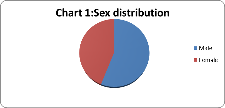
In the present study, majority of the cases were males (56%) [Chart:1]
In our study most common Histopathological findings were Diffuse pulmonary oedema in 22 cases (32%), followed by steatohepatitis in 16 cases (23%) and Myocardial infarction in 12 cases (17%) of total 69 cases.[Table no:2]
Table:2 -Case distribution on the basis of Histopathological findings:
|
Histopathological findings |
No of cases |
Percentage (%) |
|
Diffuse pulmonary oedema |
22 |
32 |
|
Steatohepatitis |
16 |
23 |
|
Myocardial infarction |
12 |
17 |
|
Hepatic congestion |
10 |
14 |
|
Splenic congestion |
9 |
13 |
|
Atherosclerosis |
7 |
10 |
|
Chronic Glomerulonephritis |
4 |
5 |
|
Products of conception |
3 |
4 |
|
Left ventricular hypertrophy |
3 |
4 |
|
Petechial hemorrhages in heart |
2 |
2 |
|
Myocarditis |
2 |
2 |
|
Pneumonitis |
2 |
2 |
|
Metastasis |
2 |
2 |
|
Tuberculosis |
2 |
2 |
|
Fungal pneumonitis |
1 |
0.5 |
|
Acute hepatic necrosis |
1 |
0.5 |
|
Chronic venous congestion of lung |
1 |
0.5 |
Note – Multiple Histopathological findings were noted per case.
In organ wise distribution of cases, Diffuse pulmonary edema, Myocardial infarction, Steatohepatitis, Splenic congestion, Chronic Glomerulonephritis emerged as the commonest histopathological findings among Lungs, Heart, Liver, Spleen and Kidney lesions respectively. [Table – 2]
In our study, 41 of 69 total cases were suspected deaths with a provisional clinical Diagnosis. 13 (32%) cases were concordant and 28(68%) cases were discordant with the clinical diagnosis. [Table -3]
Table no: 3–Cases with COD showing concordance and discordance with a provisional clinical diagnosis:
|
Clinical cause of death |
Concordance |
Discordance |
Total no of cases |
|
Myocardial infarction |
4 (57%) |
3 (43%) |
7 |
|
Poisoning /Snake bite |
7(29%) |
17(71%) |
24 |
|
RTA/Assault |
2(33%) |
4(67%) |
6 |
|
Thunder storm/Sun stroke |
- |
4(100%) |
4 |
|
Total |
13(32%) |
28(68%) |
41 |
Table no 4 - Incidental findings/ Discordant findings with clinical diagnosis (Integrated with figure):
|
Serial number |
Age /sex |
History/ indication of autopsy |
Histopathological findings |
|
1 |
47/m |
Anaphylactic shock |
Metastasis of adenocarcinoma in lungs ,kidneys and hilar lymphnodes |
|
2 |
38/f |
Strike of Thunder storm |
Myocarditis with myocardial infarction |
|
3 |
32/f |
Myocardial infarction |
Acute hepatic necrosis |
|
4 |
54/m |
Sunstroke |
Atherosclerosis with right coronary partial and left total luminal obstruction |
|
5 |
59/m |
Sunstroke |
Early myocardial infarction |
|
6 |
15/f |
Food poisoning |
Acute myocarditis |
|
7 |
23/f |
Snake bite |
Congestive changes in skin |
|
8 |
54/m |
Snake bite |
Congestive changes in skin and kidney |
|
9 |
58/m |
Myocardial infarction |
Miliary tuberculosis in lung and spleen |
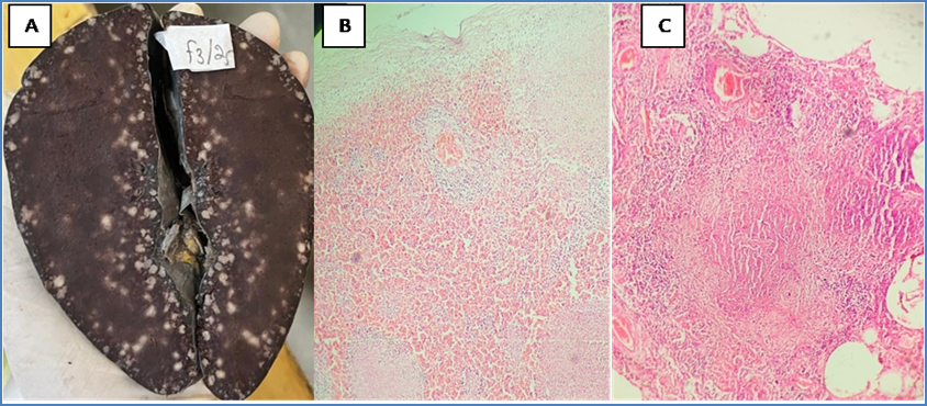
Figure 1: (A) Metastatic deposits of Adenocarcinoma in lung parenchyma (b)Renal parenchyma and Renal sinus(c)Tumor emboli in blood vessels(d)Subcapsular deposits of tumor in hilar lymphnodes. (H&E, 100X)
A 47 y/o male found deceased after getting an Intravenous injection for infection. Clinically, it was suspected to be due to anaphylactic shock but histopathology of multiple organs revealed metastatic adenocarcinoma deposits in lungs, kidney and mediastinal lymph nodes (Fig. 1).
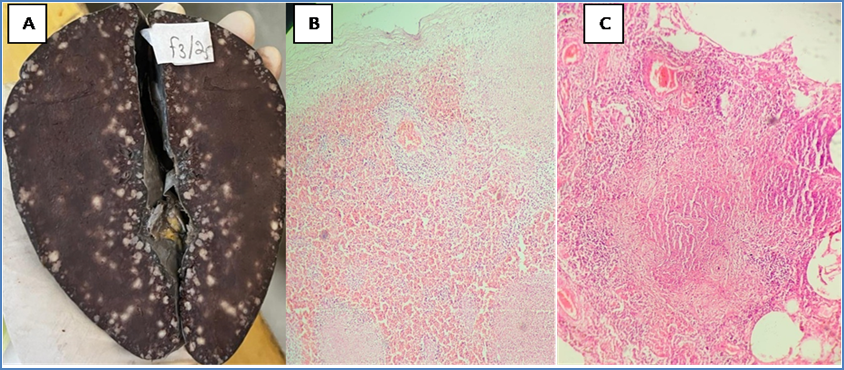
Figure 2: A.Gross and B,C microscopy of multiple caseating Granulomas in spleen and lung in a case of miliary tuberculosis. (B,C- H&E, 100X)
In a 58 yr old male with clinical diagnosis of myocardial infarction, gross examination revealed multiple tubercles and histopathology showed caseating epithelioid Granulomas in spleen, lungs and diagnosed as miliary tuberculosis (Fig-2).
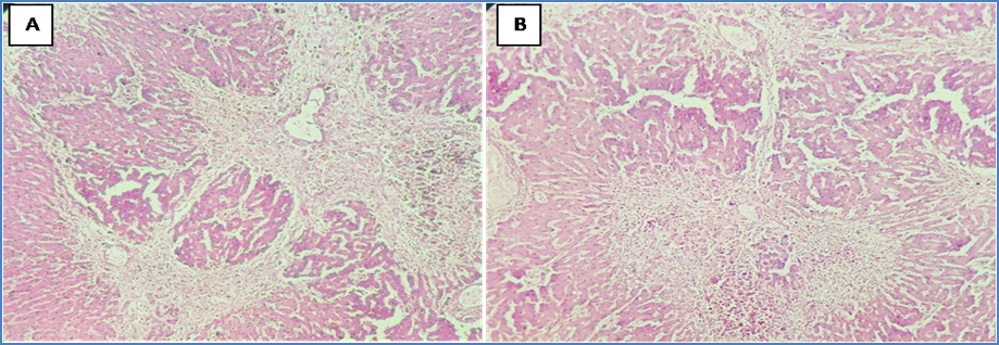
Figure 3: A & B, Hepatic parenchyma with zone 1 hepatocellular necrosis with increased eosinophilia of hepatocytes in zone ii and iii areas. (H&E, 100X)
In a 32 yr female who presented with dyspnoea, chest pain and a clinical diagnosis of myocardial infarction, histopathology of liver showed zone 1 hepatocellular necrosis and increased eosinophilia in zone ii and zone iii areas, diagnosed as acute hepatic necrosis secondary to intake of herbal medicine (Fig-3).
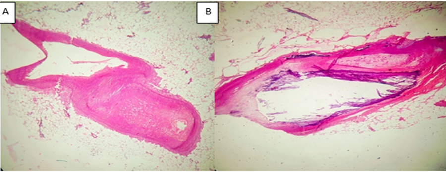
Figure 4: A & B, Partial and complete obstruction of coronaries with atherosclerotic plaques and calcification. (H&E, 100X)
Two cases with sunstroke as clinical diagnosis showed features of myocardial infarction and atherosclerosis with partial/total luminal obstruction in right and left coronary arteries (Fig-4).
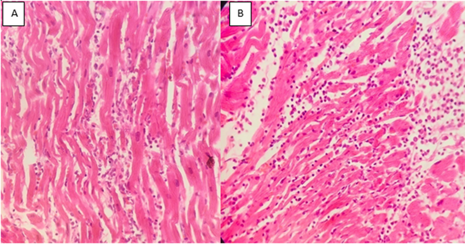
Figure 5: A&B, Mixed inflammatory cell infiltrate composed of predominantly Neutrophils and lymphocytes in case of acute Myocarditis. (H&E, 400X)
A 15 year old female, hostellite with no significant past medical history was suspected to be deceased due to acute food poisoning. Upon histopathological evaluation of heart, it revealed acute myocarditis possibly due to viral etiology (Fig-5).
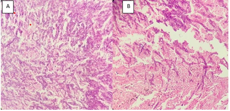
Figure 6: A&B, Acute angled septate hyphae of Aspergillus Fungi seen in a young patient. (A- H&E, B- PAS Stain, 100X)
In a 27 y/o young male with clinical diagnosis of sepsis and grade III pressure ulcer over gluteal region, histopathology showed multiple, acute angled and septate, branching hyphae of Aspergillus in lung (Fig-6).
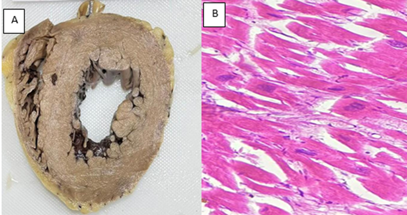
Figure 7: A Gross image of hypertrophied left ventricle with wall thickness of 3cms. B. Microscopy showing hypertrophic cardiomyocytes with nuclear enlargement, Box shaped nuclei and some binucleation. There is scant interstitial fibrosis.
A 32 year old male found dead at work place without a conclusive clinical opinion, histopathology of heart revealed hypertrophic cardiomyopathy (Fig -7).
In our study, histopathology by itself ascertained the COD in 28 cases (40%) which were lacking provisional clinical Diagnosis.
DISCUSSION:
Histopathological examination remains a cornerstone of medicolegal autopsy, particularly in cases where gross findings are equivocal or nonspecific. In the present study, microscopic evaluation provided critical insights into the underlying pathological processes and contributed to the determination of cause of death, thereby reinforcing the indispensable role of histopathology in medicolegal cases.
The commonest histopathological finding in our study was Diffuse pulmonary edema in 22 cases (32%). This correlates with the study conducted by Sulegoan. R. et al.[7] and P.N Kadam et al.[8] Pulmonary edema is frequent terminal event and may be associated with various cardiac, pulmonary or systemic conditions. Several researchers have reported pulmonary oedema as the predominant lesion in autopsy lung specimens, reflecting the lungs’ susceptibility to terminal hypoxia and circulatory failure in both natural and unnatural deaths. In a series of 100 autopsy cases, pulmonary oedema was identified as the most common histopathological alteration, occurring in over one-third of cases and underscoring its frequent involvement across diverse causes of death.[9] Similar patterns have been noted in larger retrospective analyses, where pulmonary oedema and congestion formed a significant proportion of lung pathology detected at histological examination.[10] The high prevalence of pulmonary oedema in autopsy material likely reflects its multifactorial nature. Edema may result from cardiac dysfunction, acute respiratory events, sepsis-related capillary leak, or terminal circulatory collapse, with histopathology providing a more sensitive means of detection than gross examination alone. This supports the view that lung histology can uncover subtler processes contributing to death that may not be appreciated macroscopically.[9]
Myocardial infarction (12 cases, 17%) emerged as the predominant histopathological lesion in the cardiac specimens, followed by Atherosclerosis (7 cases, 10%), left ventricular hypertrophy (3 cases, 4%), petechial hemorrhages (2 cases, 2%), and myocarditis (2 cases, 2%). Atherosclerosis emerged as one of the most significant cardiac findings, corroborating the findings of Arunalatha et.al[11]. In a six-year tertiary centre study, atherosclerotic lesions were noted as the most frequent incidental histopathological finding after pulmonary changes, observed in more than a quarter of cases.[12] Similarly, analyses focused on cardiovascular histopathology in autopsy material highlight the widespread prevalence of atherosclerotic changes, especially in the coronary arteries, among decedents subjected to postmortem examination.[13] Atherosclerosis, while often incidental to the immediate cause of death, has significant clinical and epidemiological implications. Its detection at autopsy provides insight into underlying chronic cardiovascular disease that may have been undiagnosed or underappreciated ante-mortem, and confirms that subclinical vascular pathology is common in populations undergoing medicolegal autopsy.[12]
In the present study, steatohepatitis and hepatic congestion constituted significant microscopic observations among liver lesions. Congestion of the liver is a frequent terminal phenomenon in autopsy practice, often reflecting cardiac failure, shock, or systemic hypoxia rather than primary hepatic pathology. Similar observations have been documented by Pathak and Mangal, who reported congestion as the most common hepatic lesion in medicolegal autopsies, followed by fatty change and steatohepatitis.[14]
Fatty liver and steatohepatitis detected incidentally at autopsy also reflect the growing burden of metabolic syndrome and alcohol-related liver disease in the general population. Patel et al. reported fatty liver among the most frequent incidental findings in autopsy specimens, emphasizing that such lesions may remain clinically silent during life.[12] The identification of these changes in our series reinforces the epidemiological value of autopsy histopathology in revealing subclinical chronic disease.
The case of acute hepatic necrosis identified in our study highlights the importance of microscopic evaluation in clarifying unexpected causes of death. Similar reports in the literature describe toxin-related or drug-induced hepatic necrosis diagnosed only after histopathological examination, underscoring the limitations of gross examination alone. In the present study, renal histopathological involvement was limited to glomerulonephritis and metastatic deposits, with no significant tubular or interstitial pathology observed. Although renal findings were relatively infrequent compared to other organs, their detection was diagnostically significant and consistent with observations reported in previous autopsy-based studies.
Glomerulonephritis identified in this study likely represents an underlying chronic or immune-mediated renal disease that had remained clinically silent. Similar findings have been reported by Patel et al., who documented glomerular pathology as an incidental but relevant observation in medicolegal autopsies, often unrelated to the immediate cause of death. Such lesions may not contribute directly to mortality but reflect pre-existing systemic disease, emphasizing the value of renal histopathology in autopsy practice.[12] The identification of metastatic deposits in renal tissue in our study underscores the role of histopathology in uncovering occult malignancy. Renal metastases are frequently asymptomatic and are often missed on gross examination due to their small size or multifocal distribution. Routine histopathological evaluation of renal tissue in medicolegal autopsies thus remains valuable, not only for identifying primary renal disease such as glomerulonephritis but also for detecting secondary involvement by systemic malignancies that may otherwise go unrecognized.
Gahine et al. highlighted cerebral edema as the principal microscopic alteration observed in the central nervous system.[15] However, our findings differed, as most brain specimens in the current study showed no significant histopathological abnormalities this finding itself is noteworthy. Similarly, myocarditis, fungal pneumonitis, miliary tuberculosis, and metastatic deposits identified in our study represent diagnostically significant findings that altered or refined the cause of death. Sindhura et al. and other investigators have emphasized that incidental malignancies, granulomatous inflammation, and infectious lesions are not uncommon discoveries in medicolegal autopsies.[16]
The detection of metastatic adenocarcinoma in a case clinically suspected as anaphylactic shock in our series exemplifies the decisive role of histopathology in uncovering occult malignancy. Comparable instances of unsuspected neoplasms identified only on microscopic examination have been described in autopsy literature, reinforcing the academic and medicolegal importance of routine tissue sampling.[12] This study highlights the substantial contribution of histopathology in establishing the cause of death in instances where clinical findings were inconclusive. Microscopic examination of tissues enabled the detection of not only unsuspected neoplasms and chronic disease processes that had remained undiagnosed at the time of autopsy but also in detecting Incidental and rare findings that were directly related to the cause of death.
CONCLUSION:
Diffuse pulmonary oedema constituted the predominant microscopic finding in the present series, with atherosclerotic changes representing the next most frequent lesion. The study reinforces the diagnostic significance of systematic histopathological evaluation in medicolegal autopsies, particularly in identifying incidental, silent, or previously unrecognized disease processes. Such detailed tissue examination not only aids in clarifying the cause and mechanism of death but also enhances the scientific and educational value of autopsy practice, contributing meaningfully to ongoing academic discourse and future research.Detection of subtle lesions such as early myocardial ischemia, myocarditis, infections, atherosclerosis and metastastic deposits highlights the limitations of gross examination alone.
Integration of histopathological findings with autopsy observations and circumstantial evidence enhances diagnostic accuracy and strengthens medico-legal opinions. Routine histopathological analysis should therefore be considered as an essential component of medico-legal autopsies, particularly in obscure and unexplained deaths.
LIMITATIONS
The study was conducted at a single center with a relatively small sample size, which may limit the generalizability of the findings. Only selected organs were subjected to histopathological evaluation based on clinical suspicion and autopsy findings; hence, subtle lesions in unexamined tissues may have been missed. Advanced ancillary techniques such as immunohistochemistry, toxicological correlation, and molecular analysis were not performed due to constrained resources, which might have further enhanced diagnostic precision in selected cases.
REFERENCES:
1. Rothenberg K. The autopsy through history. In: Embar-Seddon A, Pass AD, editors. Forensic science. Salem Press; 2008. p. 100–100.
2. Sharma D, et al. Role of histopathological examination in medicolegal autopsies. Natl J Lab Med. 2021 Oct;10(4):PO01–PO04.
3. Parai JL, Milroy CM. The utility and scope of forensic histopathology. Acad Forensic Pathol. 2018;8(3):426–451. doi:10.1177/1925362118797602
4. Delteil C, Tuchtan L, Torrents J, Capuani C, Piercecchi-Marti MD. Pediatric medicolegal autopsy in France: a forensic histopathological approach. J Forensic Leg Med. 2018;53:106–111. doi:10.1016/j.jflm.2017.11.010
5. Naik D, Couto F, Sousa Rocha PD, Mandrekar S, Jaladi P, Pinto RGW. Histopathological findings in autopsies – emphasising the interesting and incidental findings: a 3-year study. Indian J Appl Res. 2024;14(1):37–40.
6. Patel S. Histopathologic findings in autopsies with emphasis on interesting and incidental findings: a pathologist’s perspective. J Clin Diagn Res. 2016;10(11):EC08–EC11.
7. Medicolegal autopsies—interesting and incidental findings. Int J Forensic Sci Pathol. 2015;3:156–160. doi:10.19070/2332-287X-1500038
8. A 3-year retrospective histopathological study of autopsy findings. Indian J Forensic Med Pathol. 2017;10(1):5–9. doi:10.21088/ijfmp.0974.3383.10117.1
9. Prakasam G, Sadasivam B, Rajasekaran V, Muniraj K, Prathapan A. Histopathological spectrum of lung findings in autopsy cases. Asian Journal of Medical Sciences. 2024;15(1):204–208. doi:10.3126/ajms.v15i1.57717
10. Patel, C. B., Patel, K., Bhagat, V. M., & Shah, P. (2017). Pattern of histopathological lesions in lung autopsy. International Journal of Research in Medical Sciences, 6(1), 279–283. https://doi.org/10.18203/2320-6012.ijrms20175734
11. Arunalatha P, Sangeetha A, Devi NRS. Spectrum of histopathological findings in autopsies—highlighting the interesting and incidental findings. 2017. Available from: https://www.semanticscholar.org/
12. .Kumar S, Kumar Verma A, Kumar N, Baranwal RK, Kumar Verma R, Singh M. Coronary atherosclerosis – a postmortem histopathological study. Bratisl Lek Listy. 2012;113(4):217–219. doi:10.4149/BLL_2012_050.
13. Pathak A. Histopathological examination in medico-legal autopsy: pros and cons. J Indian Acad Forensic Med. 2010;32(2):128–131.
14. Gahine R, Joshi C, Gonnade U, Saraf M: A histopathological analysis of medico legal autopsy in cases of sudden death. JMSCR. 2018, 6:830-5.
15. Sindhura M, Vijaya Bhaskar R, Lakshmi CV. Histopathological surprises in medicolegal autopsies – a two-year experience. JMSCR. 2020;8(2):8857-8867.