Anti-MICA Antibodies, Pre-Transplant Sensitization Factors, and Post-Transplant Complications in Live-Related Kidney Transplantation.
- Dr.Ashok Sarin , Professor, Senior Consultant, Department of Nephrology, Indraprastha Apollo Hospital, New Delhi.
- Riesh Kumar Banode , Professor and Head, Department of Nephrology, MGM Medical College, Indore. E Mail ID: banode.ritesh@gmail.com.
Article Information:
Abstract:
Background: Antibodies targeting major histocompatibility-complex class I-related chain A (MICA), a non-HLA endothelial alloantigen, are increasingly identified in renal transplant candidates. However, the clinical and epidemiological factors influencing sensitization and the post-transplant complication profiles of anti-MICA-positive patients are not fully understood, especially in Indian live-donor transplant contexts. This study investigates pre-transplant sensitization factors linked to anti-MICA antibody presence and explores the range of post-transplant complications in recipients of live-related kidney transplants. Methods: This prospective case-control study (March 2016 to March 2018) at a tertiary transplant centre involved 50 live-related renal transplant recipients who tested positive for anti-MICA antibodies (cases), identified by single-antigen bead Luminex assay (MFI ≥1000). These were compared with 50 anti-MICA antibody-negative recipients (controls), all of whom had a negative CDC crossmatch and no donor-specific HLA antibodies. All demographic data, comorbidities, native kidney disease, donor profiles, dialysis duration, and blood transfusion history-related data were collected to identify factors associated with anti-MICA sensitisation. Recipients with anti-MICA antibodies underwent desensitization (rituximab, plasma exchange, intravenous immunoglobulin) prior to transplantation. Post-transplant outcomes over six months, including infections, new-onset diabetes (NODAT), rejection, graft failure, and mortality, were systematically recorded. Results: No significant differences were observed between the groups regarding age, gender, ABO blood type, comorbidities, native kidney disease patterns, or donor demographics (all p>0.05). Among the 50 anti-MICA-positive recipients, 27 (54.0%) had antibodies targeting a single MICA allele, while 23 (46.0%) had antibodies against multiple alleles. Recipients positive for anti-MICA antibodies had a significantly longer duration of pre-transplant dialysis (8.32±5.49 vs 5.20±4.58 months; p=0.001) and a higher rate of prior blood transfusions (34.0% vs 14.0%; p=0.01) than negative individuals. There were no statistically significant differences in post-transplant outcomes, including major infections, NODAT (8.0% vs 6.0%), biopsy-proven rejection (4.0% in both groups), graft failure (none), and mortality (4.0% in both groups), between the anti-MICA-positive and anti-MICA-negative groups. Conclusion: Longer dialysis duration and prior blood transfusions were identified as key, potentially modifiable, pre-transplant sensitization factors leading to anti-MICA antibody development. In contrast, demographic and donor-related factors were not significant. With pre-transplant desensitization, anti-MICA-positive recipients showed a post-transplant complication profile similar to that of non-sensitized patients. These results advocate for reducing pre-transplant blood transfusions and accelerating transplantation to decrease dialysis time for candidates awaiting live-related renal transplants. They also support adding anti-MICA screening to pre-transplant immunological risk assessments.
Keywords:
Article :
INTRODUCTION:
Sensitization to human leukocyte antigen (HLA) and non-HLA alloantigens poses a significant obstacle to successful kidney transplantation. Patients typically become sensitized through blood transfusions, pregnancy, or prior transplants, resulting in longer wait times, fewer compatible donors, and sometimes disqualification from transplantation, even when a willing living donor is available.1
Major histocompatibility complex class I-related chain A (MICA) is a non-classical, highly polymorphic class I-like glycoprotein that, unlike classical HLA, does not associate with beta-2 microglobulin or present peptides to T cells.2 Instead, MICA functions as a stress-inducible ligand for the NK-cell activating receptor NKG2D. It is expressed on vascular endothelium, dendritic cells, fibroblasts, and epithelial cells, but not on peripheral blood lymphocytes.3,4 Because MICA is expressed on donor endothelium, the principal interface between the graft and the recipient immune system, antibodies against MICA are positioned to mediate allograft injury independent of HLA sensitisation.5
While several studies have examined the association between pre-transplant anti-MICA antibodies and rejection and graft survival, relatively little attention has been paid to characterising which recipients become sensitised to MICA and why.6-11 Understanding the epidemiological and clinical factors underlying anti-MICA sensitisation, similar to how transfusion, pregnancy, and previous transplants are known to cause HLA sensitisation, could help develop strategies to reduce sensitisation risk for patients awaiting live-donor transplants. This approach is especially important in India, where most transplants are live-related and pre-emptive transplantation is often hindered by limited resources and late referrals. Similarly, while anti-MICA antibodies have been associated with rejection in various retrospective studies, detailed data on other post-transplant complications, such as infections, new-onset diabetes after transplantation (NODAT), and mortality, in anti-MICA-sensitized patients treated with modern desensitization protocols remain scarce, particularly from the Indian subcontinent.
We therefore conducted a prospective case-control study with two complementary aims: first, to identify demographic, clinical, and donor-related factors associated with anti-MICA antibody positivity in live-related renal transplant candidates; and second, to characterize the spectrum and incidence of post-transplant complications like infectious, metabolic, immunological, and mortality-related complications in anti-MICA antibody-positive recipients compared with antibody-negative recipients, all receiving live-related transplantation at a single high-volume Indian transplant centre..
MATERIALS AND METHODS:
Study design, setting, and period
This prospective case-control study was conducted at the Department of Nephrology, Indraprastha Apollo Hospital, a 700-bed multi-speciality tertiary care centre in New Delhi, India, from March 2016 to March 2018.
Study population and sample size
A total of 100 consecutive live-related renal transplant recipients were enrolled, consisting of 50 anti-MICA antibody-positive patients (cases) and 50 antibody-negative patients (controls). They were followed for six months after transplantation. The sample size was determined using the formula n = 4pq/d², which indicated a minimum of 82 patients needed at 80% power, 5% significance level, and 11% precision. To meet these criteria, 100 patients were enrolled.12
Inclusion and exclusion criteria
Cases included live-related renal transplant recipients who had a negative CDC crossmatch, negative donor-specific class I/II HLA antibodies, and a positive anti-MICA antibody during pretransplant evaluation. Controls consisted of live-related recipients negative for CDC crossmatch, donor-specific HLA antibodies, and anti-MICA antibodies. Patients were excluded if they had a positive CDC crossmatch, positive DSA, ABO-incompatible transplants, second transplants, combined solid-organ transplants, or were seropositive for HIV, HCV, or HBsAg.
Anti-MICA antibody testing and sensitization variables
Anti-MICA antibodies were identified on freshly obtained, undiluted sera using a single-antigen bead (SAB) Luminex assay (Lifecodes LSA™ MIC, Immucor, USA); a bead MFI of ≥1000 was considered positive, according to the manufacturer's guidelines. To investigate possible sensitization factors, pre-transplant variables were systematically compared between recipients who tested positive and negative for anti-MICA: recipient age and gender; recipient and donor ABO blood groups; comorbidities such as hypertension and diabetes mellitus; native kidney disease category; duration of pre-transplant dialysis; history of pre-transplant blood transfusions; and donor age, gender, and relationship to the recipient.
Desensitization and immunosuppression
Recipients positive for anti-MICA antibodies underwent pre-transplant desensitization, including rituximab 375 mg/m² (7 days before transplant), two plasma exchange sessions starting 2 days prior, intravenous immunoglobulin 100 mg/kg after plasma exchange, and early initiation of mycophenolate mofetil (MMF) 1 g twice daily for 7 days before transplant. Anti-MICA-negative controls did not receive desensitisation. All recipients received calcineurin inhibitor-based triple immunosuppression with either tacrolimus or cyclosporine, along with MMF and corticosteroids, plus prophylaxis for cytomegalovirus with valganciclovir and for Pneumocystis pneumonia with trimethoprim-sulfamethoxazole.
Post-transplant complication assessment
Recipients were monitored weekly for one month, then every two weeks for the next three months, and monthly up to six months. During each visit, complete blood counts, renal and liver function tests, and urinalysis were performed. Recorded post-transplant complications included: biopsy-confirmed rejection (using Banff criteria, with sub-classification into acute cellular rejection [ACR] or antibody-mediated rejection [ABMR], where ABMR required C4d immunohistochemistry >10%); major infections such as BK viraemia (>10,000 copies/mL via real-time PCR), CMV infection confirmed by plasma PCR, herpes zoster, urinary tract infection, pneumonia or chest infections, tuberculosis, and fungal infections diagnosed through clinical, microbiological, and radiological evaluation; NODAT according to American Diabetes Association standards in patients without pre-existing diabetes; graft failure indicated by return to dialysis; and mortality, with cause of death documented.
Statistical analysis
Categorical variables are shown as frequencies and percentages, with comparisons made using the chi-square test or Fisher's exact test when appropriate. Continuous variables are expressed as mean ± standard deviation and compared with the unpaired t-test. Relative risk (RR) and 95% confidence intervals (CIS) were calculated for binary complication outcomes. A p-value less than 0.05 (two-tailed) was deemed statistically significant. All analyses were conducted using SPSS version 16.0.
RESULTS:
Among 100 kidney transplant recipients studied, 50 tested positive for anti-MICA antibodies while 50 tested negative. The group included 71 males and 29 females.
Pre-transplant sensitization factors
The mean age of recipients was not significantly different between anti-MICA-positive and -negative groups (45.06 ± 10.77 versus 39.62 ± 13.24 years; p = 0.12). The age distribution across bands (<40, 40–50, >50 years) was also similar (p = 0.12). Male recipients were predominant in both groups, with a higher percentage in anti-MICA-positive recipients (78.0% vs 64.0%), but this was not statistically significant (p = 0.12). The distribution of ABO blood groups among recipients showed no significant difference between groups (p = 0.08), with blood group B being the most common in both (Table 1).
Table 1: Demographic and clinical factors evaluated for association with anti-MICA sensitization
|
Parameters |
Anti-MICA positive (n=50) |
Anti-MICA negative (n=50) |
p-value |
|
Mean age, years (± SD) |
45.06 ± 10.77 |
39.62 ± 13.24 |
0.12 |
|
Male recipients, n (%) |
39 (78.0) |
32 (64.0) |
0.12 |
|
Blood group B, n (%) |
20 (40.0) |
21 (42.0) |
0.08 |
|
Hypertension, n (%) |
29 (58.0) |
26 (52.0) |
0.54 |
|
Diabetes mellitus, n (%) |
18 (36.0) |
15 (30.0) |
0.52 |
|
Pre-transplant dialysis, months (mean ± SD) |
8.32 ± 5.49 |
5.20 ± 4.58 |
0.001* |
|
History of blood transfusion, n (%) |
17 (34.0) |
7 (14.0) |
0.01* |
*Statistically significant (p<0.05)
The burden of comorbidities was comparable between the groups: hypertension was found in 58.0% of anti-MICA-positive and 52.0% of anti-MICA-negative recipients (p = 0.54), while diabetes mellitus was present in 36.0% and 30.0%, respectively (p = 0.52). Regarding native kidney disease, diabetic nephropathy was the leading cause among anti-MICA-positive recipients (38.0%) and the second most common among anti-MICA-negative recipients (30.0%). Chronic glomerulonephritis was the predominant cause in the anti-MICA-negative group (32.0%). The distribution of native kidney disease types did not significantly differ between the two groups.
The variables most strongly linked to anti-MICA antibody positivity were pre-transplant dialysis duration and blood transfusion history (Tables 1 and 2). Recipients positive for anti-MICA had a significantly longer median pre-transplant dialysis duration 8 months compared to negative recipients, who had 4 months (p=0.001). Additionally, 44.0% of anti-MICA-positive recipients had a dialysis duration of 7 to 12 months, versus only 16.0% of anti-MICA-negative recipients. A pre-transplant blood transfusion was observed in 34.0% of anti-MICA-positive recipients, compared with 14.0% of anti-MICA-negative recipients (p = 0.01).
Table 2. Pre-transplant dialysis duration and blood transfusion history by anti-MICA antibody status
|
Variable |
Category |
Anti-MICA positive (n=50) |
Anti-MICA negative (n=50) |
p-value |
|
Dialysis duration |
0-6 months |
21 (42.0%) |
39 (78.0%) |
0.001 |
|
7-12 months |
22 (44.0%) |
8 (16.0%) |
||
|
>12 months |
7 (14.0%) |
3 (6.0%) |
||
|
Blood transfusion |
Yes |
17 (34.0%) |
7 (14.0%) |
0.01 |
|
No |
33 (66.0%) |
43 (86.0%) |
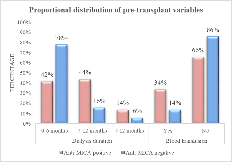
Figure 1: Proportional distribution of pre-transplant dialysis duration and blood transfusion parameters by anti-MICA antibody status
Donor characteristics showed close matching between the two groups. Donor age (p = 0.89), gender (64.0% female in both groups; p = 1.00), and ABO blood group (p = 0.56) did not differ significantly. Among anti-MICA-positive recipients, most living donors were wives (36.0%), followed by brothers (18.0%) and sisters (14.0%). In contrast, among anti-MICA-negative recipients, the most common donors were mothers (28.0%), followed by wives (20.0%) and brothers (18.0%). Of the 50 anti-MICA antibody-positive recipients, 27 (54.0%) had antibodies against a single MICA allele, while 23 (46.0%) targeted two or more alleles: 12 (24.0%) to two alleles, 6 (12.0%) to three, 1 (2.0%) to four, 2 (4.0%) to five, and 2 (4.0%) to eight alleles.
Part 2: Post-transplant complications
Graft function, measured by serum creatinine levels at discharge and at 1, 3, and 6 months, showed no significant differences between anti-MICA-positive and -negative recipients at any time point (Table 3), despite the control group not undergoing desensitisation.
Table 3. Graft function (serum creatinine, mg/dL) over the follow-up period
|
Time point |
Anti-MICA positive |
Anti-MICA negative |
p-value |
|
At discharge |
1.02 ± 0.23 |
1.06 ± 0.36 |
0.46 |
|
1 month |
1.03 ± 0.22 |
1.04 ± 0.30 |
0.90 |
|
3 months |
1.08 ± 0.21 |
1.11 ± 0.34 |
0.59 |
|
6 months |
1.11 ± 0.23 |
1.12 ± 0.29 |
0.79 |
Overall, major infectious complications were rare, occurring in less than 12% for any single infection type, with similar rates observed between groups as shown in Table 4. Urinary tract infection was the most common infection in both groups (12.0% vs 8.0%; RR 1.22, 95% CI 0.70–2.12; p = 0.50). BK viraemia was more frequent in anti-MICA-negative recipients (6.0% vs 2.0%), whereas CMV infection was more frequent in anti-MICA-positive recipients (6.0% vs 2.0%); however, these differences were not statistically significant. Additionally, tuberculosis and fungal infections were each reported in one anti-MICA-positive recipient (2.0%), with no cases in anti-MICA-negative recipients.
Table 4. Post-transplant infectious complications by anti-MICA antibody status
|
Infection |
Anti-MICA positive, n (%) |
Anti-MICA negative, n (%) |
RR (95% CI) |
p-value |
|
BK virus |
1 (2.0) |
3 (6.0) |
0.49 (0.08–2.70) |
0.30 |
|
Herpes zoster |
2 (4.0) |
1 (2.0) |
1.34 (0.59–3.07) |
0.55 |
|
CMV |
3 (6.0) |
1 (2.0) |
1.53 (0.83–2.79) |
0.30 |
|
Urinary tract infection |
6 (12.0) |
4 (8.0) |
1.22 (0.70–2.12) |
0.50 |
|
Chest infection |
2 (4.0) |
2 (4.0) |
1.00 (0.36–2.71) |
1.00 |
|
Tuberculosis |
1 (2.0) |
0 (0.0) |
2.02 (0.65–2.46) |
0.31 |
|
Fungal infection |
1 (2.0) |
0 (0.0) |
2.02 (0.65–2.46) |
0.31 |
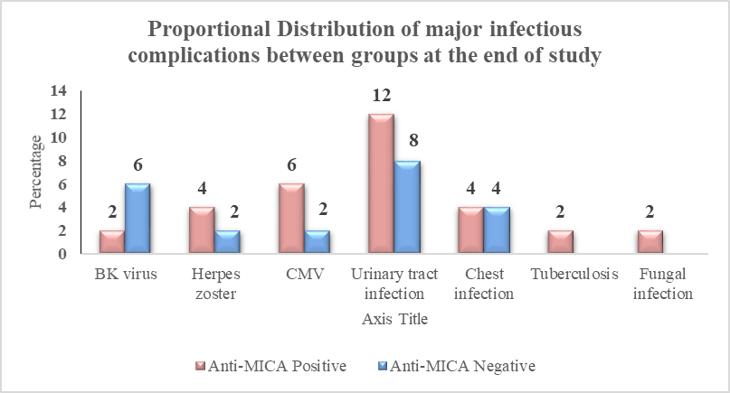
Figure 2: Proportional Distribution of major infectious complications between Anti-MICA positive and negative groups
NODAT occurred in 4 out of 50 (8.0%) anti-MICA-positive recipients and 3 out of 50 (6.0%) anti-MICA-negative recipients (RR 1.15, 95% CI 0.58–2.26; p = 0.79). Biopsy-confirmed rejection happened in 2/50 (4.0%) recipients in each group (RR 1.00, 95% CI 0.36–2.71; p = 1.00), with slight differences in rejection subtypes (anti-MICA-positive: 1 ACR, 1 combined ACR+ABMR; anti-MICA-negative: 1 ACR, 1 ABMR). These small numbers precluded meaningful statistical comparison (p = 0.99). No graft failures occurred in either group during follow-up. Mortality rates were identical (2/50, 4.0% each; RR 1.00, 95% CI 0.36–2.71; p = 1.00). Causes of death included CMV pneumonia and fungal sepsis in the anti-MICA-positive group, and bacterial sepsis and stroke in the negative group (Table 5).
Table 5. Summary of post-transplant immunological, metabolic, and survival outcomes at six months
|
Outcome after 6-months |
Anti-MICA positive (n=50) |
Anti-MICA negative (n=50) |
RR (95% CI) |
p-value |
|
Biopsy-proven rejection, n (%) |
2 (4.0) |
2 (4.0) |
1.00 (0.36–2.71) |
1.00 |
|
NODAT, n (%) |
4 (8.0) |
3 (6.0) |
1.15 (0.58–2.26) |
0.79 |
|
Graft failure, n (%) |
0 (0.0) |
0 (0.0) |
- |
- |
|
Mortality, n (%) |
2 (4.0) |
2 (4.0) |
1.00 (0.36–2.71) |
1.00 |
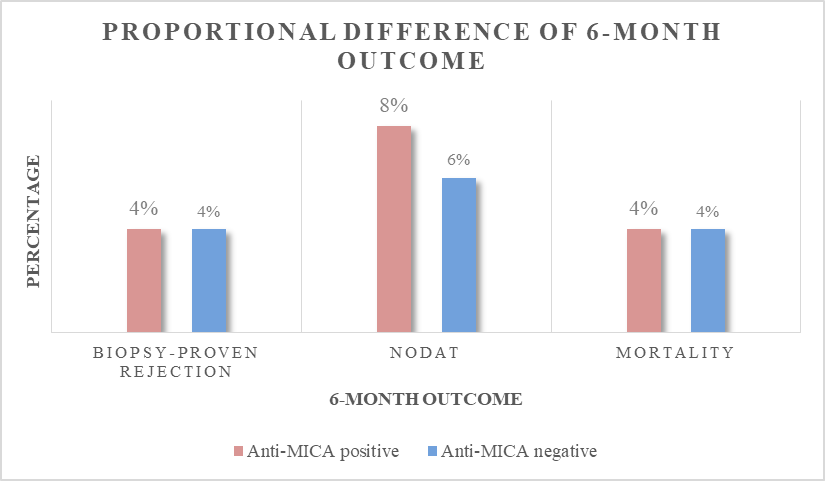
Figure 3: Proportional distribution of post-transplant immunological, metabolic, and survival outcomes at six months between the Anti-MICA positive and negative groups
DISCUSSION:
This study explored two related questions: identifying pre-transplant factors linked to anti-MICA antibody sensitisation and characterising the post-transplant complication profiles of anti-MICA-positive recipients under current desensitisation protocols. To our knowledge, it is among the first prospective studies from the Indian subcontinent to analyse both issues simultaneously within a live-related transplant cohort.
Sensitization factors
Unlike HLA sensitisation, where pregnancy is a dominant driver, particularly in female candidates, our anti-MICA-positive cohort was predominantly male (78.0%), a pattern also reported by Sánchez-Zapardiel et al. (58% male among anti-MICA-positive recipients)13 and Mohit Chowdhry et al. (71% male),10 suggesting that pregnancy is a less prominent driver of anti-MICA sensitization than of HLA sensitization, and that other exposures, mostly transfusion and dialysis-related events, may be more relevant.
Blood transfusion emerged as a significant sensitizing factor in our cohort (34.0% vs 14.0%; p = 0.01), consistent with Rui Costa, Jorge Malheiro et al., who found pre-transplant transfusion in 48.9% of anti-MICA-positive versus 33% of anti-MICA-negative recipients (p = 0.04),14 and with Lemy et al., who identified transfusion as an independent risk factor for anti-MICA antibody development.15 Transfused blood products contain leukocytes and platelets bearing MICA and other non-HLA alloantigens, providing a plausible mechanistic basis for this association, analogous to the well-recognized role of transfusion in HLA sensitization. This finding reinforces existing nephrology practice recommendations to minimize non-essential blood transfusion in patients on the transplant pathway, particularly those with an identified potential living donor.
Longer pre-transplant dialysis duration was independently associated with anti-MICA sensitisation (8.32 vs 5.20 months; p = 0.001) in our cohort, a novel finding relative to most prior literature, which has focused on transfusion and prior transplantation as sensitising events. This association may be explained by cumulative exposure to sensitising events (including occult or unrecorded transfusions) that accrue with dialysis vintage, by dialysis-associated chronic inflammation and anaemia that could upregulate stress-induced MICA expression and immune activation, or by unmeasured confounding related to healthcare exposure during prolonged dialysis. Notably, the mean dialysis duration in our anti-MICA-positive group (8.32 months) was considerably shorter than that reported by Lemy et al. (40 months)15 and by Costa/Malheiro et al. (50.5 vs 45.6 months, non-significant difference of only 4.9 months)14, both of which reflect deceased-donor transplant systems with inherently longer waiting times; the shorter absolute dialysis durations in our live-donor cohort, combined with a still-significant between-group difference, suggest that even modest increases in dialysis exposure may carry sensitization risk in settings where transplantation can otherwise occur relatively early.
In contrast to transfusion and dialysis vintage, recipient age, gender, blood group, comorbidity burden, native kidney disease category, and donor age, gender, and blood group were not significantly associated with anti-MICA sensitisation in our cohort. This pattern, in which modifiable exposure-related factors (transfusion, dialysis time) drive sensitisation risk more than fixed demographic factors, mirrors observations in the HLA-sensitization literature and suggests that strategies to reduce anti-MICA sensitisation should prioritise minimising transfusion exposure and expediting living-donor transplantation to shorten dialysis vintage, rather than targeting demographic subgroups.
Nearly half of our anti-MICA-positive recipients (46.0%) had antibodies reactive against multiple MICA alleles, indicating that broad, polyreactive sensitisation is common and is not confined to a narrow subset of highly sensitised individuals. This has practical implications for donor selection in sensitised candidates, since broad MICA reactivity may increase the likelihood of donor-directed antibody even among apparently unrelated donor–recipient pairs, though donor MICA typing was not performed in our study to confirm donor specificity.
Post-transplant complications
Despite the presence of pre-formed anti-MICA antibodies, the post-transplant complication profile of our anti-MICA-positive recipients, infection incidence, NODAT, rejection, graft failure, and mortality, closely paralleled that of anti-MICA-negative recipients. This finding is important because several retrospective series conducted without desensitization have reported worse outcomes in anti-MICA-positive recipients: Zou et al. reported reduced one-year graft survival in anti-MICA-positive recipients (88.3% vs 93.0%) in a multicentre cohort of 1910 pre-transplant sera,9 Terasaki et al. found MICA antibodies independently associated with reduced graft survival on multivariate analysis across 1329 recipients,1 and Balwani Manish et al. reported a markedly higher acute rejection rate in anti-MICA-positive recipients (47% vs 11.7%; p = 0.02) in the absence of desensitization.11 Our comparable complication rates, achieved with rituximab-, plasma exchange-, and IVIg-based desensitization, are consistent with the more favourable outcomes reported when active antibody-reduction strategies are employed, as illustrated in the case report by Narayan et al., in which MICA antibody titres and graft function normalized in parallel following plasmapheresis and IVIg for combined ACR/ABMR.16
Importantly, desensitisation did not translate into excess infectious morbidity in our cohort, consistent with Kahwaji et al., who found that rituximab combined with plasmapheresis and IVIg did not increase infectious complications in living-donor recipients.17 This is a clinically important reassurance given ongoing concerns that B-cell-depleting and antibody-reducing therapies could increase susceptibility to opportunistic infection in an already immunosuppressed population.
NODAT incidence in our overall cohort (7.0%) fell within the range reported in randomized controlled trials of tacrolimus-based immunosuppression (4–25%)18 and did not differ meaningfully by anti-MICA status, consistent with NODAT risk being driven predominantly by maintenance immunosuppression (tacrolimus) rather than by antibody sensitization status per se.
Taken together, our findings suggest a two-part clinical message: first, that specific, identifiable, and potentially modifiable exposures blood transfusion and dialysis vintage rather than fixed demographic characteristics, drive anti-MICA sensitization risk in live-related transplant candidates; and second, that once sensitization has occurred, appropriate pre-transplant desensitization can normalize the post-transplant complication trajectory relative to non-sensitized recipients, at least over a six-month observation window.
Limitations
This study's single-centre design, combined with a modest sample size and a six-month follow-up period, limits the generalizability of the findings and reduces the ability to detect less common complications or late events. In most patients, native kidney disease was primarily diagnosed based on clinical and radiological assessments rather than pre-transplant biopsy. Donor MICA typing was not performed, which prevents formal confirmation of donor specificity of detected antibodies and impacts the interpretation of sensitization factors and complication analyses. Additionally, post-desensitization anti-MICA antibody mean fluorescence intensity (MFI) was not measured, hindering the ability to correlate antibody reduction with complication risk. Possible sensitizing exposures other than transfusions and dialysis duration, such as undocumented minor procedures, infections, or subclinical prior graft exposure, were not systematically recorded and could serve as residual confounders. Furthermore, post-transplant anti-MICA antibody monitoring and protocol biopsies were not conducted, potentially leading to under-ascertainment of subclinical rejection.
CONCLUSION:
In this prospective case-control study of live-related renal transplant recipients, longer pre-transplant dialysis duration and a history of blood transfusion were significantly associated with anti-MICA antibody sensitization, while demographic and donor-related factors were not. When anti-MICA-positive recipients received structured desensitization with rituximab, plasma exchange, and intravenous immunoglobulin, their post-transplant complication profile — infections, NODAT, rejection, graft failure, and mortality — was statistically comparable to that of anti-MICA-negative recipients over six months. These findings support practical strategies to reduce anti-MICA sensitization risk — minimizing pre-transplant blood transfusion and expediting living-donor transplantation to limit dialysis exposure — and support incorporating anti-MICA antibody screening and, where positive, desensitization into standard pre-transplant immunological work-up for live-related renal transplant candidates. Larger, multicentre, longer-term prospective studies incorporating donor MICA typing and post-transplant antibody monitoring are warranted to confirm and extend these findings.
REFERENCES:
1. Terasaki, P.I., Ozawa, M. and Castro, R. (2007), Four-year Follow-up of a Prospective Trial of HLA and MICA Antibodies on Kidney Graft Survival. American Journal of Transplantation, 7: 408-415.
2. Bahram S, Bresnahan M, Geraghty DE, Spies T. A second lineage of mammalian major histocompatibility complex class I genes. Proc Natl Acad Sci USA. 1994;91(14):6259–63.
3. Jinushi M, Takehara T, Kanto T, et al. Critical role of MHC class I-related chain A and B expression on IFN-alpha-stimulated dendritic cells in NK cell activation: impairment in chronic hepatitis C virus infection. J Immunol. 2003;170:1249–56.
4. Bauer S, Groh V, Wu J, Steinle A, Phillips JH, Lanier LL, Spies T. Activation of NK cells and T cells by NKG2D, a receptor for stress-inducible MICA. Science. 1999;285(5428):727–9.
5. Zwirner NW, Dole K, Stastny P. Differential surface expression of MICA by endothelial cells, fibroblasts, keratinocytes, and monocytes. Hum Immunol. 1999;60(4):323–30.
6. Sumitran-Holgersson S, Wilczek HE, Holgersson J, Söderström K. Identification of the non-classical HLA molecules, MICA, as targets for humoral immunity associated with irreversible rejection of kidney allografts. Transplantation. 2002;74(2):268–77.
7. Mizutani K, Terasaki PI, Shih RN, Pei R, Ozawa M, Lee J. Frequency of MIC antibody in rejected renal transplant patients without HLA antibody. Hum Immunol. 2006;67(3):223–9.
8. Zou Y, Heinemann FM, Grosse-Wilde H, et al. Detection of anti-MICA antibodies in patients awaiting kidney transplantation, during the post-transplant course, and in eluates from rejected kidney allografts by Luminex flow cytometry. Hum Immunol. 2006;67(3):230–7.
9. Zou Y, Stastny P, et al. Multicentre study of MICA sensitisation and renal graft survival in 1910 pre-transplant sera. Hum Immunol. 2007 (as cited).
10. Mohit Chowdhry, R.N. Makroo, Mandhata Singh, Manoj Kumar, Yogit Thakur, Jasuja Sanjiv, P164 Role of anti-mica antibodies in graft survival of renal transplant recipients of India: A single centre experience, Human Immunology, Volume 78, Supplement, 2017, Page 179, ISSN 0198-8859, https://doi.org/10.1016/j.humimm.2017.06.224.
11. Balwani, M. R., Pasari, A. S., Bawankule, C., Sejpal, K., Gurjar, P., Dubey, S., Kashiv, P., Malde, S., Gupta, S., & J, V. (2024). 410.3: Anti-MICA antibodies and graft outcomes in kidney transplant recipients.Transplantation, 108(9S).
12. Charan, J., & Biswas, T. (2013). How to calculate sample size for different study designs in medical research? Indian Journal of Psychological Medicine, 35(2), 121–126.
13. Sánchez-Zapardiel, E., Castro-Panete, M. J., Mancebo, E., Morales, P., Laguna-Goya, R., Morales, J. M., … Paz-Artal, E. (2016). Early renal graft function deterioration in recipients with preformed anti-MICA antibodies: Partial contribution of complement-dependent cytotoxicity. Nephrology Dialysis Transplantation, 31(1), 150–160.
14. Costa, Rui & Malheiro, Jorge & Tafulo, Sandra & Santos, Clara & Almeida, Manuela & Pedroso, Sofia & Martins, Lasalete & Dias, Leonídio & Henriques, António. (2015). Impact of pre-transplant anti-MICA sensitization in graft rejection and survival. Port J Nephrol Hypert 2015; 29(2): 130-138.
15. Lemy, A., Andrien, M., Wissing, K. M., Ryhahi, K., Vandersarren, A., Racapé, J., Heylen, C., Ghisdal, L., Broeders, E., Vereerstraeten, P., Toungouz, M., & Abramowicz, D. (2010). Major histocompatibility complex class 1 chain-related antigen a antibodies: sensitizing events and impact on renal graft outcomes. Transplantation, 90(2), 168–174.
16. Narayan, S., Tsai, E.W., Zhang, Q., Wallace, W.D., Reed, E.F. and Ettenger, R.B. (2011), Acute rejection associated with donor-specific anti-MICA antibody in a highly sensitized pediatric renal transplant recipient. Pediatric Transplantation, 15: E1-E7.
17. Turza, K. C., Shafique, M., Lobo, P. I., Sawyer, R. G., Keith, D. S., Brayman, K. L., & Agarwal, A. (2014). Infectious complications in living-donor kidney transplant recipients undergoing multi-modal desensitization. Surgical infections, 15(3), 182–186.
18. Sharif A, Hecking M, de Vries AP, et al. Proceedings from an international consensus meeting on posttransplantation diabetes mellitus: recommendations and future directions. Am J Transplant. 2014;14(9):1992–2000.