A Prospective Study For Analysis Of Functional And Radiological Outcomes Of Benign Cystic Lesions In The Proximal Femur Managed By Different Modalities.
- Shashi Kumar , Senior Resident, Department of Orthopaedics, Motilal Nehru (MLN) Medical College, Prayagraj, Uttar Pradesh, India.
- Vyas Narayan Shukla , Assistant Professor, Department of Orthopaedics, Motilal Nehru (MLN) Medical College, Prayagraj, Uttar Pradesh, India.
- Ankur Gupta , Senior Resident, Department of Orthopaedics, Motilal Nehru (MLN) Medical College, Prayagraj, Uttar Pradesh, India.
- Arun Kumar Yadav , Assistant Professor, Department of Orthopaedics, Government Medical College (GMC), Azamgarh, Uttar Pradesh, India.
Article Information:
Abstract:
Background: Aim: To evaluate and compare the functional and radiological outcomes of these treatment modalities. Methods: A prospective study was conducted at Moti Lal Nehru Medical College, involving 48 patients diagnosed with benign cystic lesions of the proximal femur. Participants were equally divided into three treatment groups: Fixation, Sclerotherapy, and Conservative management. Outcomes were assessed using the Musculoskeletal Tumor Society (MSTS) score and Harris Hip Score at multiple follow-up intervals. Radiological stability, pain relief, mobility, and complications were also evaluated. Results: The fixation group demonstrated superior outcomes, with 87.5% achieving radiological stability, compared to 62.5% in the sclerotherapy group and 50% in the conservative group. Early mobilization was most frequent in the fixation group (81.25%). Functional scores, including MSTS and Harris Hip Score, were significantly higher in the fixation group across all follow-ups, indicating better pain relief and mobility. Complication rates, such as infection, non-union, and recurrence, were similar across all groups. Conclusion: Surgical fixation appears to offer the best outcomes for patients with benign cystic lesions in the proximal femur, providing enhanced functional recovery, early mobilization, and greater radiological stability compared to sclerotherapy and conservative management. Further research with larger sample sizes is needed to confirm these findings and explore long-term outcomes.
Keywords:
Article :
INTRODUCTION:
Bone cysts are commonly detected incidentally on radiographs and may remain asymptomatic, although pain and pathological fractures can occur. The most common benign cystic lesions include simple/unicameral bone cysts (UBCs) and aneurysmal bone cysts (ABCs). UBCs are solitary, fluid-filled lesions occurring predominantly in the proximal humerus and proximal femur, particularly in children and adolescents. Approximately two-thirds may present with pathological fracture, while lesions in flat bones are often asymptomatic. UBCs tend to become active during periods of skeletal growth and may resolve spontaneously after skeletal maturity. [1]
ABCs are benign, blood-filled, locally aggressive lesions that predominantly affect adolescents and young adults. They commonly involve the metaphysis of long bones, particularly the distal femur, proximal tibia, proximal humerus, and spine. Pain is the most common clinical manifestation, while rapidly expanding lesions may cause significant bone destruction and neurological symptoms when located in the spine. [2] In the WHO classification, UBCs are considered benign lesions, whereas ABCs are classified as intermediate, locally aggressive lesions. [3]
The pathogenesis of UBC is considered reactive rather than neoplastic, with venous stasis, increased intraosseous pressure, bone resorption, and inflammatory mediators contributing to cyst formation. In contrast, primary ABCs are now recognized to have a neoplastic basis, with recurrent chromosomal translocations involving the USP6 gene identified in approximately 70% of cases. Secondary ABC-like changes may occur in other benign or malignant bone lesions and generally lack these characteristic translocations. [4,5]
UBCs are relatively common, with approximately 85% occurring during the second decade of life and a male predominance. ABCs are uncommon, accounting for approximately 9.1% of bone tumors, with around 80% occurring during the second decade and a slight female predominance. [6]
Benign cystic lesions of the proximal femur, including UBCs, ABCs, and other benign lesions such as fibrous dysplasia, can pose important clinical challenges because of the weight-bearing role of the proximal femur. Patients may present with pain, limp, swelling, or pathological fracture, although many lesions are incidentally detected. Radiographs typically demonstrate well-defined osteolytic lesions with variable cortical thinning and expansion. [7]
Radiographs are the initial imaging modality for assessing lesion location, size, and morphology, while computed tomography (CT) and magnetic resonance imaging (MRI) provide additional information regarding lesion extent, cortical involvement, adjacent structures, and soft-tissue extension. Biopsy may be required when the diagnosis is uncertain or malignancy cannot be excluded. [8] The primary goals of treatment are symptom relief, prevention of pathological fracture, restoration of bone integrity, and preservation of function. Radiological assessment includes reduction or resolution of the lesion and restoration of normal bone architecture, whereas functional outcomes include pain relief, return to activities, and overall functional recovery. Recurrence remains an important consideration, particularly in ABCs, necessitating appropriate follow-up. [9]
Despite advances in the management of benign cystic lesions of the proximal femur, evidence regarding the comparative effectiveness of different treatment modalities remains limited. Most available studies are retrospective or involve small patient cohorts, with limited information on long-term functional outcomes, recurrence, and patient-reported outcomes. Therefore, further prospective studies are required to evaluate both radiological and functional outcomes following treatment. The present study was undertaken prospectively to evaluate the functional and radiological outcomes of benign cystic lesions of the proximal femur treated using various modalities.
MATERIALS AND METHODS:
This was a single-center, randomized control trial conducted at Moti Lal Nehru Medical College and SRN Hospital, Department of Orthopaedics which is a tertiary care center and one of the largest hospitals in Prayagraj, after taking permission from ethical committee of M.L.N. Medical College Prayagraj. The study was carried out according to the principles of the Helsinki declaration. To conduct this study ethical approved was obtained from the Institutional Board of the MLN medical college. Patients were selected and followed up for period of 1 year (August 2023 to July 2024).
Sample size: 48 (n=16 in each group)
Sample Size Calculation:
Assumptions:
1. Significance Level (α) = 0.05 (two-tailed)
2. Power (1-β) = 0.80 (80%)
3. Effect size (Cohen's d) = 0.7 (Moderate effect size, based on prior studies or pilot data)
4. Allocation Ratio (N2/N1) = 1 (Equal allocation of patients in both groups)
Calculation:
1. Using a formula for the sample size for three independent groups:
n = 3×[(Zα/2+Zβ)^2×σ^2]/Δ^2
where,
n = sample size per group
Zα/2 = 1.96 for α = 0.05 (two-tailed)
Zβ = 0.84 for 80% power
σ^2 = population variance (assumed)
Δ = difference in means of the three groups (effect size)
For Cohen's d = 0.7, we have:
Δ = 0.7×σ
n = 3×[(1.96+0.84)^2×σ^2]/(0.7×σ)^2
n = 3×[(2.8)^2×σ^2]/(0.49×σ^2)
n = 3×[7.84×σ^2]/0.49×σ^2
n = 3×16
n = 48 (approximately)
Thus, the calculated sample size was 48 (n=16 in each group).
Inclusion criteria
Benign tumor of proximal femur:
1. With impending pathological fracture.
2. Tumors causing pain and limp.
3. Tumors with expansile natural cause.
4. Persons (>15 years) with active life style and no associated comorbidities.
Exclusion criteria
1. Primary malignant or metastatic tumors of bone or any other origin.
2. Synovial lesion such as pigmented villonodular synovitis and synovial chondromatosis.
3. Avascular necrosis of femoral head, osteomyelitis, or developmental disorders.
4. Any systemic disease like sickle cell anaemia, Hemophilia.
Data collection: After explaining the procedures, all subjects were made sign an informed consent and underwent a complete history taking and examination.
Following detailed history was obtained from the patients:
1. Name
2. Age and sex
3. Residence
4. Educational status
5. Socioeconomic status
6. Chief complaints
7. History of presenting illness
8. Past history
9. Family history
10. Personal history
11. Treatment history
12. Medical history including history of Hypertension, Diabetes Mellitus
All patients included in this study was randomly distributed into three groups. One group for conservative Management, second group for sclerotherapy and third group for Fixation.
Following examinations was done:
• Base line investigations: Hb, TLC, DLC, RBS, BT, CT, Serum electrolytes, blood urea, serum creatinine, HIV 1 and 2, HBsAg, Anti HCV, ECG.
• Basic radiological evaluation: Anteroposterior view and Lateral view of involved hip.
• Diagnosis: Clinical and as well as radiological.
• Informed written consent for the surgical procedure.
• Routine antibiotics, analgesics/anti-inflammatory drugs was administered.
• Post-procedure evaluation by clinical and radiological examination.
• Post-procedure evaluation for complication, if any was done.
• Follow up was done at 1, 3, 6, 12 and 24 weeks and 1year post-procedure.
Statistical analysis: Data was entered in Microsoft Excel and analysed using statistical software SPSS version 26 (SPSS Inc., Chicago, IL, USA). The continuous variables were evaluated by mean (standard deviation) value when required. The dichotomous variables was presented in number/frequency and was analysed using the Chi-square test. To compare the means between the two or more groups, analysis by ANOVA Test was used. A p-value of <0.05 or 0.001 was regarded as significant.
RESULTS:
The mean age is around 27-29 years across all groups, with no significant differences noted. Age distribution is fairly consistent, with most participants being in the younger age groups, but a few are over 50. Gender distribution is balanced, with a slightly higher percentage of males. The majority of participants reside in urban areas, and socioeconomic and educational status are comparable across the groups. Chief complaints and the duration of illness are also similar, indicating that the groups are well-matched (table 1).
Table 1: Baseline Characteristics
|
Parameter |
Fixation [n=16] |
Sclerotherapy [n=16] |
Conservative [n=16] |
P-value |
|
Age |
27.12 ± 4.45 |
28.89 ± 3.67 |
27.33±9.42 |
F=0.2782 p=0.7584 |
|
Gender |
|
|
||
|
Male |
9 (56.25%) |
10 (62.50%) |
8 (50.00%) |
X=0.5079 p=0.7757 |
|
Female |
7 (43.75%) |
6 (37.50%) |
8 (50.00%) |
|
|
Residence |
|
|
|
|
|
Urban |
11 (68.75%) |
9 (56.25%) |
9 (56.25%) |
X=0.6969 p=0.7058 |
|
Rural |
5 (31.25%) |
7 (43.75%) |
7 (43.75%) |
|
|
Socioeconomic Status |
|
|
||
|
Upper |
4 (25.00%) |
5 (31.25%) |
4 (25.00%) |
x=0.3986 p=0.9826 |
|
Middle |
8 (50.00%) |
7 (43.75%) |
7 (43.75%) |
|
|
Lower |
4 (25.00%) |
4 (25.00%) |
5 (31.25%) |
|
|
Educational Status |
|
|
|
|
|
Graduate |
6 (37.50%) |
5 (31.25%) |
4 (25.00%) |
X=1.218 p=0.8751 |
|
High School |
7 (43.75%) |
8 (50.00%) |
7 (43.75%) |
|
|
Primary |
3 (18.75%) |
3 (18.75%) |
5 (31.25%) |
|
|
Chief Complaints |
|
|
|
|
|
Pain |
12 (75.00%) |
13 (81.25%) |
11 (68.75%) |
X=0.667 p=0.7165 |
|
Limping |
4 (25.00%) |
3 (18.75%) |
5 (31.25%) |
|
|
Duration of Presenting Illness (months) |
4.20 ± 1.78 |
5.10 ± 2.14 |
4.76±2.38 |
F=0.7391 p=0.4832 |
Chondroblastoma was the most common lesion, occurring in 37.50% of the fixation and conservative groups and 31.25% of the sclerotherapy group. GCT was observed in 25.00% of the fixation group and 31.25% of both sclerotherapy and conservative groups. ABCs were present in 12.50%, 12.50%, and 18.75% of the fixation, sclerotherapy, and conservative groups, respectively. Simple bone cysts and fibrous dysplasia were less frequent. The distribution of lesions did not differ significantly among the three groups (p = 0.995) as shown in graph 1.
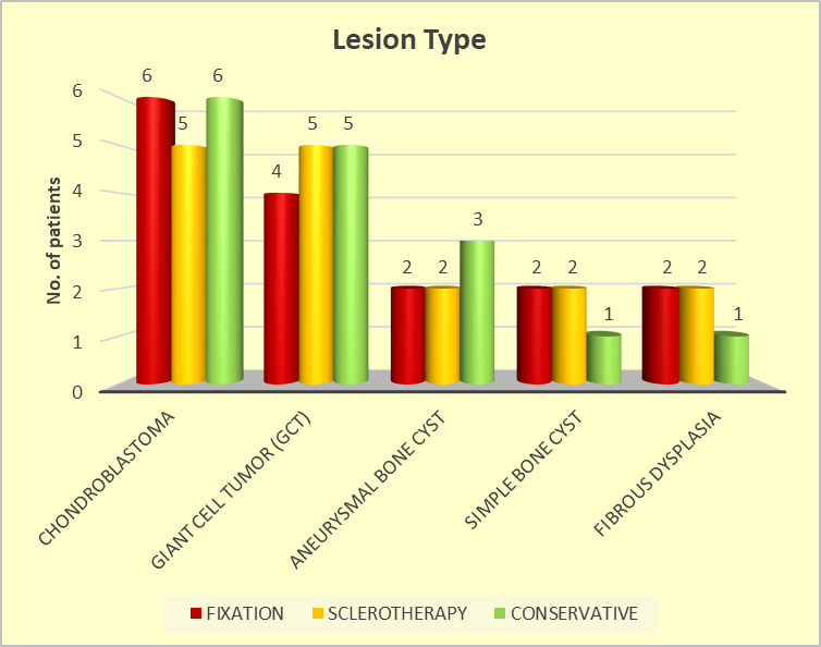
Graph 1: Graphical representation of Incidence and Prevalence of Benign Lesions (Clinical and Radiological)
Radiological stability was highest in the fixation group (87.5%), followed by sclerotherapy (62.5%) and conservative management (50%), although the difference was not statistically significant (p = 0.724). Early mobilization was also more frequent with fixation (81.25%) than sclerotherapy (56.25%) and conservative management (50%), without statistical significance (p = 0.1546). Overall, fixation showed a favorable trend for stability and early mobilization (table 2).
Table 2: Postoperative Outcomes
|
Parameter |
FIXATION [n=16] |
SCLEROTHERAPY [n=16] |
CONSERVATIVE [n=16] |
P-VALUE |
|
Radiological Stability |
|
|
|
|
|
Stable |
14 |
10 |
8 |
X=5.250 p=0.724 |
|
Unstable |
2 |
6 |
8 |
|
|
Early Mobilization |
|
|
|
|
|
Achieved |
13 |
9 |
8 |
X=3.733 p=0.1546 |
|
Delayed |
3 |
7 |
8 |
At 1 week, the fixation group consistently had higher scores across all subcomponents, with significant differences in pain, function, emotional acceptance, use of walking support, gait, and walking ability compared to the other groups (p<0.0001 for most parameters). The total MSTS score was also significantly higher in the fixation group (90.77 ± 7.55) than in the sclerotherapy (82.26 ± 8.61) and conservative groups (79.36 ± 6.79), with a p-value of 0.001. This trend continued at 3, 6, 12, 24 weeks, and 1 year, with the fixation group consistently outperforming the other groups in all subcomponents and the overall MSTS score, showing statistically significant differences across the board (p<0.0001 for most parameters). By 1 year, the total MSTS score in the fixation group was 106.26 ± 6.14, compared to 97.57 ± 7.72 in the sclerotherapy group and 92.63 ± 6.73 in the conservative group, indicating that the fixation group had the best functional outcomes throughout the study period (table 3).
Table 3: Functional Outcome (MSTS Score)
|
MSTS Score |
Fixation (n=16) |
Sclerotherapy (n=16) |
Conservative (n=16) |
p-value |
|
1 Week |
90.77 ± 7.55 |
82.26 ± 8.61 |
79.36 ± 6.79 |
F = 8.591, p = 0.001* |
|
3 Weeks |
94.24 ± 7.20 |
86.12 ± 8.36 |
85.25 ± 7.54 |
F = 5.146, p = 0.001* |
|
6 Weeks |
97.28 ± 6.92 |
88.28 ± 8.20 |
85.62 ± 7.72 |
F = 14.26, p < 0.0001* |
|
12 Weeks |
100.26 ± 6.62 |
91.25 ± 8.04 |
87.62 ± 7.27 |
F = 12.52, p < 0.0001* |
|
24 Weeks |
103.26 ± 6.44 |
94.52 ± 7.85 |
89.53 ± 6.62 |
F = 13.86, p < 0.0001* |
|
1 Year |
106.26 ± 6.14 |
97.57 ± 7.72 |
92.63 ± 6.73 |
— |
Fixation treatment consistently resulted in better hip function over time compared to sclerotherapy and conservative management (table 4).
Table 4: Harris Hip score at follow-up
|
Time Point |
Fixation [n=16] |
Sclerotherapy [n=16] |
Conservative [n=16] |
P-value |
|
1 Week |
65.50 ± 5.25 |
60.75 ± 6.80 |
50.09 ± 8.55 |
F=40.514, p<0.0001* |
|
3 Weeks |
70.25 ± 6.40 |
65.15 ± 7.21 |
58.43 ± 7.81 |
F=12.09, p<0.0001* |
|
6 Weeks |
75.80 ± 7.12 |
68.22 ± 8.50 |
63.69 ± 9.06 |
F=8.888, p=0.001 |
|
12 Weeks |
80.43 ± 7.50 |
72.85 ± 9.10 |
68.15 ± 9.48 |
F=6.707, p=0.003 |
|
24 Weeks |
85.75 ± 8.20 |
78.31 ± 9.80 |
74.02 ± 9.04 |
F=8.592, p=0.001 |
|
1 Year |
90.12 ± 8.75 |
82.45 ± 10.30 |
78.54 ± 9.41 |
F=4.121, p=0.023 |
At all the intervals, fixation group showed better outcomes in pain relief, pain-free status, and recovery compared to the other groups (table 5).
Table 5: Follow-up outcomes with statistical analysis
|
Time point |
Outcome |
Fixation [n=16] |
Sclerotherapy [n=16] |
Conservative [n=16] |
P-value |
|
1 Week |
Pain Relief |
12 (75%) |
10 (62.50%) |
6 (37.50%) |
X=4.8, p=0.091 |
|
Swelling |
4 (25%) |
6 (37.50%) |
10 (62.50%) |
||
|
3 Weeks |
Pain Relief |
13 (81.25%) |
11 (68.75%) |
7 (43.75%) |
X=5.101, p=0.078 |
|
Swelling |
3 (18.75%) |
5 (31.25%) |
9 (56.25%) |
||
|
6 Weeks |
Pain-Free |
15 (93.75%) |
11 (68.75%) |
7 (43.75%) |
X=9.214, p=0.01 |
|
Instability |
1 (6.25%) |
5 (31.25%) |
9 (56.25%) |
||
|
12 Weeks |
Full Mobility |
14 (87.50%) |
10 (62.50%) |
8 (50.00%) |
X=5.25, p=0.072 |
|
Stiffness |
2 (12.50%) |
6 (37.50%) |
8 (50.00%) |
||
|
24 Weeks |
Complete Recovery |
14 (87.50%) |
11 (68.75%) |
9 (56.25%) |
X=3.832, p=0.147 |
|
Stiffness |
2 (12.50%) |
5 (31.25%) |
7 (43.75%) |
||
|
1 Year |
Full Recovery |
15 (93.75%) |
12 (75%) |
10 (62.50%) |
X=4.482, p=0.106
|
|
Instability |
1 (6.25%) |
4 (25%) |
6 (37.50%) |
Complications were infrequent and did not differ significantly among groups (p=0.622). Infection occurred in 6.25% of fixation, 12.50% of sclerotherapy, and none of the conservative cases. Non-union and recurrence were most frequent in the conservative group (18.75% and 12.50%, respectively), while AVN occurred only in 6.25% of conservative cases. Mechanical failure was reported in 6.25% of the sclerotherapy group. Overall, no complication showed a statistically significant difference between treatment modalities (graph 2).
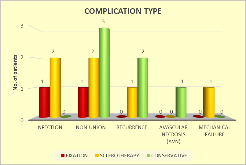
Graph 2: Graphical representation of Complication type.
DISCUSSION:
Management of benign cystic lesions of the proximal femur requires balancing effective lesion control with preservation of function and mobility. This prospective study evaluates the functional and radiological outcomes of different treatment modalities, including conservative management, sclerotherapy, and surgical fixation. By assessing functional recovery, pain relief, healing, recurrence, and complications, the study aims to identify the most effective and safe treatment strategy. The findings may help guide individualized clinical decision-making and optimize long-term patient outcomes.
The present study showed a relatively comparable demographic profile across the three treatment groups, with similar age, gender distribution, socioeconomic status, presenting complaints, and duration of illness. This similarity indicates adequate baseline comparability and minimizes the influence of demographic factors on treatment outcomes. Shi et al. [10] reported a mean age of 21.2 years among patients with benign bone lesions, with males accounting for 58% of cases. Their findings, including the broad age distribution and male predominance, were broadly comparable with the demographic characteristics observed in our study.
The fixation group demonstrated consistently better functional outcomes than the sclerotherapy and conservative groups. MSTS scores were significantly higher in the fixation group from the first week and remained superior throughout the one-year follow-up (p<0.0001). At the final assessment, the mean MSTS score was 106.26 ± 6.14 in the fixation group compared with 97.57 ± 7.72 in the sclerotherapy group and 92.63 ± 6.73 in the conservative group. These findings indicate better pain relief, gait, walking ability, emotional acceptance, and overall function following fixation. Shi et al. [10] reported acceptable functional outcomes with different curettage techniques, whereas Wijsbek et al. [11] supported hip replacement as a treatment option. In contrast, Sakayama et al. [12] recommended preservation of the native joint whenever possible. Our findings suggest that fixation can provide satisfactory functional recovery while avoiding more extensive procedures.
Radiological stability was highest in the fixation group (87.5%), followed by the sclerotherapy (62.5%) and conservative groups (50%), although the difference was not statistically significant (p=0.724). Similarly, early mobilization was more frequent after fixation (81.25%) than after sclerotherapy (56.25%) or conservative treatment (50%), but this difference was also not significant (p=0.1546). Preoperative laboratory parameters were comparable among the groups, suggesting similar baseline clinical status. Complications such as infection, non-union, recurrence, AVN, and mechanical failure showed numerical differences but none reached statistical significance. Krishna et al. [13] emphasized early weight-bearing, radiological assessment, and functional evaluation using the modified Harris Hip Score. Their findings of non-union and screw-related complications further highlight the importance of appropriate fixation and postoperative monitoring.
Chondroblastoma was the most frequently observed lesion in the present study, followed by giant cell tumor (GCT) and aneurysmal bone cyst (ABC). Chondroblastoma accounted for 37.5% of patients in the fixation and conservative groups and 31.25% in the sclerotherapy group. Strong et al. [14] reported recurrence following transcervical curettage for femoral-head chondroblastoma, while Suneja et al. [15] observed a higher risk of recurrence among younger patients with an open physis. GCT of the proximal femur has also been associated with increased recurrence following curettage, with pooled recurrence rates consistent with previous reports [15,16,17]. These findings emphasize the importance of lesion characteristics, skeletal maturity, surgical access, and choice of treatment technique in preventing recurrence.
The fixation group showed significantly better Harris Hip Scores at all follow-up intervals. The mean score increased from 65.50 ± 5.25 at one week to 90.12 ± 8.75 at one year, with statistically significant differences compared with the other groups. Krishna et al. [13] similarly reported favorable functional outcomes following fixation, although 6.66% of patients developed non-union requiring reoperation. Reoperation rates have varied among other studies, with Kumar et al. [18] and Halder et al. [16] reporting different rates. The functional outcome in our study was comparable to the results of Hu et al. [17], while being better than those reported by Shah et al [19]. The consistently higher Harris Hip Scores therefore suggest that fixation may facilitate earlier and better functional recovery.
The fixation group demonstrated progressive improvement in pain relief and functional recovery during follow-up. At six weeks, 93.75% of patients in the fixation group were pain-free compared with 75% in the sclerotherapy group and 43.75% in the conservative group (p=0.01). Similarly, full mobility at 12 weeks was achieved in 87.5% of fixation patients compared with 62.5% and 50% in the sclerotherapy and conservative groups, respectively. By one year, complete recovery was reported in 93.75%, 75%, and 62.5% of the respective groups, although the difference was not statistically significant (p=0.106). Kapoor et al. [20] reported sustained pain relief, unassisted ambulation, and full hip movement during follow-up. Rahman et al. [21] and Wai et al. [22] also reported favorable long-term outcomes and low recurrence following appropriate surgical management.
Limitations
1. In this study limited number of participants may reduce the generalizability of the findings and limit the statistical power to detect differences between treatment groups.
2. The follow-up period may be insufficient to fully capture long-term outcomes and potential late complications associated with each treatment modality.
3. If participants were not randomly assigned to treatment groups or if there were differences in baseline characteristics, the results might be influenced by selection bias.
4. Variability in treatment protocols, surgical techniques, and postoperative care among participants could introduce inconsistencies in the outcomes, making it difficult to isolate the effects of each treatment modality.
CONCLUSION:
Overall, fixation demonstrated a consistent advantage in terms of functional recovery, MSTS and Harris Hip Scores, radiological stability, early mobilization, pain relief, and long-term recovery. Although several differences in complications and follow-up outcomes were not statistically significant, the overall trend favored fixation over sclerotherapy and conservative management. These findings support fixation as a potentially effective treatment option for benign cystic lesions of the proximal femur. However, larger studies with longer follow-up are required to establish the statistical significance and long-term superiority of fixation.
REFERENCES:
1. Mascard E, Gomez-Brouchet A, Lambot K. Bone cysts: unicameral and aneurysmal bone cyst. Orthop Traumatol Surg Res. 2015 Feb;101(1 Suppl):S119-27.
2. Rapp TB, Ward JP, Alaia MJ. Aneurysmal bone cyst. J Am Acad Orthop Surg. 2012 Apr;20(4):233-41.
3. Fletcher C, Bridge JA, Hogendoorn PC, Mertens F. WHO classification of tumours of soft tissue and bone: WHO classification of tumours, vol. 5. World Health Organization; 2013.
4. Warren M, Xu D, Li X. Gene fusions PAFAH1B1-USP6 and RUNX2-USP6 in aneurysmal bone cysts identified by next generation sequencing. Cancer Genet. 2017 Apr;212-213:13-18.
5. Oliveira AM, Perez-Atayde AR, Dal Cin P, Gebhardt MC, Chen CJ, Neff JR, Demetri GD, Rosenberg AE, Bridge JA, Fletcher JA. Aneurysmal bone cyst variant translocations upregulate USP6 transcription by promoter swapping with the ZNF9, COL1A1, TRAP150, and OMD genes. Oncogene. 2005 May 12;24(21):3419-26.
6. Cottalorda J, Bourelle S. Current treatments of primary aneurysmal bone cysts. J Pediatr Orthop B. 2006 May;15(3):155-67.
7. Tomaszewski R, Rutz E, Mayr J, Dajka J. Surgical treatment of benign lesions and pathologic fractures of the proximal femur in children. Archives of orthopaedic and trauma surgery. 2020 Nov 24:1-0.
8. DOğAnAvşArgİL B, Ayhan E, Argin M, Pehlivanoğlu B, Keçeci B, Sezak M, Başdemir G, Öztop F. Cystic bone lesions: histopathological spectrum and diagnostic challenges. Turk Patoloji Derg. 2015 May 1;31(2):95-103.
9. Noordin S, Ahmad T, Umer M, Allana S, Hilal K, Uddin N, Hashmi P. Aneurysmal bone cyst of the pelvis and extremities: Contemporary management. IJS Oncology. 2019 Jan 1;4:e711-2.
10. Shi J, Zhao Z, Yan T, Guo W, Yang R, Tang X, et al. Surgical treatment of benign osteolytic lesions in the femoral head and neck: a systematic review. BMC Musculoskelet Disord. 2021 Jun 16;22(1).
11. Wijsbek AE, Vazquez-Garcia BL, Grimer RJ, Carter SR, Abudu AA, Tillman RM, et al. Giant cell tumour of the proximal femur: is joint-sparing management ever successful? Bone Joint J. 2014;96-B(1):127–31.
12. Sakayama K, Sugawara Y, Kidani T, Miyawaki J, Fujibuchi T, Kamei S, et al. Diagnostic and therapeutic problems of giant cell tumor in the proximal femur. Arch Orthop Trauma Surg. 2007;127(10):867–72.
13. Krishna V, Venkatesan A, Singh AK. Functional and Radiological Outcomes of Unstable Proximal Femur Fractures Fixed With Anatomical Proximal Locking Compression Plate. Cureus. 2022 May 11;14(5):e24903.
14. Strong DP, Grimer RJ, Carter SR, Tillman RM, Abudu A. Chondroblastoma of the femoral head: management and outcome. Int Orthop. 2010;34(3):413–7.
15. Suneja R, Grimer RJ, Belthur M, Jeys L, Carter SR, Tillman RM, et al. Chondroblastoma of bone: long-term results and functional outcome after intralesional curettage. J Bone Joint Surg Br. 2005;87(7):974–8.
16. Halder SC. J. The Gamma nail for peritrochanteric fractures. Bone Joint Surg Br. 1992;74:340–344.
17. Hu SJ, Zhang SM, Yu GR. Treatment of femoral subtrochanteric fractures with proximal lateral femur locking plates. Acta Ortop Bras. 2012;20:329–333.
18. Kumar N, Kataria H, Yadav C, Gadagoli BS, Raj R. J Evaluation of proximal femoral locking plate in unstable extracapsular proximal femoral fractures: Surgical technique & mid term follow up results. Clin Orthop Trauma. 2014;5:137–145.
19. Shah MD, Kapoor CS, Soni RJ, Patwa JJ, Golwala PP. Evaluation of outcome of proximal femur locking compression plate (PFLCP) in unstable proximal femur fractures. J Clin Orthop Trauma. 2017;8:308–312.
20. Kapoor C, Shah M, Soni R, Patwa J, Merh A, Golwala P. Aneurysmal Bone Cyst of the Proximal Femur and Its Management - A Case Report. Cureus. 2017 Jan 23;9(1):e991.
21. Rahman MA, El Masry AM, Azmy SI. Review of 16 cases of aneurysmal bone cyst in the proximal femur treated by extended curettage and cryosurgery with reconstruction using autogenous nonvascularized fibula graft. J Orthop Surg (Hong Kong). 2018;26(2):2309499018783905.
22. Wai EK, Davis AM, Griffin A, Bell RS, Wunder JS. Pathologic fractures of the proximal femur secondary to benign bone tumors. Clin Orthop Relat Res.2001;393:279–86.