To study higher order aberrations in post Phacoemulsification and Small incision cataract surgery in a tertiary health care centre.
- Ramanna , Assistant Professor Department of Ophthalmology Raichur Institute of Medical Sciences, Raichur, Karnataka, India
- Mukesh Kumar Saini , Assistant Professor Department of Ophthalmology Shri Bhausaheb Hire Government Medical College, Dhule, Maharashtra, India
- Shruthi , Assistant Professor Department of Pathology Navodaya Medical College Hospital and Research Centre, Raichur, Karnataka, India
Article Information:
Abstract:
Background: Higher-order aberrations (HOAs) are important determinants of visual quality and may contribute to glare, halos, reduced contrast sensitivity, and impaired night vision despite satisfactory postoperative visual acuity. Cataract surgery may alter corneal optical characteristics and consequently influence HOAs. This study evaluated changes in visual, refractive, keratometric, and corneal higher-order aberration parameters following cataract surgery. Materials and Methods: This prospective, single-centre observational study included 200 eyes of 200 patients undergoing cataract surgery at an urban tertiary care hospital. Demographic characteristics, spherical equivalent, logMAR visual acuity, keratometric parameters, astigmatic axis, and total corneal higher-order aberrations (cHOA) were assessed. Preoperative and postoperative measurements were compared using appropriate statistical tests. A p value <0.05 was considered statistically significant. Results: The mean age was 54.22 ± 5.30 years; 68% of participants were aged >55 years, and 60% were males. Mean spherical equivalent improved from −0.89 ± 1.70 D preoperatively to −0.55 ± 1.10 D postoperatively (p=0.0009). Mean logMAR visual acuity improved significantly from 0.31 ± 0.10 to 0.02 ± 0.01 (p=0.0009). Flat keratometry decreased from 41.8 ± 2.1 D to 41.2 ± 1.9 D (p<0.0001), while steep keratometry showed no significant change (43.6 ± 2.1 vs. 43.8 ± 2.2 D; p=0.19). The astigmatic axis changed significantly from 72.5 ± 93.1° to 104.2 ± 92.3° (p=0.0001). Total cHOA increased significantly from 0.28 ± 0.10 µm to 0.36 ± 0.10 µm (p<0.001). Conclusion: Cataract surgery significantly improved visual acuity and refractive status but was associated with measurable changes in corneal higher-order aberrations. Assessment of HOAs may complement conventional measures when evaluating postoperative visual quality.
Keywords:
Article :
INTRODUCTION:
The human eye is a complex optical system in which visual quality is influenced not only by refractive errors but also by various optical aberrations. Optical quality is limited by aberrations, diffraction, and intraocular light scatter. The air–cornea interface represents the first and most important refractive surface of the eye and contributes substantially to the total refractive power because of the marked difference in refractive indices between air and the cornea.[1]Ocular aberrations are broadly classified into lower-order and higher-order aberrations (HOAs). Lower-order aberrations, including myopia, hypermetropia, and regular astigmatism, account for more than 90% of total wavefront aberrations and can generally be corrected using conventional spectacles or contact lenses.[5] In contrast, HOAs, such as spherical aberration, coma, and trefoil, cannot be adequately corrected with conventional spherical or cylindrical lenses.[6] Although they constitute a smaller proportion of total ocular aberrations, HOAs can significantly impair retinal image quality and may manifest clinically as glare, halos, blurred vision, starbursts, diplopia, and reduced night vision. Increasing interest in wavefront-guided refractive surgery,[2] customized contact lenses,[3] and wavefront-based intraocular lenses (IOLs)[4] has emphasized the importance of assessing these aberrations.The optical characteristics of the eye also change with age. In younger individuals, positive spherical aberration of the anterior cornea is partially compensated by the negative spherical aberration of the crystalline lens. With increasing age, lenticular spherical aberration increases, while corneal aberrations remain relatively stable, resulting in deterioration of overall optical quality. Cataract formation further alters the optical characteristics of the crystalline lens. Following cataract surgery, replacement of the natural crystalline lens with an IOL may modify the balance of ocular aberrations. Conventional spherical IOLs may contribute positive spherical aberration, thereby influencing postoperative visual quality. Modern wavefront technology permits quantitative assessment of these aberrations using Zernike polynomials and root mean square (RMS) values.[7]
Phacoemulsification and manual small-incision cataract surgery (MSICS) are widely used techniques for cataract management. MSICS, initially described by Blumenthal, provides rapid visual rehabilitation with relatively low cost and has become particularly important in resource-limited settings.[8] Phacoemulsification, pioneered by Charles D. Kelman, uses ultrasonic energy to emulsify and aspirate the crystalline lens through a small incision and has become an established technique for modern cataract surgery.[9]Although both procedures provide satisfactory postoperative visual acuity, conventional measures of visual outcome may not completely reflect the quality of vision. Evaluation of HOAs may therefore provide a more comprehensive assessment of postoperative optical performance. Hence, the present study was undertaken to evaluate and compare higher-order aberrations following phacoemulsification and small-incision cataract surgery at a tertiary care centre.
MATERIALS AND METHODS:
This prospective, single-centre, observational study was conducted in the Department of Ophthalmology at an urban tertiary care hospital. The study was designed to evaluate and compare higher-order aberrations in patients following phacoemulsification and small-incision cataract surgery (SICS).
Study Duration
The study was conducted over a period of 18 months following approval from the Institutional Ethics Committee (IEC).
Study Population
Patients undergoing cataract surgery at the Department of Ophthalmology during the study period were screened for eligibility. A total of 200 eyes of 200 patients fulfilling the predefined selection criteria were enrolled after obtaining written informed consent.
Sample Size
The study included a total of 200 eyes of 200 patients.
Inclusion Criteria
Patients were included if they fulfilled the following criteria:
- Patients of either sex, aged 35–60 years, who were candidates for cataract surgery and had an otherwise normal ocular examination.
- Patients with cataractous media requiring surgical intervention.
- Patients who were willing to participate in the study and provided written informed consent.
Exclusion Criteria
Patients were excluded if they had:
- A previous history of ocular or corneal surgery, ocular trauma, or corneal scarring.
- Pathological myopia or severe chorioretinal atrophy that could interfere with vision or wavefront measurements.
- Best spectacle-corrected visual acuity (BSCVA) of 20/40 or worse attributable to ocular pathology other than cataract.
- Any other ocular condition likely to interfere with accurate assessment of higher-order aberrations.
- Unwillingness to participate or provide written informed consent.
Study Procedure
A total of 200 eligible patients attending the Department of Ophthalmology were enrolled in the study after application of the inclusion and exclusion criteria and obtaining written informed consent.Demographic characteristics and relevant clinical information were recorded using a predefined case record form. All patients underwent a detailed ocular examination. Eligible patients subsequently underwent cataract surgery by either phacoemulsification or small-incision cataract surgery according to the planned surgical procedure.Following cataract surgery, patients underwent postoperative ophthalmological assessment. Higher-order aberrations were evaluated using wavefront analysis. Parameters related to higher-order aberrations, including spherical aberration, coma, trefoil, and total higher-order aberrations, were recorded wherever applicable. Wavefront aberrations were expressed using Zernike coefficients and root mean square (RMS) values.
The postoperative findings were documented and compared between patients who underwent phacoemulsification and those who underwent small-incision cataract surgery to determine differences in postoperative higher-order aberrations.
Data Collection
Demographic, clinical, ophthalmological, surgical, and wavefront aberration data were recorded in the case record form. The collected data were subsequently entered into Microsoft Excel for compilation, coding, and analysis. Results were presented in appropriate tabular and graphical formats.
Ethical Considerations
The study was initiated after obtaining approval from the Institutional Ethics Committee. Written informed consent was obtained from all participants before enrolment. The confidentiality and privacy of patient information were maintained throughout the study, and the collected data were used exclusively for the stated research objectives. Any deviation or modification in the approved study methodology was undertaken only after appropriate approval from the Institutional Ethics Committee.
Statistical Analysis
Statistical analysis was performed using GraphPad Prism version 6.0 (GraphPad Software Inc., La Jolla, CA, USA). Continuous variables were summarized as mean ± standard deviation or median with interquartile range, as appropriate, while categorical variables were expressed as frequencies and percentages.The distribution of continuous variables was assessed before selecting the appropriate statistical test. For non-normally distributed continuous variables, the Mann–Whitney U test was used to compare independent groups, including the phacoemulsification and SICS groups. Appropriate statistical tests were applied for categorical variables wherever required. All statistical tests were two-sided, and a p value <0.05 was considered statistically significant.
RESULTS:
A total of 200 participants were included in the study. The majority of participants were aged >55 years, accounting for 68.0% of the study population, followed by those aged 45–55 years (22.0%) and 35–45 years (10.0%). Males constituted 60.0% of the participants, while females accounted for 40.0%, showing a male predominance in the study population (Table 1).The mean preoperative spherical equivalent was −0.89 ± 1.70 D, which improved to −0.55 ± 1.10 D postoperatively. This change was statistically significant (p=0.0009). Similarly, mean visual acuity improved from 0.31 ± 0.10 logMAR preoperatively to 0.02 ± 0.01 logMAR postoperatively, and this improvement was also statistically significant (p=0.0009) (Table 2).With regard to keratometric parameters, mean flat keratometry decreased significantly from 41.8 ± 2.1 D preoperatively to 41.2 ± 1.9 D postoperatively (p<0.0001). Mean steep keratometry increased slightly from 43.6 ± 2.1 D to 43.8 ± 2.2 D; however, this difference was not statistically significant (p=0.19). Cylinder keratometry decreased from 41.8 ± 2.1 preoperatively to 41.2 ± 1.9 postoperatively, with a statistically significant difference (p=0.0004) (Table 3, Figure 1).The mean astigmatic axis changed from 72.5 ± 93.1° preoperatively to 104.2 ± 92.3° postoperatively. This postoperative change in astigmatic axis was statistically significant (p=0.0001) (Table 4, Figure 2).The mean total corneal higher-order aberration (cHOA) increased from 0.28 ± 0.10 µm preoperatively to 0.36 ± 0.10 µm postoperatively. The observed increase was statistically significant (p<0.001), indicating a significant postoperative change in corneal higher-order aberrations (Table 5, Figure 3).Overall, significant postoperative improvements were observed in spherical equivalent and visual acuity. Flat keratometry showed a significant reduction, whereas the change in steep keratometry was not statistically significant. The astigmatic axis showed a significant postoperative shift, while total corneal higher-order aberrations increased significantly after surgery. A summary of the major postoperative visual and optical changes is presented in Table 6.
Table 1. Demographic characteristics of the study participants (N=200)
|
Characteristic |
Frequency (n) |
Percentage (%) |
|
Age group (years) |
||
|
35–45 |
20 |
10.0 |
|
45–55 |
44 |
22.0 |
|
>55 |
136 |
68.0 |
|
Sex |
||
|
Male |
120 |
60.0 |
|
Female |
80 |
40.0 |
|
Total |
200 |
100.0 |
Table 2. Comparison of preoperative and postoperative spherical equivalent and visual acuity
|
Parameter |
Preoperative Mean ± SD |
Postoperative Mean ± SD |
p value |
Significance |
|
Spherical equivalent (D) |
−0.89 ± 1.70 |
−0.55 ± 1.10 |
0.0009 |
Significant |
|
Visual acuity (logMAR) |
0.31 ± 0.10 |
0.02 ± 0.01 |
0.0009 |
Significant |
Table 3. Comparison of preoperative and postoperative keratometric parameters
|
Keratometric parameter |
Preoperative Mean ± SD |
Postoperative Mean ± SD |
p value |
Significance |
|
Flat keratometry (D) |
41.8 ± 2.1 |
41.2 ± 1.9 |
<0.0001 |
Significant |
|
Steep keratometry (D) |
43.6 ± 2.1 |
43.8 ± 2.2 |
0.19 |
Not significant |
|
Cylinder keratometry* |
41.8 ± 2.1 |
41.2 ± 1.9 |
0.0004 |
Significant |
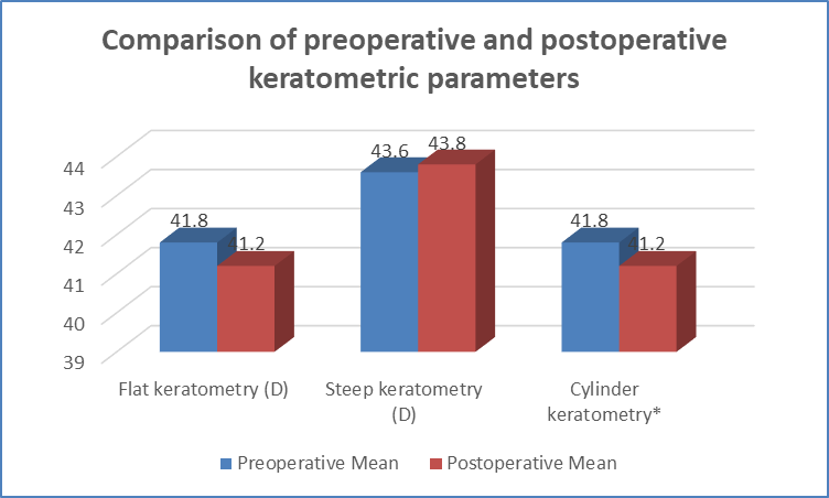
Figure 1 Comparison of preoperative and postoperative keratometric parameters
Table 4. Comparison of preoperative and postoperative astigmatic axis
|
Parameter |
Preoperative Mean ± SD |
Postoperative Mean ± SD |
p value |
Significance |
|
Astigmatic axis (degrees) |
72.5 ± 93.1 |
104.2 ± 92.3 |
0.0001 |
Significant |
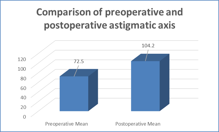
Figure 2 Comparison of preoperative and postoperative astigmatic axis
Table 5. Comparison of preoperative and postoperative total corneal higher-order aberrations
|
Parameter |
Preoperative Mean ± SD |
Postoperative Mean ± SD |
p value |
Significance |
|
Total corneal higher-order aberration (cHOA), µm |
0.28 ± 0.10 |
0.36 ± 0.10 |
<0.001 |
Significant |
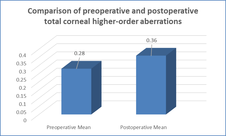
Figure 3 Comparison of preoperative and postoperative total corneal higher-order aberrations
Table 6. Summary of changes in major postoperative visual and optical parameters
|
Parameter |
Preoperative Mean |
Postoperative Mean |
Direction of change |
p value |
|
Spherical equivalent (D) |
−0.89 |
−0.55 |
Improved toward emmetropia |
0.0009 |
|
Visual acuity (logMAR) |
0.31 |
0.02 |
Improved |
0.0009 |
|
Flat keratometry (D) |
41.8 |
41.2 |
Decreased |
<0.0001 |
|
Steep keratometry (D) |
43.6 |
43.8 |
Slightly increased |
0.19 |
|
Astigmatic axis (°) |
72.5 |
104.2 |
Changed |
0.0001 |
|
Total cHOA (µm) |
0.28 |
0.36 |
Increased |
<0.001 |
DISCUSSION:
The present study evaluated demographic characteristics, refractive and keratometric parameters, visual acuity, and corneal higher-order aberrations (cHOAs) in 200 patients undergoing cataract surgery. Overall, cataract surgery resulted in significant improvement in refractive status and visual acuity, although measurable postoperative changes in corneal keratometry and higher-order aberrations were also observed.In the present study, 68% of patients were older than 55 years, 22% were aged 45–55 years, and 10% were aged 35–45 years, with a mean age of 54.22 ± 5.3 years. Yasushi Inoue et al.[10] reported a considerably higher mean age of 72.6 ± 8.0 years, whereas Kasturi Bhattacharjee et al.[11] reported a mean age of 56.53 ± 9.06 years, comparable to the present study. In contrast, Ferial M Al-Zeraid et al.[12] studied a much younger population with a mean age of 25.6 ± 4.9 years. These variations may be related to differences in patient selection and study populations. Males constituted 60% of the present cohort, whereas female predominance was reported by Inoue et al.[10] and Bhattacharjee et al.[11]The mean spherical equivalent improved significantly from −0.89 ± 1.70 D preoperatively to −0.55 ± 1.10 D postoperatively (p=0.0009), indicating a postoperative refractive shift toward emmetropia. Dominika et al.[13] similarly reported an improvement from −0.95 D to −0.54 D. Mean logMAR visual acuity also improved significantly from 0.31 ± 0.10 to 0.02 ± 0.01 (p=0.0009), closely corresponding with the improvement from 0.33 to 0.02 logMAR reported by Dominika et al.[13] Haldipurkar et al.[14], however, reported no significant intergroup difference in best-corrected distance visual acuity (p=0.48), possibly reflecting differences in study design and assessment methods.Flat keratometry decreased significantly from 41.8 ± 2.1 D to 41.2 ± 1.9 D (p<0.0001), whereas Dominika et al.[13] observed only a minimal change from 42.8 D to 42.77 D. In contrast, steep keratometry showed a nonsignificant increase from 43.6 ± 2.1 D to 43.8 ± 2.2 D (p=0.19), comparable to the findings of Dominika et al.[13] The astigmatic axis changed significantly from 72.5 ± 93.1° to 104.2 ± 92.3° (p=0.0001), indicating that cataract surgery may influence corneal astigmatic orientation through incision-related biomechanical changes.Importantly, total cHOA increased significantly from 0.28 ± 0.10 µm preoperatively to 0.36 ± 0.10 µm postoperatively (p<0.001). Dominika et al.[13] reported nearly identical findings, with an increase from 0.27 µm to 0.35 µm. Increased HOAs may adversely affect visual quality by contributing to glare, halos, reduced contrast sensitivity, and impaired night vision despite satisfactory postoperative visual acuity.Overall, cataract surgery produced significant refractive and visual improvement but was accompanied by changes in corneal keratometry and higher-order aberrations. These findings, particularly those concerning spherical equivalent, visual acuity, steep keratometry, and cHOAs, were broadly consistent with Dominika et al.[13] and emphasize the importance of evaluating postoperative optical quality in addition to conventional visual acuity.
CONCLUSION:
The present study demonstrated significant improvement in visual acuity and spherical equivalent following cataract surgery. Significant postoperative changes were also observed in flat keratometry and astigmatic axis, whereas steep keratometry remained relatively stable. Total corneal higher-order aberrations increased significantly after surgery, indicating measurable changes in postoperative corneal optical quality. These findings emphasize that assessment of higher-order aberrations, along with conventional visual acuity and refractive outcomes, may provide a more comprehensive evaluation of visual quality following cataract surgery.
LIMITATIONS
The present study was a single-centre study, which may limit the generalizability of the findings to other populations. The sample size was relatively limited, and long-term postoperative changes in higher-order aberrations were not assessed. Factors such as pupil size, intraocular lens characteristics, and other variables influencing postoperative optical quality were not evaluated in detail. Larger multicentre studies with longer follow-up are required to validate these findings.
REFERENCES:
1. Anbar M, Mohamed Mostafa E, Elhawary AM, Awny I, Farouk MM, Mounir A, et al. Evaluation of corneal higher-order aberrations by Scheimpflug–Placido topography in patients with different refractive errors: a retrospective observational study. J Ophthalmol. 2019;2019:5640356.
2. Kim A, Chuck RS. Wavefront-guided customized corneal ablation. Curr Opin Ophthalmol. 2008;19(4):314-320.
3. Marsack JD, Parker KE, Applegate RA. Performance of wavefront-guided soft lenses in three keratoconus subjects. Optom Vis Sci. 2008;85(12):E1172-E1178.
4. Nochez Y, Favard A, Majzoub S, Pisella PJ. Measurement of corneal aberrations for customisation of intraocular lens asphericity: impact on quality of vision after micro-incision cataract surgery. Br J Ophthalmol. 2010;94(4):440-444.
5. Lombardo M, Lombardo G. Wave aberration of human eyes and new descriptors of image optical quality and visual performance. J Cataract Refract Surg. 2010;36(2):313-331.
6. Thibos LN. Retinal image quality for virtual eyes generated by a statistical model of ocular wavefront aberrations. Ophthalmic Physiol Opt. 2009;29(3):288-291.
7. Hayashi K, Yoshida M, Hayashi H. Correlation of higher-order wavefront aberrations with visual function in pseudophakic eyes. Eye (Lond). 2008;22(12):1476-1482.
8. Ang M, Evans JR, Mehta JS. Manual small incision cataract surgery (MSICS) with posterior chamber intraocular lens versus extracapsular cataract extraction (ECCE) with posterior chamber intraocular lens for age-related cataract. Cochrane Database Syst Rev. 2014;(11):CD008811.
9. Buratto L, Apple DJ, Zanini M, editors. Phacoemulsification: Principles and Techniques. Thorofare, NJ: SLACK Incorporated; 2003.
10. Inoue Y, Ochi S. Effects of 3% diquafosol sodium ophthalmic solution on higher-order aberrations in patients diagnosed with dry eye after cataract surgery. Clin Ophthalmol. 2017;11:87-93.
11. Bhattacharjee K, Misra D, Singh M, Deori N. Long-term changes in contrast-sensitivity, corneal topography and higher-order aberrations after upper eyelid blepharoplasty: a prospective interventional study. Indian J Ophthalmol. 2020;68(12):2906-2910.
12. Al-Zeraid FM, Osuagwu UL. Induced higher-order aberrations after laser in situ keratomileusis (LASIK) performed with wavefront-guided IntraLase femtosecond laser in moderate to high astigmatism. BMC Ophthalmol. 2016;16:29.
13. Pohlmann D, Pilger D, Bertelmann E, von Sonnleithner C. Corneal higher-order aberrations after cataract surgery: manual phacoemulsification versus femtosecond-laser assisted technique. Eur J Ophthalmol. 2021;31(6):2955-2961.
14. Haldipurkar SS, Shetty V, Shah D, Haldipurkar T, Kashelkar P, Khatib Z, et al. Comparison of post-cataract surgery visual outcomes and quality of life in patients bilaterally implanted with multifocal intraocular lenses. Ophthalmol Ther. 2021;10(1):101-113.