Study of Low Dose Atropine in Prevention of Progression of Myopia in Children.
- Ramanna , Assistant Professor Department of Ophthalmology Raichur Institute of Medical Sciences, Raichur, Karnataka, India
- Karishma Munoli , Associate Professor Raichur Institute of Medical Sciences, Raichur, Karnataka, India
- Shruthi , Assistant Professor Department of Pathology Navodaya Medical College Hospital and Research Centre, Raichur, Karnataka, India
- Garlapati Vardhan Viishhnu , Senior Resident Department of Ophthalmology Raichur Institute of Medical Sciences, Raichur, Karnataka, India
Article Information:
Abstract:
Background: Childhood myopia is increasing worldwide and can lead to serious ocular complications. This study evaluated the effectiveness and safety of topical 0.01% atropine in controlling myopia progression in children. Methods: This prospective interventional study included 100 children aged 6–12 years, randomly allocated to an atropine group (n=50) or non-atropine control group (n=50). The atropine group received one drop of 0.01% atropine daily at bedtime. Mean spherical equivalent (MSE) was assessed at baseline and at 3, 6 and 9 months. The atropine group was reassessed after a three-month atropine-free interval to evaluate rebound progression. Results: At nine months, the mean progression in MSE was significantly lower in the atropine group than in the control group (−0.256 ± 0.050 D versus −0.732 ± 0.433 D; p<0.0001), representing an approximately 65% reduction in myopia progression. After the three-month atropine-free interval, the change in MSE was minimal and statistically insignificant (−0.001 D; p=0.659). Photophobia occurred in 4% of atropine-treated children, with no persistent adverse effects. Conclusion: Topical 0.01% atropine was effective and well tolerated in reducing childhood myopia progression. No significant short-term rebound was observed after treatment discontinuation.
Keywords:
Article :
INTRODUCTION:
Myopia is among the most prevalent refractive errors worldwide, with its frequency increasing rapidly, particularly in developed nations and East Asian populations. In some Asian countries, more than 90% of adolescents and young adults are affected. Myopia frequently begins during the early school years, commonly between 6 and 8 years of age, and may continue to progress throughout childhood and adolescence. This increasing prevalence represents a major public health concern because childhood-onset myopia allows a longer period for progression and increases the likelihood of developing high myopia later in life.[1]
Myopia is not merely an optical problem requiring refractive correction. Progressive axial elongation of the eye increases the lifetime risk of sight-threatening complications, including retinal detachment, glaucoma, cataract and myopic macular degeneration. The risk of these complications generally increases with the severity of myopia; therefore, early identification and effective control of myopic progression are essential for preserving long-term visual health.[1]
Various optical and pharmacological strategies have been evaluated for controlling childhood myopia.[2,3] Conventional single-vision spectacles and contact lenses provide clear vision but have little effect on the underlying progression of myopia. Rigid gas-permeable contact lenses and progressive-addition spectacle lenses have also demonstrated limited or inconsistent long-term benefits in controlling refractive progression and axial elongation.[4,5]
Recent investigations have reported encouraging results with interventions such as orthokeratology and topical atropine.[6–9] Bifocal and multifocal soft contact lenses incorporating peripheral-defocus designs have also emerged as promising options for myopia control.[10] In addition, greater outdoor exposure has been associated with a reduced risk of myopia development, although its effectiveness in slowing the progression of established myopia is less certain.[11] Despite the availability of these approaches, the optimal method of myopia control remains a subject of continuing investigation.
Among pharmacological interventions, topical atropine has demonstrated consistent effectiveness in reducing childhood myopia progression. However, conventional or higher atropine concentrations may produce concentration-dependent adverse effects, including photophobia, blurred near vision, reduced accommodation, pupillary dilatation and allergic reactions. These undesirable effects can interfere with daily activities, reduce treatment adherence and restrict the long-term use of atropine in children.[12,13] Consequently, lower concentrations of atropine have been investigated to achieve an appropriate balance between therapeutic effectiveness and tolerability.Low-dose atropine, particularly 0.01% atropine eye drops, has gained considerable attention as a practical approach to childhood myopia control. It may reduce the rate of refractive progression while producing fewer ocular adverse effects than higher concentrations. Nevertheless, variations in treatment response have been reported, and evidence regarding its long-term effectiveness, safety, optimal duration of therapy and rebound progression after discontinuation remains incomplete.The growing burden of myopia also has substantial medical, economic and psychosocial implications. Repeated ophthalmic examinations, frequent replacement of spectacles or contact lenses and treatment of associated ocular complications contribute to healthcare expenditure. Myopia may additionally affect children’s academic activities, outdoor participation, self-image and overall quality of life.[14–16] Therefore, a safe, acceptable and effective intervention for controlling childhood myopia would have considerable clinical and public health value.Although previous studies have demonstrated promising results with atropine therapy, further evidence is required regarding the effectiveness and adverse-effect profile of 0.01% atropine in different paediatric populations. Therefore, the present study was conducted to evaluate the effectiveness of low-dose atropine (0.01%) in preventing the progression of myopia in children and to assess the adverse effects associated with its use.
MATERIALS AND METHODS:
Study Design and Setting
This prospective, single-centre, interventional study was conducted in the Department of Ophthalmology at an urban tertiary-care hospital. The study evaluated the effectiveness of topical low-dose atropine (0.01%) in controlling the progression of myopia among children.
Study Population and Sample Size
A total of 100 children with myopia were enrolled. Participants were randomly allocated by the lottery method into two groups:
- Atropine group: 50 children
- Control group: 50 children
Ethical Considerations
The study was conducted after obtaining approval from the Institutional Ethics Committee. Written informed consent was obtained from the parents or legal guardians before enrolment. The confidentiality of the participants and their clinical information was maintained throughout the study.
Selection Criteria
Inclusion Criteria
Children fulfilling the following criteria were included:
- Children of either sex aged 6–12 years.
- Children diagnosed with simple myopia.
- Myopic refractive error of at least −0.50 dioptre.
- Willingness of the parent or legal guardian to provide informed consent.
Exclusion Criteria
Children with the following conditions were excluded:
- Pathological myopia.
- Previous intraocular surgery.
- Amblyopia.
- Known hypersensitivity to atropine eye drops.
- Significant cardiac or respiratory disease.
- Systemic disorders associated with myopia, such as Marfan syndrome or Stickler syndrome.
- Refusal to provide informed consent.
Randomisation and Group Allocation
Eligible participants were randomly assigned in a 1:1 ratio using the lottery method. Children in the atropine group received topical 0.01% atropine, whereas those in the control group did not receive atropine.
Study Intervention
Children in the atropine group were advised to instil one drop of 0.01% atropine into each myopic eye once daily at bedtime. Children in the control group received appropriate refractive correction but were not prescribed atropine eye drops.
Baseline Ophthalmic Evaluation
At enrolment, participants in both groups underwent the following examinations:
- Visual acuity using a Snellen chart
- Autorefraction
- Best-corrected visual acuity
- Dilated autorefraction
- Anterior-segment examination
- Posterior-segment examination
Follow-Up Assessment
Participants in both groups were reviewed at three-month intervals for nine months. Evaluations were performed at baseline and at 3, 6 and 9 months. At each follow-up visit, visual acuity, autorefraction, best-corrected visual acuity and dilated autorefraction were recorded.
The atropine group was also assessed for treatment-related adverse effects, including photophobia, blurred near vision, ocular irritation, redness and allergic reactions.
Assessment of Rebound Progression
Atropine eye drops were discontinued after nine months of treatment. Children in the atropine group were reassessed three months after discontinuation, at the 12-month visit, to evaluate rebound progression of myopia during the atropine-free interval.
Outcome Measures
Primary Outcome
The primary outcome was the change in myopic refractive error from baseline to nine months in the atropine group compared with the control group.
Secondary Outcomes
The secondary outcomes included:
- Change in visual acuity and best-corrected visual acuity.
- Frequency of adverse effects associated with 0.01% atropine.
- Rebound myopic progression after discontinuation of atropine.
Materials Used
The following materials and instruments were used:
- Atropine eye drops (0.01%)
- Snellen visual acuity chart
- Autorefractometer
- Trial frame
- Trial lens set
Data Collection
Clinical findings were documented using a structured case-record form. The collected data were entered into Microsoft Excel and checked for completeness and accuracy before analysis.
Statistical Analysis
Quantitative variables were expressed as mean ± standard deviation, while categorical variables were presented as frequencies and percentages. Mean values between the atropine and control groups were compared using the independent-samples Student’s t-test. Changes between two time points within the same group were analysed using the paired Student’s t-test. Categorical variables were compared using the chi-square test or Fisher’s exact test, as appropriate. A two-tailed p-value <0.05 was considered statistically significant.
RESULTS:
A total of 100 children were included in the study, with 50 participants each in the atropine and non-atropine groups. In the atropine group, 38.0% of participants were aged 6–8 years, 30.0% were aged 8–10 years, and 32.0% were aged 10–12 years. The corresponding proportions in the non-atropine group were 32.0%, 26.0%, and 42.0%, respectively. The mean age was 9.18 ± 1.96 years in the atropine group and 9.64 ± 2.04 years in the non-atropine group. Males constituted 48.0% of the atropine group and 52.0% of the non-atropine group, indicating a nearly equal sex distribution in both groups (Table 1 and Figure 1).
At baseline, the mean spherical equivalent (MSE) was −2.336 ± 0.465 D in the atropine group and −1.775 ± 0.711 D in the non-atropine group. A progressive myopic shift was observed in both groups during follow-up. At three months, the mean MSE was −2.545 ± 0.488 D and −1.988 ± 0.777 D in the atropine and non-atropine groups, respectively. At six months, the corresponding values were −2.580 ± 0.484 D and −2.288 ± 0.713 D, while at nine months they were −2.592 ± 0.481 D and −2.507 ± 0.772 D, respectively. The trend demonstrated substantially slower progression after three months in the atropine group, whereas the non-atropine group continued to show a marked myopic shift throughout follow-up (Table 2 and ).
From baseline to nine months, the mean MSE changed from −2.336 ± 0.465 D to −2.592 ± 0.481 D in the atropine group, corresponding to a mean progression of −0.256 ± 0.050 D. In comparison, the mean MSE in the non-atropine group changed from −1.775 ± 0.711 D to −2.507 ± 0.772 D, resulting in a substantially greater mean progression of −0.732 ± 0.433 D (Table 3).
The between-group comparison showed that the atropine group had 0.476 D less myopic progression than the non-atropine group over nine months. This difference was statistically significant (p<0.0001, independent-samples t-test), demonstrating that atropine treatment was associated with significantly reduced myopic progression (Table 4).
Following discontinuation of atropine at nine months, the mean MSE remained nearly unchanged during the subsequent three-month atropine-free interval. It changed marginally from −2.592 ± 0.481 D at nine months to −2.593 ± 0.483 D after treatment discontinuation, representing a mean change of only −0.001 D. This change was not statistically significant (p=0.659, paired Student’s t-test), indicating no evidence of rebound myopic progression during the three-month atropine-free period (Table 5 and Figure 2).
Table 1. Baseline demographic characteristics of the study participants
|
Characteristic |
Atropine group (n=50) |
Non-atropine group (n=50) |
|
Age group, n (%) |
||
|
6–8 years |
19 (38.0) |
16 (32.0) |
|
8–10 years |
15 (30.0) |
13 (26.0) |
|
10–12 years |
16 (32.0) |
21 (42.0) |
|
Mean age (years), mean ± SD |
9.18 ± 1.96 |
9.64 ± 2.04 |
|
Sex, n (%) |
||
|
Male |
24 (48.0) |
26 (52.0) |
|
Female |
26 (52.0) |
24 (48.0) |
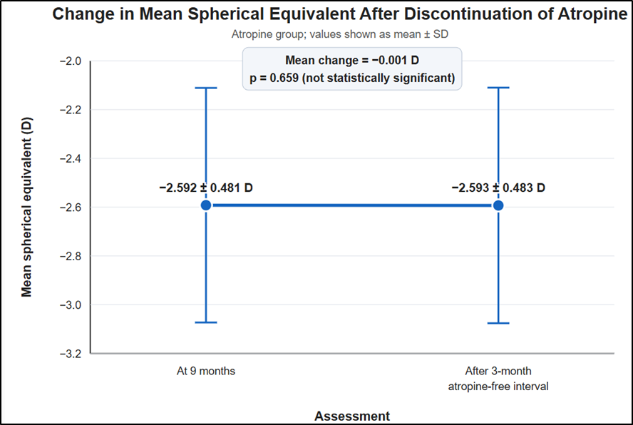
Figure 1. Baseline demographic characteristics of the study participants
Table 2. Comparison of mean spherical equivalent between the study groups during follow-up
|
Assessment time |
Atropine group, mean ± SD (D) |
Non-atropine group, mean ± SD (D) |
|
Baseline |
−2.336 ± 0.465 |
−1.775 ± 0.711 |
|
3 months |
−2.545 ± 0.488 |
−1.988 ± 0.777 |
|
6 months |
−2.580 ± 0.484 |
−2.288 ± 0.713 |
|
9 months |
−2.592 ± 0.481 |
−2.507 ± 0.772 |
Table 3. Change in mean spherical equivalent from baseline to nine months
|
Group |
Baseline MSE, mean ± SD (D) |
Nine-month MSE, mean ± SD (D) |
Mean progression ± SD (D) |
|
Atropine group |
−2.336 ± 0.465 |
−2.592 ± 0.481 |
−0.256 ± 0.050 |
|
Non-atropine group |
−1.775 ± 0.711 |
−2.507 ± 0.772 |
−0.732 ± 0.433 |
Table 4. Comparison of myopia progression between the atropine and non-atropine groups
|
Outcome |
Atropine group (n=50) |
Non-atropine group (n=50) |
Mean difference (D) |
p-value |
|
MSE progression from baseline to nine months, mean ± SD (D) |
−0.256 ± 0.050 |
−0.732 ± 0.433 |
0.476 |
<0.0001 |
|
Independent T test |
||||
Table 5. Change in mean spherical equivalent after discontinuation of atropine
|
Group |
MSE at 9 months, mean ± SD (D) |
MSE after 3-month atropine-free interval, mean ± SD (D) |
Mean change (D) |
p-value |
|
Atropine group |
−2.592 ± 0.481 |
−2.593 ± 0.483 |
−0.001 |
0.659 |
|
Paired Student’s t-test |
||||
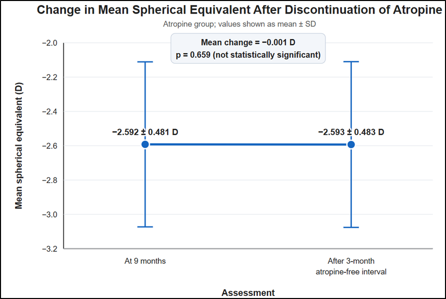
Figure 3. Change in mean spherical equivalent after discontinuation of atropine
DISCUSSION:
The present prospective interventional study evaluated the effectiveness and safety of topical 0.01% atropine in controlling myopia progression among children aged 6–12 years. Compared with the non-atropine group, children receiving low-dose atropine demonstrated significantly less progression of mean spherical equivalent (MSE) over nine months. No significant rebound progression was observed during the three-month atropine-free period, and treatment-related adverse effects were infrequent.
A total of 100 children were studied, with 50 participants in each group. The mean age was 9.18 ± 1.96 years in the atropine group and 9.64 ± 2.04 years in the non-atropine group. Both groups had a nearly equal distribution of males and females. Chia et al. conducted the ATOM2 study in a comparable paediatric age group and used similar criteria for evaluating myopia progression.[17] Sacchi et al. included a wider age range of 5–16 years but applied comparable eligibility criteria.[18] Thus, the demographic profile of the present study was broadly consistent with previous investigations of low-dose atropine.The baseline MSE was −2.336 ± 0.465 D in the atropine group and −1.775 ± 0.711 D in the non-atropine group. Therefore, children allocated to atropine had comparatively greater myopia at enrolment. This baseline difference should be considered when interpreting the final refractive values; consequently, the change in MSE from baseline provides a more appropriate measure of treatment effectiveness than the absolute MSE at follow-up.Sacchi et al. reported baseline MSE values of −3.00 ± 2.23 D in the atropine group and −2.63 ± 2.68 D in the control group.[18] Differences in baseline refractive error across studies may be attributable to variations in age distribution, inclusion criteria, ethnicity and clinical setting. The present investigation had a nine-month intervention period, whereas the ATOM2 study was a randomised, double-masked study with follow-up extending to five years, and the study by Sacchi et al. used a retrospective design with a 12-month follow-up period. [17,18]Over nine months, the mean change in MSE was −0.256 ± 0.050 D in the atropine group compared with −0.732 ± 0.433 D in the non-atropine group. This difference was statistically significant (p<0.0001), indicating that 0.01% atropine was effective in slowing myopia progression. The observed absolute reduction in progression was 0.476 D, corresponding to an approximately 65% reduction relative to the control group.Chia et al. reported an overall MSE progression of −1.38 ± 0.98 D in children treated with 0.01% atropine over five years.[17] Sacchi et al. observed a 12-month progression of −0.54 ± 0.61 D in the atropine group compared with −1.09 ± 0.64 D in the control group, with a statistically significant difference between the groups.[18] Although direct numerical comparison is limited by differences in study duration and design, these findings support the present observation that low-dose atropine reduces childhood myopia progression.After completing nine months of atropine therapy, the mean MSE changed from −2.592 ± 0.481 D to −2.593 ± 0.483 D during the subsequent three-month atropine-free interval. The mean change was only −0.001 D and was not statistically significant (p=0.659). Thus, no appreciable rebound progression was detected during the short period following atropine withdrawal.
In contrast, Chia et al. reported an MSE progression of −0.28 ± 0.33 D during a one-year washout period, indicating a significant rebound effect after treatment discontinuation.[17] This apparent difference may be related to the substantially shorter washout period in the present study. A three-month observation period may be inadequate to identify delayed rebound progression; therefore, longer post-treatment follow-up is required before concluding that rebound does not occur with 0.01% atropine.Low-dose atropine was generally well tolerated. At the three-month follow-up, photophobia was reported in 4% of children in the atropine group, corresponding to two participants. No persistent or additional adverse effects were documented during subsequent visits. Sacchi et al. reported photophobia in 9.6% of children treated with low-dose atropine.[18] Although the frequency observed in the present study was lower, differences in follow-up duration, assessment methods and reporting practices should be considered
CONCLUSION:
Topical 0.01% atropine significantly reduced myopia progression in children aged 6–12 years compared with no atropine treatment. It was well tolerated, with photophobia reported in only 4% of participants and no persistent adverse effects. No significant rebound progression was observed during the three-month atropine-free follow-up period.
Limitations
The study was single-centre, had a relatively small sample size and included only nine months of treatment with a three-month washout period. Axial length was not assessed, and the difference in baseline refractive error between the groups may have influenced the findings.
REFERENCES:
1. Leo S-W, Young TL. An evidence-based update on myopia and interventions to retard its progression. J AAPOS. 2011;15:181–9. doi:10.1016/j.jaapos.2010.09.020.
2. Walline JJ. Myopia Control: a review. Eye Contact Lens. 2016;42:3–8. doi:10.1097/ICL.0000000000000207.
3. Aller TA. Clinical management of progressive myopia. Eye (Lond). 2014;28:147–53. doi:10.1038/eye.2013.259.
4. Walline JJ, Lindsley K, Vedula SS, Cotter SA, Mutti DO, Twelker JD. Interventions to slow progression of myopia in children. Cochrane Database Syst Rev. 2011;12:CD004916. doi:10.1002/14651858.CD004916.pub3.
5. Saw SM, Shih-Yen EC, Koh A, Tan D. Interventions to retard myopia progression in children: an evidence-based update. Ophthalmology. 2002;109:415–21.
6. Shih KC, Chan TC-Y, Ng AL-K, Lai JS-M, Li WW-T, Cheng AC-K, et al. Use of atropine for prevention of childhood myopia progression in clinical practice. Eye Contact Lens. 2016;42:16–23. doi:10.1097/ICL.0000000000000189.
7. Si J-K, Tang K, Bi H-S, Guo D-D, Guo J-G, Wang X-R. Orthokeratology for myopia control: a meta-analysis. Optom Vis Sci. 2015;92:252–7.
8. Smith MJ, Walline JJ. Controlling myopia progression in children and adolescents. Adolesc Health Med Ther. 2015;6:133–40. doi:10.2147/AHMT.S55834.
9. Liu YM, Xie P. The safety of orthokeratology—a systematic review. Eye Contact Lens. 2016;42:35–42. doi:10.1097/ICL.0000000000000219.
10. Li SM, Kang MT, Wu SS, Meng B, Sun YY, Wei SF, et al. Studies using concentric ring bifocal and peripheral add multifocal contact lenses to slow myopia progression in school-aged children: a meta-analysis. Ophthalmic Physiol Opt. 2017;37:51–9. doi:10.1111/opo.12332.
11. Sherwin JC, Reacher MH, Keogh RH, Khawaja AP, Mackey DA, Foster PJ. The association between time spent outdoors and myopia in children and adolescents: a systematic review and meta-analysis. Ophthalmology. 2012;119:2141–51. doi:10.1016/j.ophtha.2012.04.020.
12. Iribarren R, Morgan I. Myopia - EyeWiki. 2017. http://eyewiki.aao.org/w/index.php?title=Myopia&printable=yes. Accessed 19 Mar 2017.
13. Wu P-C, Huang H-M, Yu H-J, Fang P-C, Chen C-T. Epidemiology of Myopia. Asia-Pacific J Ophthalmol (Philadelphia, Pa). 2016;5:386–93. doi:10.1097/APO.0000000000000236.
14. Foster PJ, Jiang Y. Epidemiology of myopia. Eye (Lond). 2014;28:202–8. doi:10.1038/eye.2013.280.
15. Fricke T, Holden B, Wilson D, Schlenther G, Naidoo K, Resnikoff S, et al. Global cost of correcting vision impairment from uncorrected refractive error. Bull World Health Organ. 2012;90:728–38. doi:10.2471/BLT.12.104034.
16. Zheng YF, Pan CW, Chay J, Wong TY, Finkelstein E, Saw SM. The economic cost of myopia in adults aged over 40 years in Singapore. Investig Ophthalmol Vis Sci. 2013;54:7532–7. doi:10.1167/iovs.13-12795.
17. Chia A, Lu Q-S, Tan D. Five-Year Clinical Trial on Atropine for the Treatment of Myopia 2: Myopia Control with Atropine 0.01% Eyedrops. Ophthalmology. 2016 Feb;123(2):391–9.
18. Sacchi M, Serafino M, Villani E, Tagliabue E, Luccarelli S, Bonsignore F, et al. Efficacy of atropine 0.01% for the treatment of childhood myopia in European patients. Acta Ophthalmol. 2019 Dec;97(8):e1136–40.